
Achelidelphys papuensis, Boxshall, Geoff A. & Marchenkov, Andrey, 2007
|
publication ID |
https://doi.org/ 10.5281/zenodo.176361 |
|
DOI |
https://doi.org/10.5281/zenodo.5661748 |
|
persistent identifier |
https://treatment.plazi.org/id/C03D8785-0440-FFA8-FF2D-FF49FDBAFC1C |
|
treatment provided by |
Plazi |
|
scientific name |
Achelidelphys papuensis |
| status |
sp. nov. |
Achelidelphys papuensis n. sp.
Type material: Holotype female, 6 paratype females. Registration nos MNHN-Cp2411 (holotype in alcohol), MNHN-Cp2412 (3 paratype females in alcohol), BMNH 2007.1 (1 paratype female in alcohol), BMNH 2007.2–3 (2 paratype females mounted on SEM stubs).
Type Locality: 02°39.49’S 150°25.56’E off Papua New Guinea, 18 m depth; 2 July 2003.
Host: Didemnum sp.
Locality in host: within the common tunic between the zooids (F. Monniot, pers. comm.).
Etymology: the species name refers to the type locality.
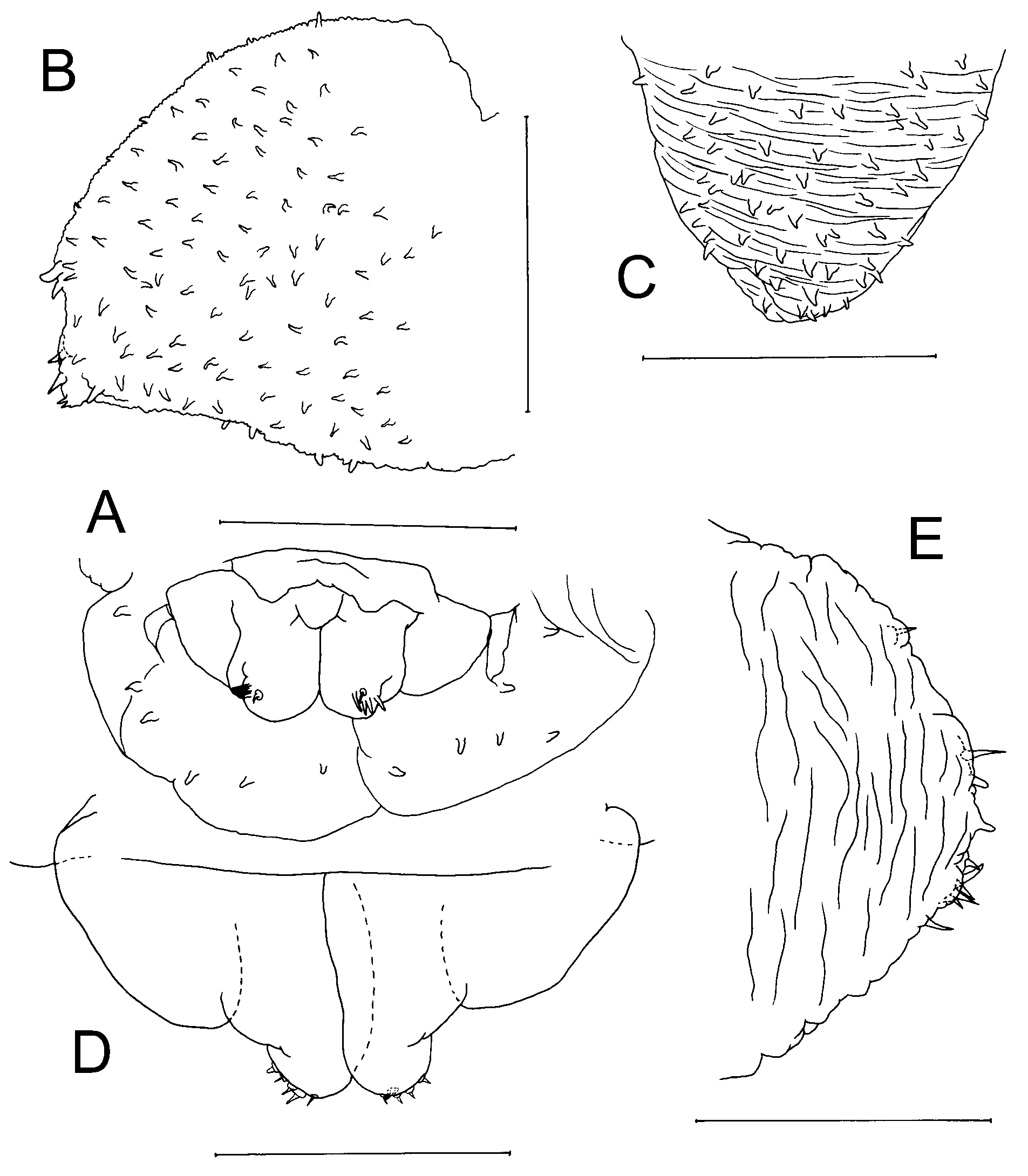
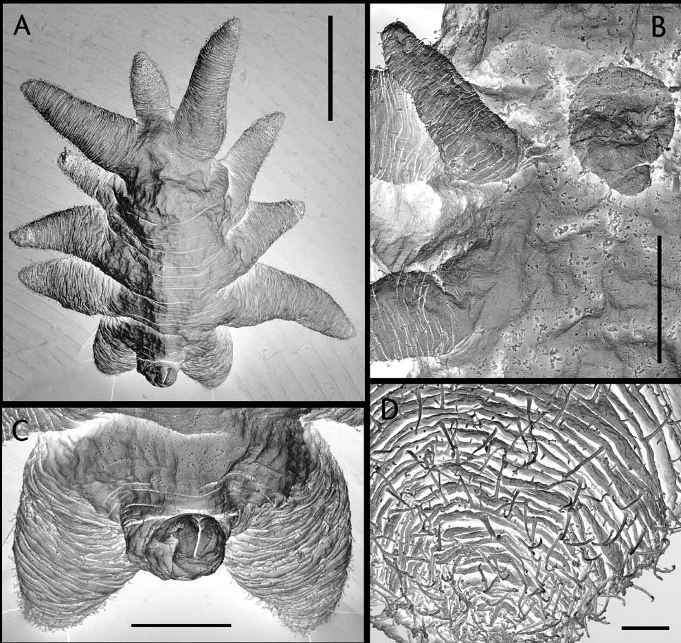
Description: Body highly transformed, stellate ( Figs 10 View FIGURE 10 , 11 View FIGURE 11 A); segmentation indistinct with segmental boundaries marked by superficial folds. Cephalosome with frontal margin merging into tapering anterolaterally-directed antennulary lobes. Rostrum simple, elongate, frontally-directed lobe, without accessory median lobe. Post-rostral median lobe absent. Labrum forming short, tapering, posteriorly-directed lobe ( Fig. 11 View FIGURE 11 B). Lateral margin of cephalosome not produced into ridge-like swellings. Antennomedial processes absent. Metasome truncated, with urosome hardly extending posterior to origin of leg 4. Legs 1 to 4 transformed, originating laterally, each occupying entire margin of somite and produced laterally, giving body stellate appearance. Mid-ventral metasomal processes absent. Urosome vestigial, located terminally and typically directed dorsally ( Fig. 11 View FIGURE 11 C); bearing partly incorporated caudal rami. Surface of body, rostrum, labrum, cephalosomic processes, and legs densely ornamented with surface setules: surface of rostrum, antennules and legs 1 to 4 densely folded, with deep folds orientated around circumference of process ( Fig. 11 View FIGURE 11 D).
Antennules forming bluntly-rounded lobe on either side of frontal margin of cephalosome, with vestigial apical setae ( Figs 10 View FIGURE 10 , 11 View FIGURE 11 A). Antenna to maxilliped entirely lacking. Legs 1 to 3 biramous; rami represented by unsegmented, tapering lobes; exopodal lobes laterally-directed, with broad base, carrying smaller endopodal lobes ventrally ( Figs 10 View FIGURE 10 , 11 View FIGURE 11 B). Leg 4 uniramous comprising short, posterolaterally-directed lobe representing exopod; lobe strongly tapering, conical in shape, ranging from 0.90–1.38 times longer than width at base. Exopodal lobes of legs 2–4 each housing internal expansion of multilobate uterus, containing eggs visible through body wall. Leg 5 absent.
Body length of female 1.36–1.93 mm, measured from base of rostrum to posterior extremity of urosome (based on 6 specimens). Male unknown.
Remarks: The new species lacks any median ventral processes on the metasome between the legs. The lack of such processes serves to distinguish it from A. steinitzi which has two processes, one located between each of the second and third leg pairs, and from A. drachi and A. nigra which have a single process located between the second legs. It differs from A. stellata and A. reducta , both of which have a strongly convex frontal margin to the head which merges laterally into the incorporated antennulary lobes. It differs from A. ampla in the shape of the uterus, which is compact and does not extend into the exopodal lobes of the legs in A. ampla but is multilobate and extends into the exopodal lobes in the new species.
Achelidelphys papuensis n. sp. most closely resembles A. chengae and these two species are also similar in body size, but they can be distinguished by the relative length of the paired lobes representing the fourth legs. In all specimens of the new species this lobe is short and conical, ranging from 0.9 to 1.4 times longer than the width at its base, whereas the lobes of A. chengae are elongate, about three times longer than the basal width. Although in these highly modified endoparasites overall shape appears to vary according to the degree of contraction or expansion of the limbs and rostrum, we believe that the difference in proportions of leg 4 exceeds the level that can be attributed to intraspecific variation. In addition, in their description of A. chengae, Lafargue and Laubier (1978b) state “L’ornementation cuticulaire est particulièrement réduite par rapport aux autres espèces du genre: elle se compose de minuscules spinules mesurant 1 à 2 microns de longeur.” In contrast, the surface setules of the new species are an order of magnitude longer, often exceeding 20 microns in length. Finally, Lafargue and Laubier (1978b) describe the tip of the antennule of A. chengae as bearing some fine setae accompanied by very short conical-cylindrical setae, the presence of which we have confirmed ( Fig. 9 View FIGURE 9 E), and they show these surrounded by very sparse surface setules. In the new species, the tip of the antennule lacks such short conical-cylindrical setae and the entire surface of the antennule is densely covered with long setules ( Fig. 11 View FIGURE 11 D).

No known copyright restrictions apply. See Agosti, D., Egloff, W., 2009. Taxonomic information exchange and copyright: the Plazi approach. BMC Research Notes 2009, 2:53 for further explanation.
|
Kingdom |
|
|
Phylum |
|
|
Class |
|
|
Order |
|
|
Family |
|
|
Genus |
Achelidelphys papuensis
| Boxshall, Geoff A. & Marchenkov, Andrey 2007 |
A. chengae
| Lafargue and Laubier 1978 |