
Achelidelphys stellata ( Lafargue and Laubier, 1977 ) Lafargue and Laubier, 1977
|
publication ID |
https://doi.org/ 10.5281/zenodo.176361 |
|
DOI |
https://doi.org/10.5281/zenodo.5661750 |
|
persistent identifier |
https://treatment.plazi.org/id/C03D8785-044E-FFA9-FF2D-FC04FAEFFF44 |
|
treatment provided by |
Plazi |
|
scientific name |
Achelidelphys stellata ( Lafargue and Laubier, 1977 ) |
| status |
comb. nov. |
Achelidelphys stellata ( Lafargue and Laubier, 1977) n. comb.
Syn: Cephalodelphys stellata Lafargue and Laubier, 1977
Material examined: Holotype female, reg. no. ZMA CO. 106.630.
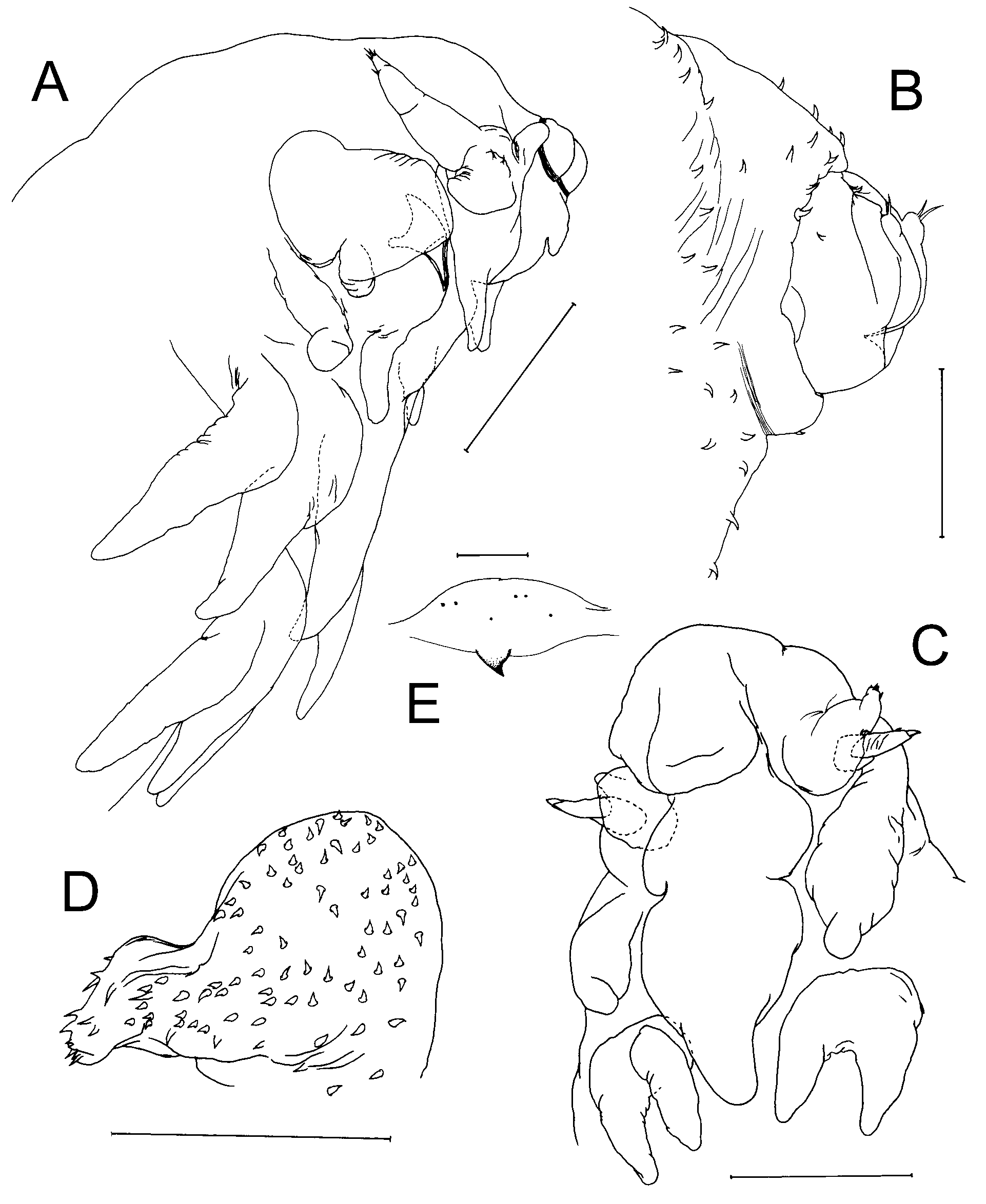
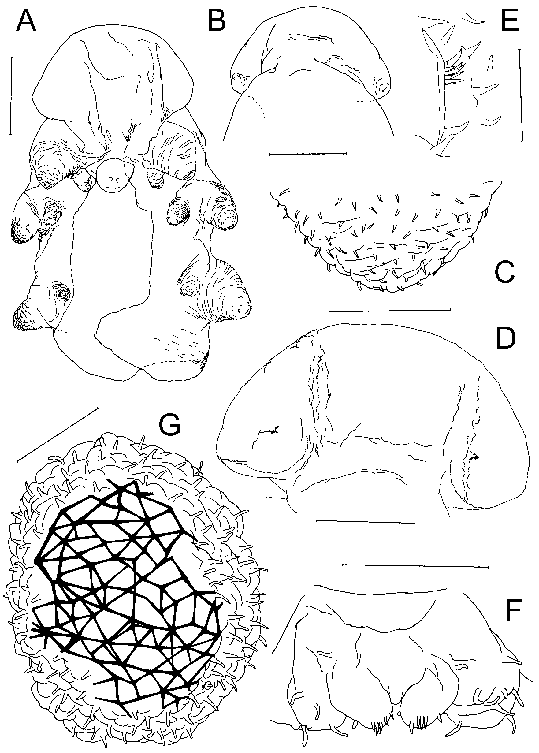
Differential Diagnosis: Body highly transformed, stellate ( Fig. 12 View FIGURE 12 A); segmentation indistinct with segmental boundaries marked by superficial folds. Cephalosome with broad, strongly-convex frontal margin merging laterally into large, ventrally-directed antennulary lobes ( Fig. 12 View FIGURE 12 B). Rostrum ventrally-directed; simple rounded lobe. Post-rostral median lobe present. Labrum forming elongate, posteriorly-directed lobe. Lateral margin of cephalosome not produced into ridge-like swellings. Antenno-medial swellings also absent. Metasome truncated, with urosome hardly extending posterior to origin of leg 4. Legs 1–4 transformed, originating laterally, each occupying entire margin of somite, produced laterally giving body a stellate appearance. Mid-ventral metasomal processes between legs lacking. Urosome reduced, located terminally; incorporating caudal rami. Surface of body, rostrum, labrum, cephalosomic processes, and legs densely ornamented with surface setules ( Fig. 12 View FIGURE 12 C).
Antennules forming large, rounded lobe on either side of convex frontal margin of cephalosome ( Fig. 12 View FIGURE 12 D). Oral region with 3 pairs of tiny lobes, arranged either side of labrum; positions of lobes suggesting possible homology with mouthparts (mandibles to maxillae?). Legs 1 to 3 biramous; rami represented by unsegmented, rounded lobes; exopodal lobe laterally-directed, with broad base, carrying smaller endopodal lobe ventrally. Leg 4 uniramous, comprising short, posterolaterally-directed lobe representing exopod. Exopodal lobes of legs 2–4 each housing internal expansion of uterus, containing eggs visible through body wall. Leg 5 absent.
Body length of female 0.87–2.30 mm. Male unknown.
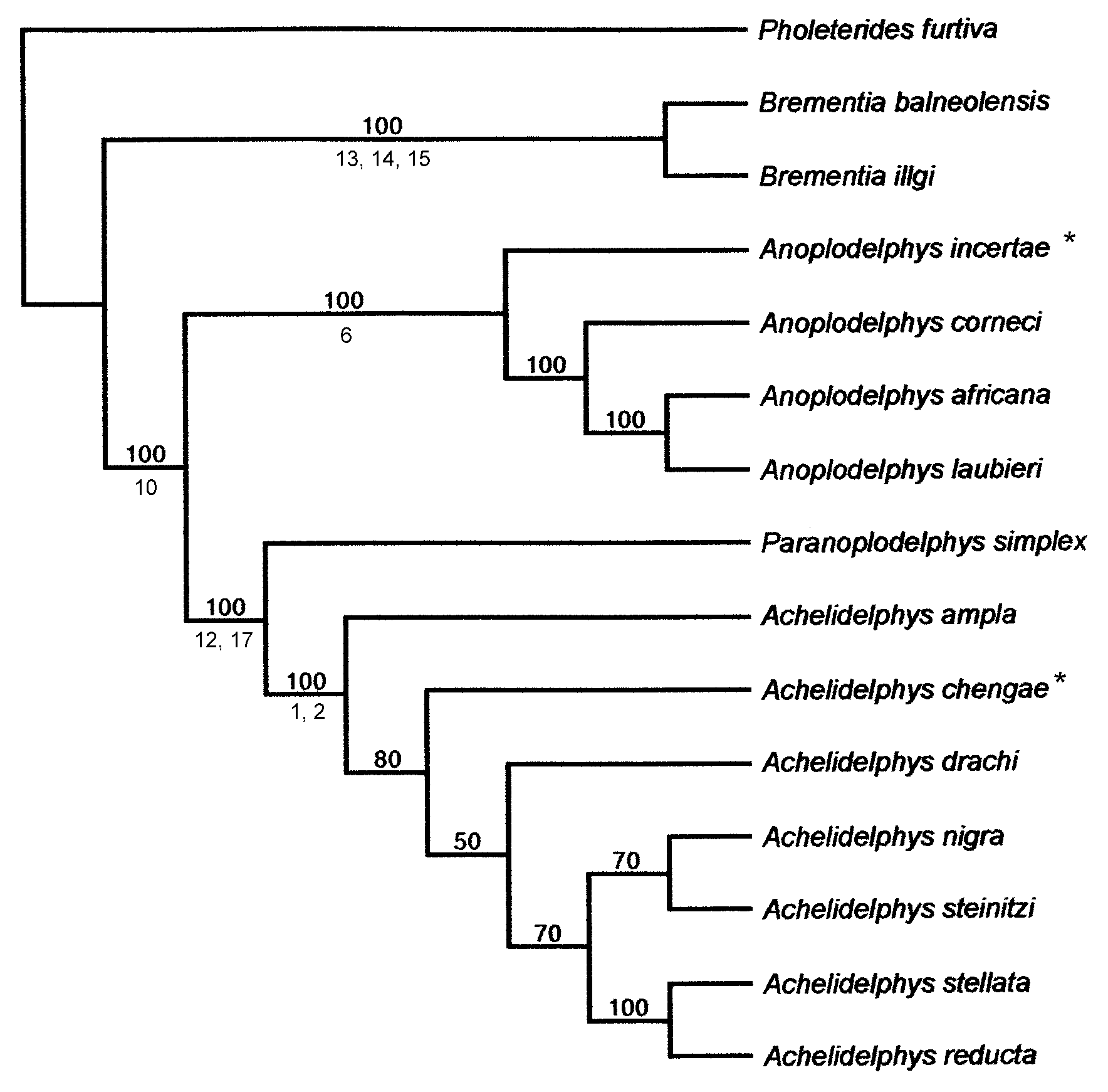
Remarks: This species has a strongly convex frontal margin to the cephalosome, but retains a rostrum. The phylogenetic analysis places this species within the Achelidelphys group of species ( Fig. 14 View FIGURE 14 ) and does not support the retention of Cephalodelphys as a separate genus. Lafargue and Laubier (1977: Fig. 4 View FIGURE 4 A) figured a small median ventral process (just posterior to the three pairs of oral lobes) concealed beneath the tip of the labrum but we were unable to confirm the presence of this structure in our re-examination of the holotype.
| ZMA |
Universiteit van Amsterdam, Zoologisch Museum |
No known copyright restrictions apply. See Agosti, D., Egloff, W., 2009. Taxonomic information exchange and copyright: the Plazi approach. BMC Research Notes 2009, 2:53 for further explanation.
|
Kingdom |
|
|
Phylum |
|
|
Class |
|
|
Order |
|
|
Family |
|
|
Genus |