
Notophlebia ganeshi Kluge, 2014
|
publication ID |
https://doi.org/ 10.11646/zootaxa.3760.4.2 |
|
publication LSID |
lsid:zoobank.org:pub:A180D4A6-78B7-4FFB-A291-728B2FAF3495 |
|
DOI |
https://doi.org/10.5281/zenodo.5040939 |
|
persistent identifier |
https://treatment.plazi.org/id/C0488791-BA78-AC3C-F5EA-F82670A7080C |
|
treatment provided by |
Felipe |
|
scientific name |
Notophlebia ganeshi Kluge |
| status |
sp. nov. |
Notophlebia ganeshi Kluge sp. n.
( Figs 1–38 View FIGURES 1–3 View FIGURES 4–10 View FIGURES 11–20 View FIGURES 23–25 View FIGURES 26–30 View FIGURES 31–45 )
Material. Holotype: L-S-I ♂ {specimen [XV] (20)}: INDIA, state Karnataka, Shivamogga district, tributaries of rivers Agumbe-hole and Yele-hole near Agumbe , 1.II.2013, coll. N. Kluge, L. Sheyko. Paratypes: the same locality, 11.I.–1.II.2013: 2 L-S-I ♂, 2 L-S-I ♀, 54 larvae. Part of larvae were collected in river Yele-hole above Onake Abbe Falls , about 4 km WNW of Agumbe. Most part of larvae were collected in a small mountain forest stream—tributary of river Agumbe-hole between village Malandur and the road Agumbe—Sringeri , about 4 km ESE of Agumbe; there is a confluence of two streams, at which Ganesha mini temple is situated (in honor of which this species is named). Imagos were reared in cages, and larvae from these two localities have been mixed .
Larva. CUTICULAR COLORATION: Ocher-brownish, without contrasting markings; head, labrum, lateral parts of mandibles, pronotum, mesonotum, thoracic pleurites and abdominal terga darker; thoracic and abdominal sterna lighter. Legs ocher-brownish; fore femur with diffusive, large, roundish lighter blank in proximal half; middle and hind femora with diffusive, longitudinal, arched blank parallel to outer margin.
HYPODERMAL COLORATION: Pronotum and mesonotum at most ocher, with lateral margins darker brownish. Femora either entirely ocher, or with darker brown macula at apex. Abdomen ocher. Both lamellae of each tergalius dark gray.
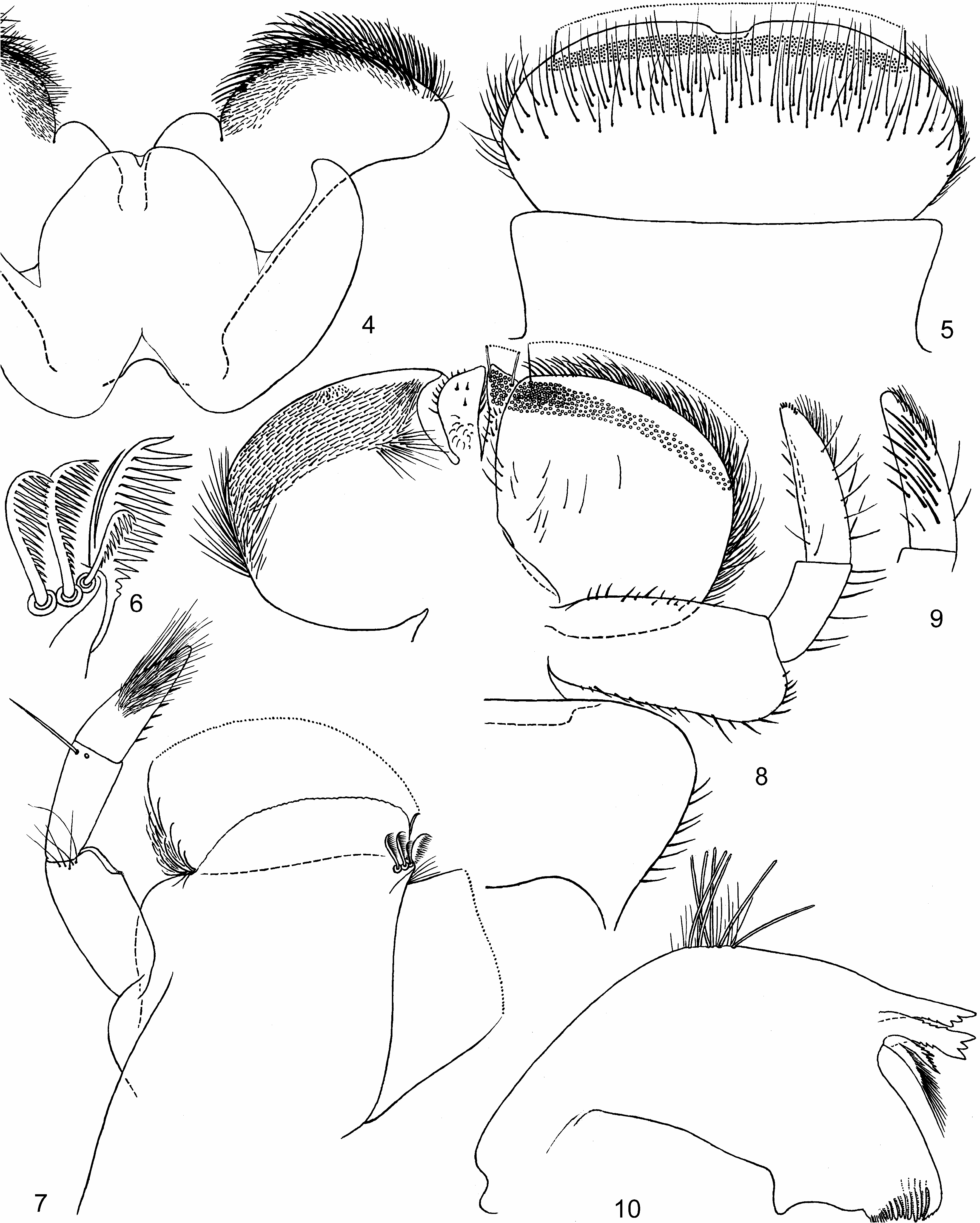
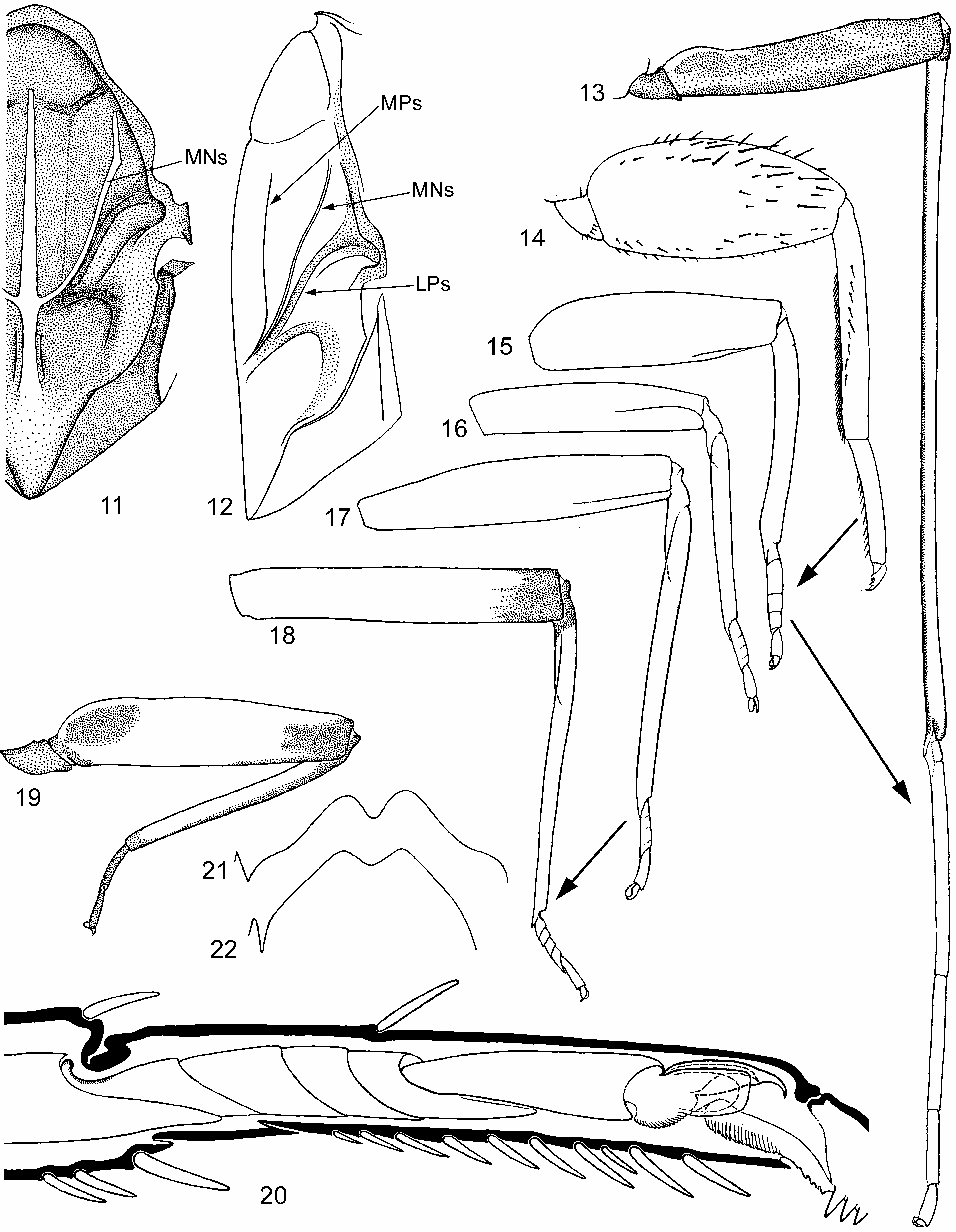
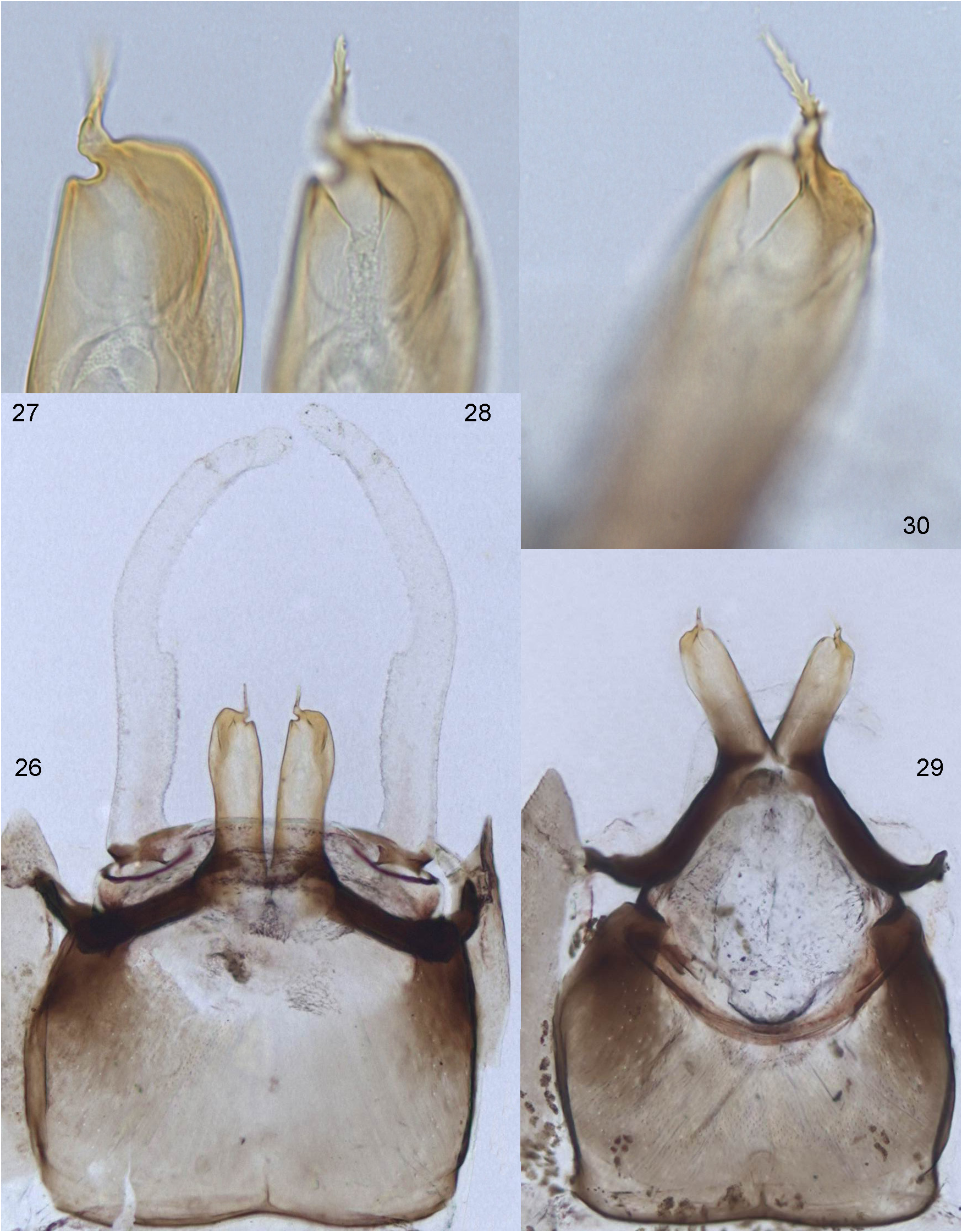
SHAPE AND SETATION: Labrum ( Fig. 5 View FIGURES 4–10 ) as wide as clypeus, widest in proximal half; fore margin convex; median incision small, with smooth shelf instead of denticles (unlike N. jobi , whose median incision and shelf are lost); distal transverse setal row dense and irregular, its setal bases are situated close one to another and form a stripe 5–6 setal bases width; instead of proximal transverse setal row, a wide field of irregularly situated setae [see Notophlebia (1)]. Mandibles ( Fig. 10 View FIGURES 4–10 ) with outer margin moderately convex, with a few setae not forming arched field (unlike N. jobi ). Hypopharynx and superlinguae not widened (unlike N. jobi ), inner lobes of superlinguae projected distad of hypopharynx ( Fig. 4 View FIGURES 4–10 ) [see Notophlebia (2)]. Maxilla ( Fig. 7 View FIGURES 4–10 ) without tusk, with well-developed apical flange (see Kluge 2012: Fig. 3 View FIGURES 1–3 ), dentiseta and subapical row of pectinate setae (unlike N. jobi ); number of subapical pectinate setae reduced to 3, with marginal one smaller than two others ( Fig. 6 View FIGURES 4–10 ) [see Iscini (1)]. 2 nd segment of maxillary palp with one long straight pointed stout seta near apex on ventro-lateral side, and with one placoid sensilla mediad of this seta. 3 rd (apical) segment of maxillary palp with numerous, moderately long, slender setae, situated densely and irregularly; their bases occupy apical 2/3 of ventral side ( Fig. 7 View FIGURES 4–10 ) and apical 1/3 of dorsal side of the 3 rd segment (in contrast to N. jobi , they do not form regular rows); inner margin with longitudinal row of 3–6 shorter spine-like setae. Labium ( Fig. 8 View FIGURES 4–10 ) with glossae small and not projected dorsad or ventrad of paraglossae; paraglossae moderately widened, roundish (unlike greatly widened in N. jobi ). Labial palp non-specialized; 2 nd segment not shortened; 3 rd segment less than twice longer than 2 nd segment; filtering setae on dorsal side of 3 rd segment moderately long and directed apically-inward ( Fig. 9 View FIGURES 4–10 ) (unlike very long and directed apically-outward in N. jobi ); besides these setae, 1 st, 2 nd and 3 rd segments bear a longitudinal row of sparse setae on outer margins.
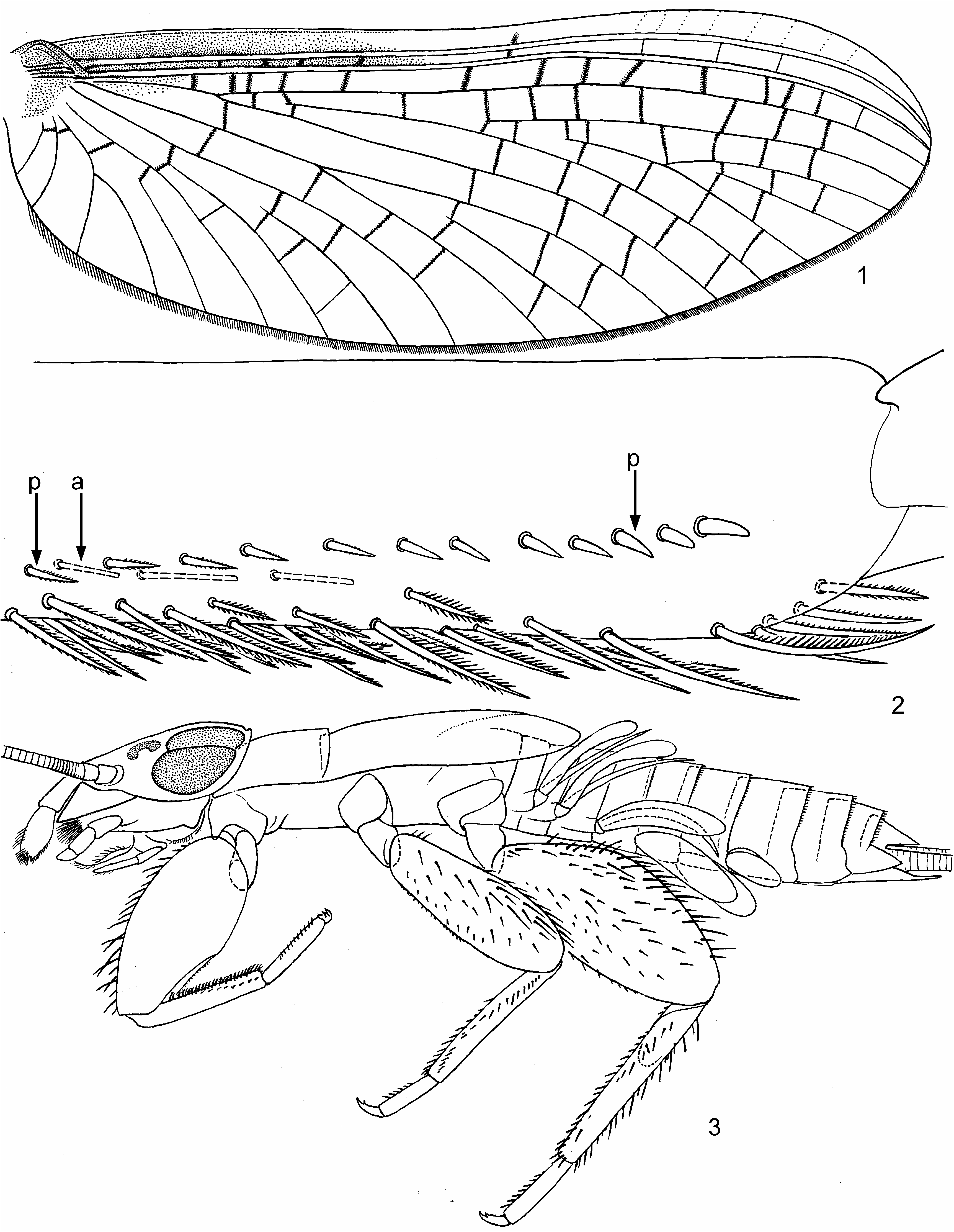
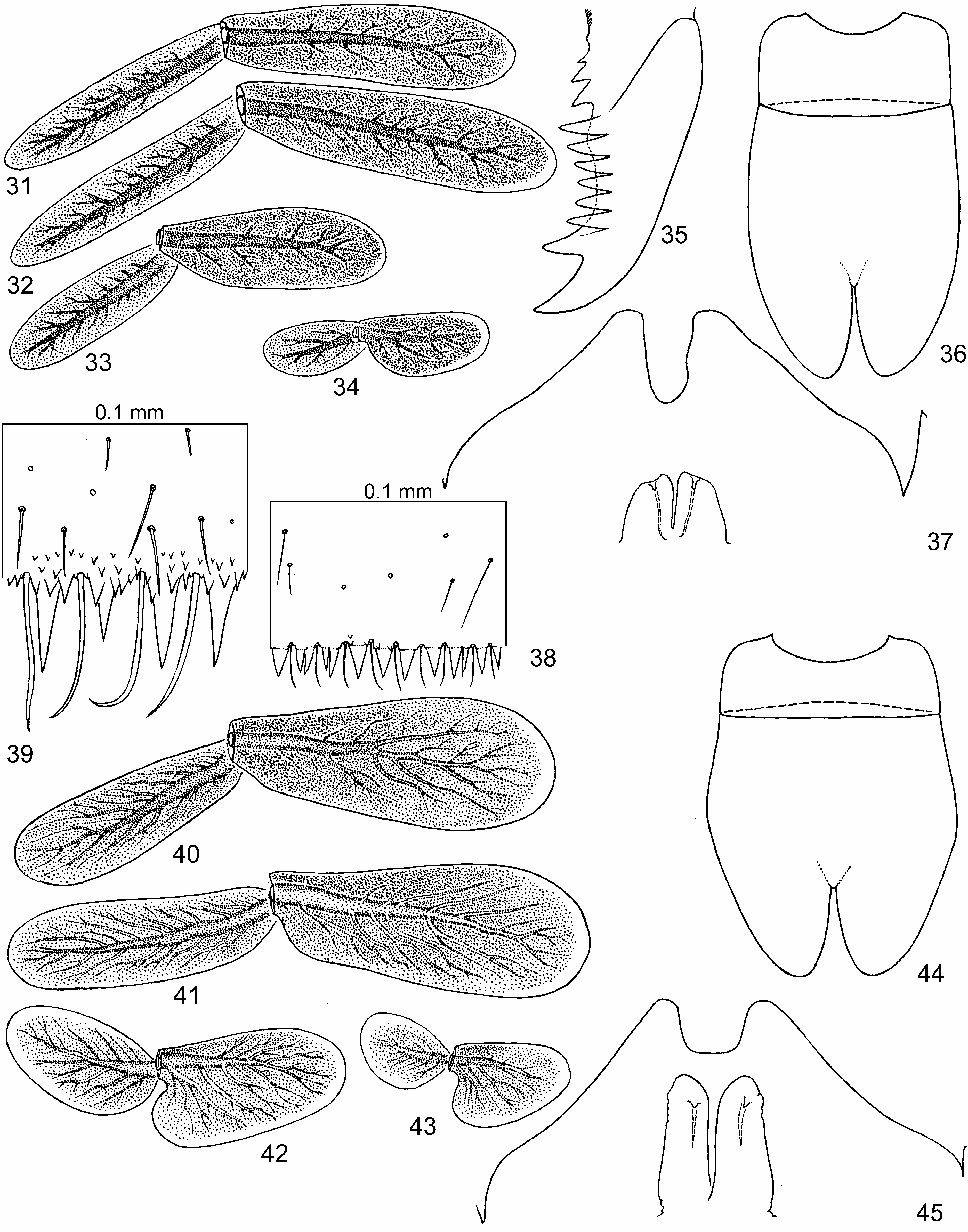
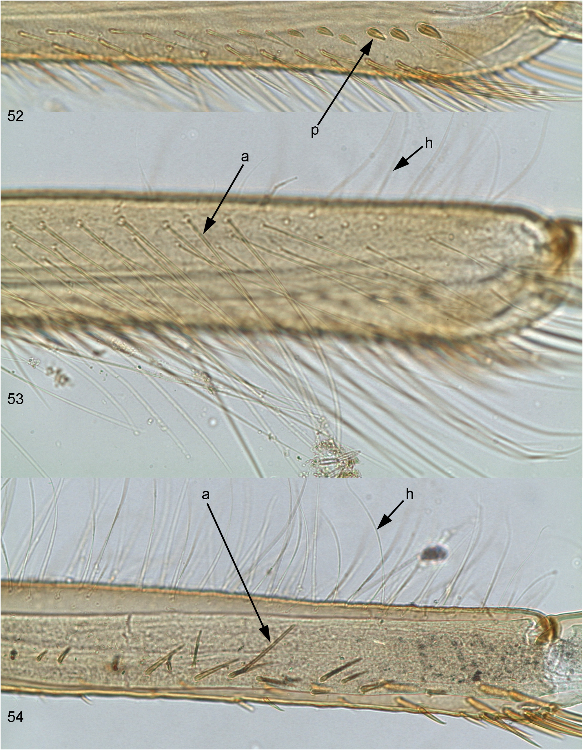
Thorax ( Fig. 36 View FIGURES 31–45 ) much narrower than in N. jobi ; fore protoptera brought together, in intact position their tornoapical margins contiguous up to tips. Hind legs much larger than fore and middle legs; fore and hind femora much thicker than middle femur ( Fig. 3 View FIGURES 1–3 ). Femora wide, narrowed toward apex; fore femur wide, widest in middle part ( Fig. 14 View FIGURES 11–20 ); middle femur less wide, widest in distal half; hind femur wide, widest near middle—in younger larva proximad of middle ( Fig. 3 View FIGURES 1–3 ), in mature larva distad of middle. All femora with irregularly arranged stout long blunt setae mainly on outer and dorsal sides, and with long slender hair-like setae on outer side (not shown on Figs 3 View FIGURES 1–3 and 14 View FIGURES 11–20 ). Patella-tibial suture absent on fore and middle legs, present on hind leg ( Fig. 3 View FIGURES 1–3 ) [see Iscini (3)]. Anterior (dorsal) side of fore, middle and hind tibia with a longitudinal row of stout elongate blunt setae ( Fig. 2a View FIGURES 1–3 ; 14 View FIGURES 11–20 ) [see Notophlebia (3)]; fore tibia with dense pointed bipectinate stout setae on inner side and very stout setae in distal part of posterior (ventral) side ( Fig. 2p View FIGURES 1–3 ) [see Iscini (2)]; middle tibia with spine-like setae on inner side; hind tibia with spine-like setae on inner side and stout elongate blunt setae on outer side ( Fig. 3 View FIGURES 1–3 ); middle and hind tibiae with numerous long slender hair-like setae on outer side (as in Fig. 54 View FIGURES 52–54 ), fore tibia with a few such setae (not shown in Fig. 3 View FIGURES 1–3 ). Tarsus of each leg with row of spine-like setae on inner side ( Fig. 20 View FIGURES 11–20 ). Claw with 2–4 denticles on proximal portion, 5–8 denticles in anterior row and 1 larger denticle apically-posteriorly ( Fig. 35 View FIGURES 31–45 ).
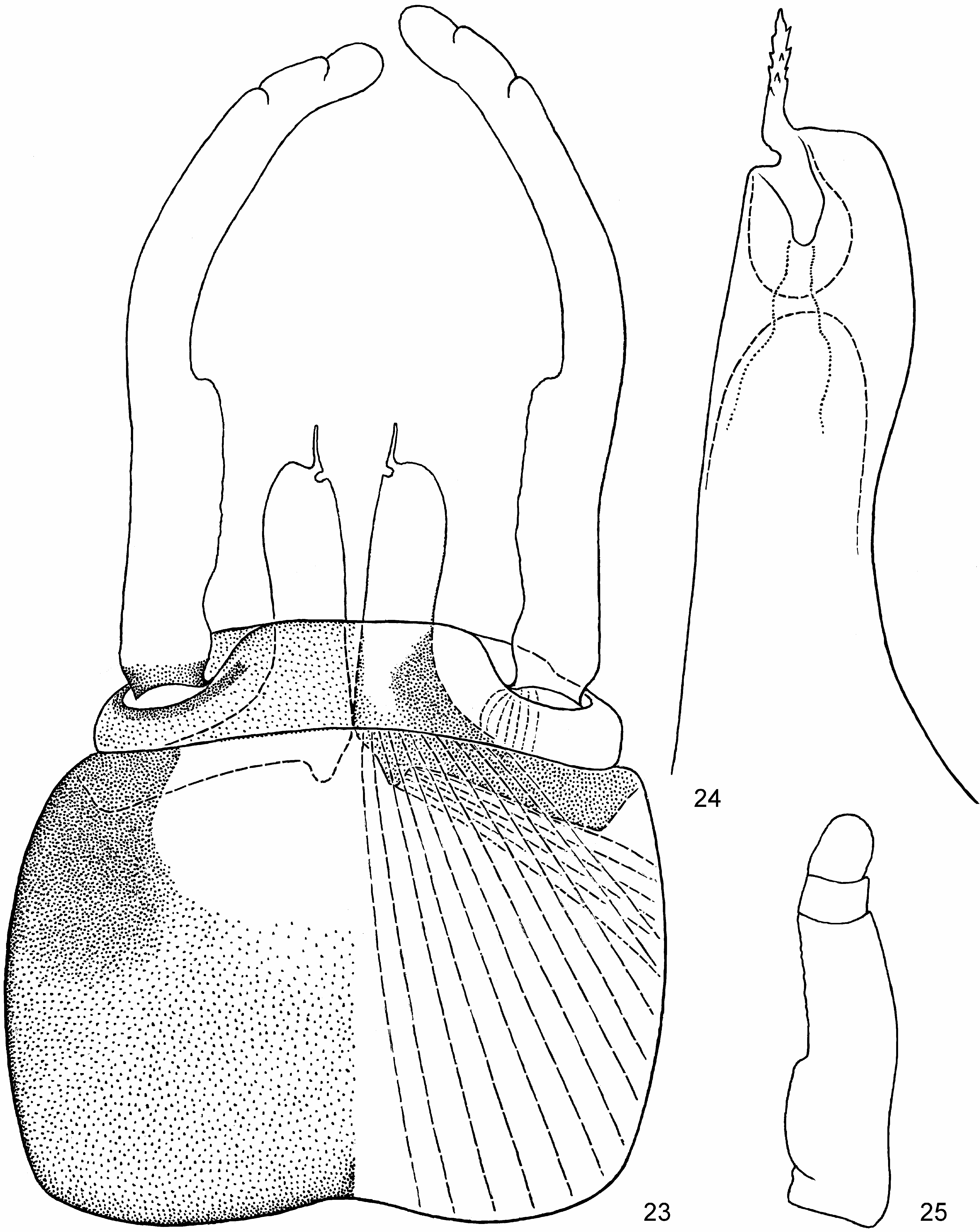
Abdomen has characteristic form [see Iscini (5)], lacks posterolateral projections on terga I– VI, have short blunt projections on terga VII and VIII and long blunt projections on tergum IX [see Notophlebia (4)] ( Fig. 3 View FIGURES 1–3 ). Posterior margins of abdominal terga with a regular row of pointed denticles alternated with setae ( Fig. 38 View FIGURES 31–45 ); denticles become longer from tergum I to tergum IX. Posterior margins of abdominal sterna without denticles. All tergalii I– VI bilamellate, with both lamellae blunt; tergalius I a little shorter than tergalius II; tergalii from II to V gradually shorter; tergalius VI much smaller [see Notophlebia (5)] ( Figs 31–34 View FIGURES 31–45 ); all tergalii narrower than in N. jobi . Protopenis of mature male larva much shorter than subimaginal penis, with gonopore located dorsally and lined with cuticle ( Fig. 37 View FIGURES 31–45 ); before molt to subimago, most part of subimaginal penis projects proximad of cuticular larval protopenis. Female larva with small shallow incision on posterior margin of sternum IX (Fig. 21). Cerci longer than body, paracercus 1.5 times longer than cerci.
Subimago. CUTICULAR COLORATION: Cuticle nearly entirely colorless, thoracic sutures and legs partly diffusively washed with light brownish. Mesonotum without pigmented areas present in most Leptophlebiidae ; lateral portion of mesonotal suture does not serve as boundary of pigmented area, but has a form of longitudinal suture running between medioparapsidal and lateroparapsidal sutures ( Fig. 12 View FIGURES 11–20 ).
SHAPE AND TEXTURE: Fore femur widened proximally, middle and hind femora narrower ( Figs 15–17 View FIGURES 11–20 ). On fore leg of male tibia and tarsus as short as on other legs, tarsus with swollen 1 st –4 th segments ( Fig. 15 View FIGURES 11–20 ). Tarsi of all legs of male and female covered with microtrichiae, like other parts of legs and body. Caudalii 5–6 times shorter than body, cerci shorter than paracercus.
Imago, male. Head ocher with brown. Upper eyes not elevated, brownish-orange. Thorax with light brown and ocher maculation; mesonotum with brown pigmented areas and ocher stripes between them [see Iscini (7)] ( Fig. 11 View FIGURES 11–20 ). On fore wing ( Fig. 1 View FIGURES 1–3 ) proximal part of subcostal field and partly proximal part of costal fields brown, remainder membrane colorless; longitudinal veins ocher; cross veins in anterior-proximal part of wing bordered with brown, in posterior and distal parts of wing colorless. Oblique cross veins of pterostigma either simple, or branched, or incomplete; either bordered by brown, or colorless, or invisible. Fore leg: femur brown with ocher; tibia white with brownish line on inner side, 2.5 times longer than femur; tarsus white, nearly 2 times longer than femur ( Fig. 13 View FIGURES 11–20 ). Middle and hind legs: femur white with brown apex; tibia white with brown base; tarsus white ( Fig. 18 View FIGURES 11–20 ). Middle and hind legs with patella-tibial suture well-developed; tarsus secondarily 5-segmented [see Iscini (10)] ( Fig. 20 View FIGURES 11–20 ). Abdominal terga I–VII white, with blackish dots on lateral tracheal trunks; pale gray median maculae can be present on terga III–VII or on most posterior of them. Terga VIII–X dark brown with anterior margin light. Sterna I–VIII white. Sternum IX brown, with darker posterior-lateral areas and light posterior-median area ( Figs 23 View FIGURES 23–25 , 26, 29 View FIGURES 26–30 ). Styliger brown, gonostyli white with brown bases. Penial arms dark brown, external penis lobes ocher. Styliger short. Each penis lobe with straight pointed serrate apical projection [see Notophlebia (7)]; laterad of it, penis lobe forms convexity; mediad of it, penis lobe has a small sharp incision; gonopore locates on dorsal side of penis lobe, just dorsad of the serrate apical projection and the incision ( Figs 32 View FIGURES 31–45 , 24 View FIGURES 23–25 , 26–29 View FIGURES 26–30 ). Caudalii white, without markings; cerci less than ½ of body length.
Imago, female. Thorax as in male; mesonotum with the same brown pigmented areas separated by ocher stripes (as in Fig. 11 View FIGURES 11–20 ). Wings as in male. Fore femur widened proximally, middle and hind femora narrower (as in subimago). Fore femur light brownish, with darker brown maculae near base and at apex ( Fig. 19 View FIGURES 11–20 ). Middle and hind femora with larger longitudinal brown maculae. Tibiae of all legs with light inner side and brown outer side ( Fig. 19 View FIGURES 11–20 ). Tarsi of all legs brownish. Abdominal terga I–X uniformly brown; sides lighter, with blackish dots on lateral tracheae. Abdominal sterna I–VIII lighter brownish, sternum IX brown. Posterior margin of sternum IX with small shallow incision (Fig. 22).
Egg (extracted from mature female larva). Oval, 0.13 mm length; surface with irregular rugosity.
Molts and transformations. Molt from subimago to imago occurs in both sexes (unlike Isca , whose female subimago does not molt). All 5 individuals reared in cages (3 males and 2 females) molted from larva to subimago late at night (later than other mayflies, which do it at beginning of darkness) and molted from subimago to imago at the same night (earlier than most other mayflies in the same conditions), so that in morning I found only larval and subimaginal exuviae and dead imagos; female imagos had no eggs. I was unable to see subimagos alive and don't know duration of subimaginal stage, but can state that it is much shorter than in most mayflies. Unlike shed subimaginal skin of other mayflies (whose cuticle of wings is crumpled and can't be spread), on shed subimaginal skin of Notophlebia ganeshi cuticle of each wing has a form of soft sack and can be easily spread. Subimaginal legs retain size and proportions of larval legs, and male fore legs undergo great changes in course of molt from subimago to imago: tarsus of subimaginal fore leg has peculiar swollen shape ( Fig. 15 View FIGURES 11–20 ); after molt to imago, tibia and tarsus become several times longer ( Fig. 13 View FIGURES 11–20 ) [see Iscini (9)]. Subimaginal caudalii are 3–4 times shorter than larval caudalii; when larva molts to subimago, each subimaginal caudalius develops from proximal 1/7–1/5 of larval caudalius, while in most part of larval caudalius tissues degenerate; when subimago molts to imago, caudalius elongates 2–3 times thanks to its crumpling condition under subimaginal cuticle [see Iscini (13)].
Dimensions. Fore wing length (and approximated body length) 6–7 mm.
| VI |
Mykotektet, National Veterinary Institute |
| V |
Royal British Columbia Museum - Herbarium |
No known copyright restrictions apply. See Agosti, D., Egloff, W., 2009. Taxonomic information exchange and copyright: the Plazi approach. BMC Research Notes 2009, 2:53 for further explanation.