
Otiothops kathiae, Piacentini, Luis N., Ávila Calero, Sergio L., Pérez, Marianela E. & Grismado, Cristian J., 2013
|
publication ID |
https://doi.org/ 10.11646/zootaxa.3619.4.6 |
|
publication LSID |
lsid:zoobank.org:pub:11CC1FA2-A09A-4ED9-89E3-3706ABE2A091 |
|
DOI |
https://doi.org/10.5281/zenodo.6150244 |
|
persistent identifier |
https://treatment.plazi.org/id/C05E87AD-FFA8-8A55-FF6F-DC4345C3FD30 |
|
treatment provided by |
Plazi |
|
scientific name |
Otiothops kathiae |
| status |
sp. nov. |
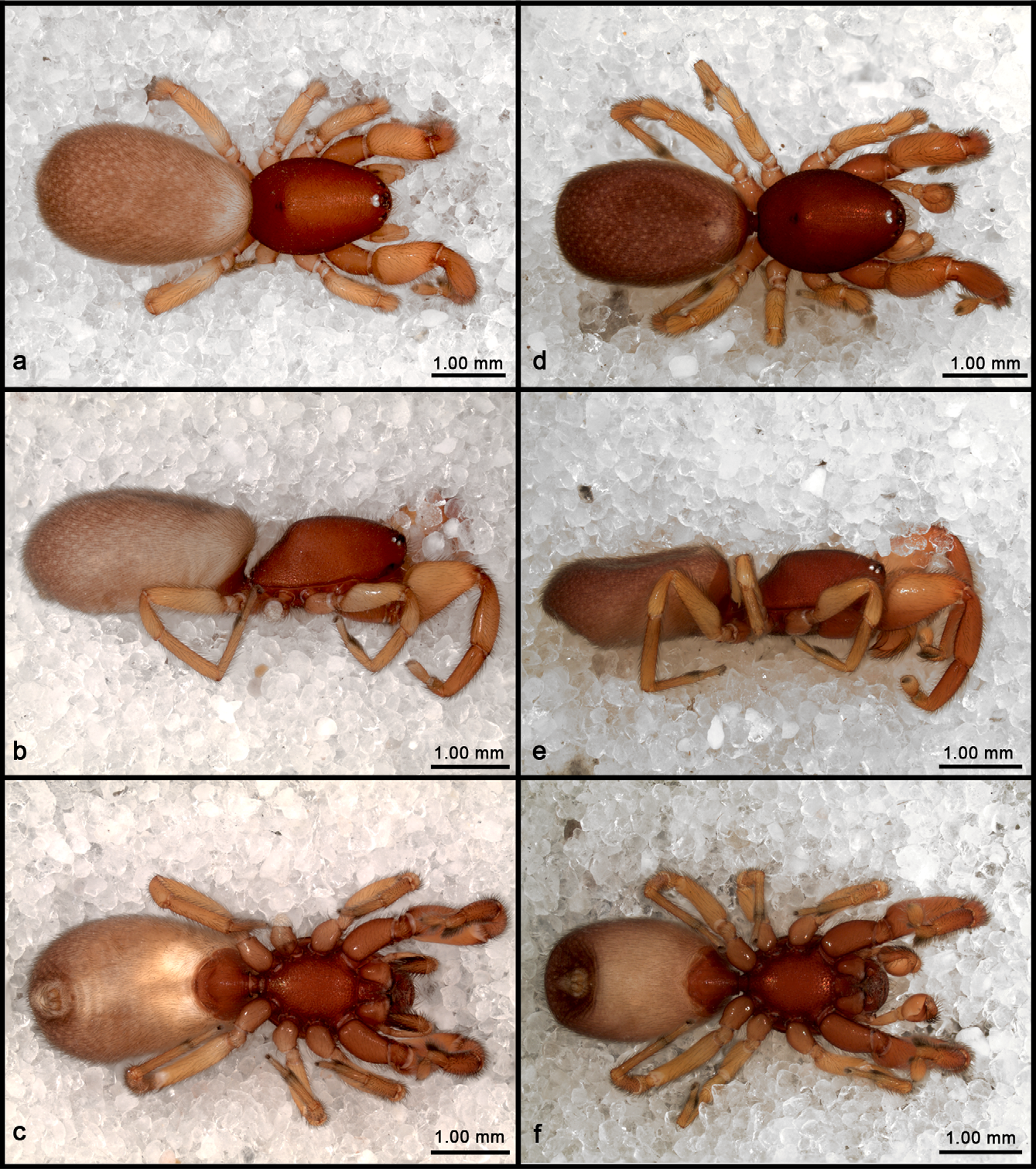
Otiothops kathiae View in CoL n. sp.
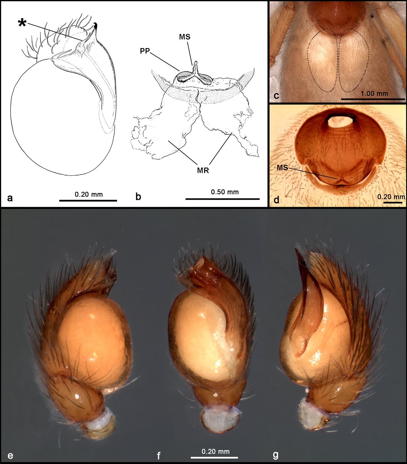
( Figs 1–2 View FIGURE 1 View FIGURE 2 )
Type material. BOLIVIA: Santa Cruz: holotype male, Yabaré (Estación de la Univ. A. Gabriel R. Moreno), N. of Tres Cruces, Prov. Chiquitos, 16°26'30"S, 62°10'21"W, 20-24 October 2010, transition between Chaco and Chiquitano forests, sifting litter, elev. 260 m, C. Grismado, S. Ávila Calero, M. Pérez (CBF). Paratypes: 1 female, same data as holotype (MACN-Ar 28905).
Other material examined. BOLIVIA: Santa Cruz: 1 juvenile, same data as holotype (MACN-Ar 28904).
Etymology. The specific name is a patronym in honour of Kathia Rivero, researcher of the Museo de Historia Natural Noel Kempff Mercado (Santa Cruz de la Sierra) in recognition of her fundamental contribution to the organization, logistics and success of the expedition to Bolivia in 2010.
Diagnosis. The male is very similar to that of O. naokii n. sp. in the shape of the embolus, which is distally flattened and widened, with a flattened projection on the anteromedian margin, but differs by having the embolus more closely adpressed against the tegulum (see ventral view, Fig. 2 View FIGURE 2 f), and by the different shape of the distal elements of the embolus ( Figs 2 View FIGURE 2 a, 2e–g). The female resembles those O. payak Grismado & Ramírez, 2002 and O. hoeferi Brescovit & Bonaldo, 1993 in having large, paired, posteriorly directed receptacula ( Figs 2 View FIGURE 2 b–c; see Grismado and Ramírez 2002, fig. 4; Brescovit and Bonaldo 1993, fig. 10), but differs by having smaller, almost contiguous anterior poreplates, and by having a median sclerite on the anterior part of the atrium (MS in Figs 2 View FIGURE 2 b, 2d).
Description. Male (holotype). Total length 4.16. Carapace 1.74 long, 1.24 wide. Femur I 1.14 long, 0.60 high. Posterior median eyes touching. Paturon with inconspicuous lateral ridge. Sclerotized portions of cephalothorax orange brown ( Figs 1 View FIGURE 1 d–f); abdominal scutum orange brown, darker at booklungs, unsclerotized portion of abdomen purplish brown with numerous small, yellowish dots on dorsum ( Figs 1 View FIGURE 1 d–f); venter uniformly yellowish ( Fig. 1 View FIGURE 1 f). Pedipalpal femur not thickened, tibia smaller than bulb, nearly cup-shaped; bulb globose, embolus distally flattened, widened, with pointed, flattened, retrolaterally directed lobe near sperm outlet area ( Fig. 2 View FIGURE 2 a), distal retrolateral extension with curved tip, distal prolateral extension with a thin lamella running parallel to embolar margin ( Figs 2 View FIGURE 2 a, 2e–g).
Female (paratype). Total length 2.46. Carapace 1.98 long, 1.32 wide. Femur I 1.20 long, 0.64 high. Eyes and chelicerae as in male. Cephalothorax slightly lighter than in male ( Figs 1 View FIGURE 1 a–c). Abdominal scutum small, entire, less sclerotized than male, curved, with entire sclerite present behind epigastric furrow ( Fig. 2 View FIGURE 2 c). Abdominal pattern as in male, but on light brown background; anterodorsal area uniformly yellowish, without spots ( Figs 1 View FIGURE 1 a–c). Female internal genitalia ( Fig. 2 View FIGURE 2 b) composed of large, paired, globose, posterodorsally directed median receptacula, with no visible posterior extension of bursa; each receptaculum with small sclerotized poreplates situated anteriorly, nearly contiguous to each other. The median receptacula were nearly oval in situ, slightly pointed posteriorly (approximately as reconstructed in Fig. 2 View FIGURE 2 c), but collapsed during KOH digestion ( Fig. 2 View FIGURE 2 b). Anterior wall of atrium with a median sclerite (MS) bearing anterior pointed projection and two posterolateral arms.
Distribution. Known only from the type locality.
No known copyright restrictions apply. See Agosti, D., Egloff, W., 2009. Taxonomic information exchange and copyright: the Plazi approach. BMC Research Notes 2009, 2:53 for further explanation.