
Chaetonotus (Chaetonotus) polyspinosus Greuter, 1917
|
publication ID |
https://doi.org/ 10.11646/zootaxa.3701.5.3 |
|
publication LSID |
lsid:zoobank.org:pub:472882BF-6499-47D3-A242-A8D218BE2DFD |
|
DOI |
https://doi.org/10.5281/zenodo.5632015 |
|
persistent identifier |
https://treatment.plazi.org/id/C1146C7C-4C31-FF8E-02CD-C6311F5FFD5F |
|
treatment provided by |
Plazi |
|
scientific name |
Chaetonotus (Chaetonotus) polyspinosus Greuter, 1917 |
| status |
|
Chaetonotus (Chaetonotus) polyspinosus Greuter, 1917 View in CoL
( Fig. 9 View FIGURE 9 )
Synonyms: Chaetonotus annulatus Martin, 1981 [syn: Kisielewski, 1997]; Chaetonotus brevisetosus Roszczak, 1936 [syn: Kisielewski, 1981].
Localities: Lake Ånnsjön, Jämtland (N 63º 15’ 65’’; E 12º 27’ 03’’), July 8, 2008; Storlien E, Jämtland (N 63º 18’ 42’’; E 12º 06’ 43’’), July 10, 2008.
Material: 2 specimens.
TL, 183–197 µm; FL, 25–27 µm; AL, 13–15 µm; PhL, 57–60 µm; MD, 10 µm; TNC, 37–40; DC, 25–28; VLC, 12; HS, 1 x 1 µm; NS, 1– 2 x 1–2 µm; DS, 2 x 2 µm; HSp, 1 µm; NSp, 1 µm; DSp, 1–3 µm; VC, 9; VTS, 10 x NA µm.
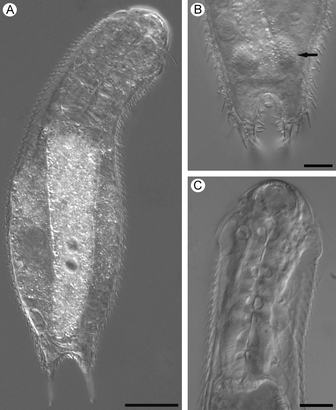
Head clearly five-lobed with two pairs of cephalic sensory ciliary tufts. Anterior pair of sensory tufts shorter than posterior pair. Cephalion and pleurae well developed. Hypostomium with two teeth. Anterior and posterior dorsal sensory cilia present, posterior pair emerging from small sub-triangular double keeled scales. Furca straight with adhesive tubes constituting half to 3/5 of the total furca length. Dorsal surface covered by very small threelobed scales with short straight simple spines. Scales and spines increase only slightly in size towards the posterior end. Five to six longer simple spines, 8–12 µm in length, at the dorsal base of the furcal appendages.
Ventrolateral scales and spines similar to those of the dorsal surface. Ventral interciliary area covered by rounded to oval smooth scales; towards the posterior end they become keeled and rounded rectangular in shape. Ventral ciliation in two longitudinal bands that can merge on the head.
Mouth subterminal. Pharynx muscular, widens towards the posterior end. PhIJ at U31–34. Intestine straight with anus at approximately U87.
The Swedish specimens fall well within the morphometric ranges previously reported in the literature. However, according to Schwank (1990) the ventral ciliation does not merge on the head, but Swedish specimens show a clear transverse row of cilia, that connects the two longitudinal bands behind the hypostomium.
Previously reported from Denmark (Grilli et al. 2010), France (d’Hondt 1967), Great Britain (Martin 1981), Italy (Balsamo 1983), Poland (Kisielewski 1981), Romania (Rudescu 1967), Russia (Preobrajenskaja 1926), Sweden (Kånneby et al. 2009), Switzerland (Greuter 1917), Brazil (Kisielewski 1991), Canada (Schwank 1990), Israel (Kisielewski 1999) and Korea (Lee & Chang 2000).
No known copyright restrictions apply. See Agosti, D., Egloff, W., 2009. Taxonomic information exchange and copyright: the Plazi approach. BMC Research Notes 2009, 2:53 for further explanation.