
Taeniogonalos Schulz, 1906
|
publication ID |
https://doi.org/10.11646/zootaxa.5361.4.6 |
|
publication LSID |
lsid:zoobank.org:pub:B9FF2182-22E2-4EAC-8872-32E15BC54E7B |
|
DOI |
https://doi.org/10.5281/zenodo.10167929 |
|
persistent identifier |
https://treatment.plazi.org/id/C12087E6-FFD6-DA48-E6CA-FDFF6193B13F |
|
treatment provided by |
Plazi |
|
scientific name |
Taeniogonalos Schulz |
| status |
|
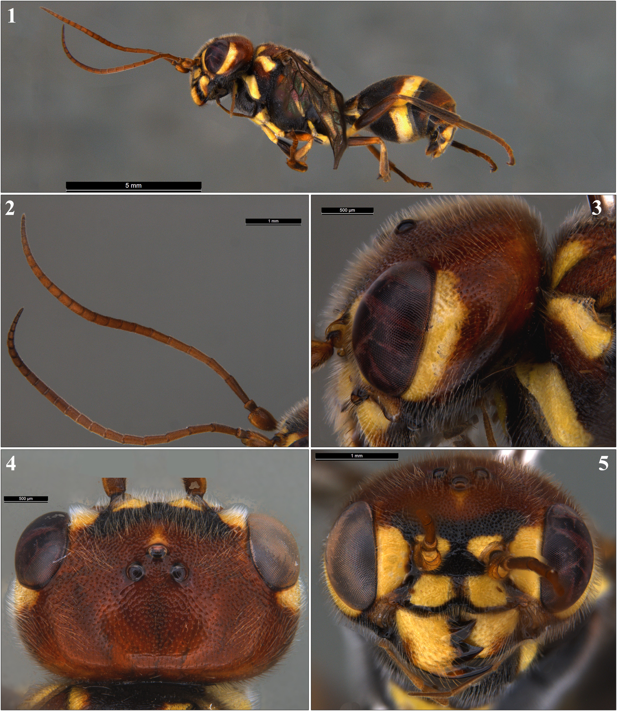
Key to Indian species of Taeniogonalos Schulz (based on females; modified from Binoy et al. 2022)
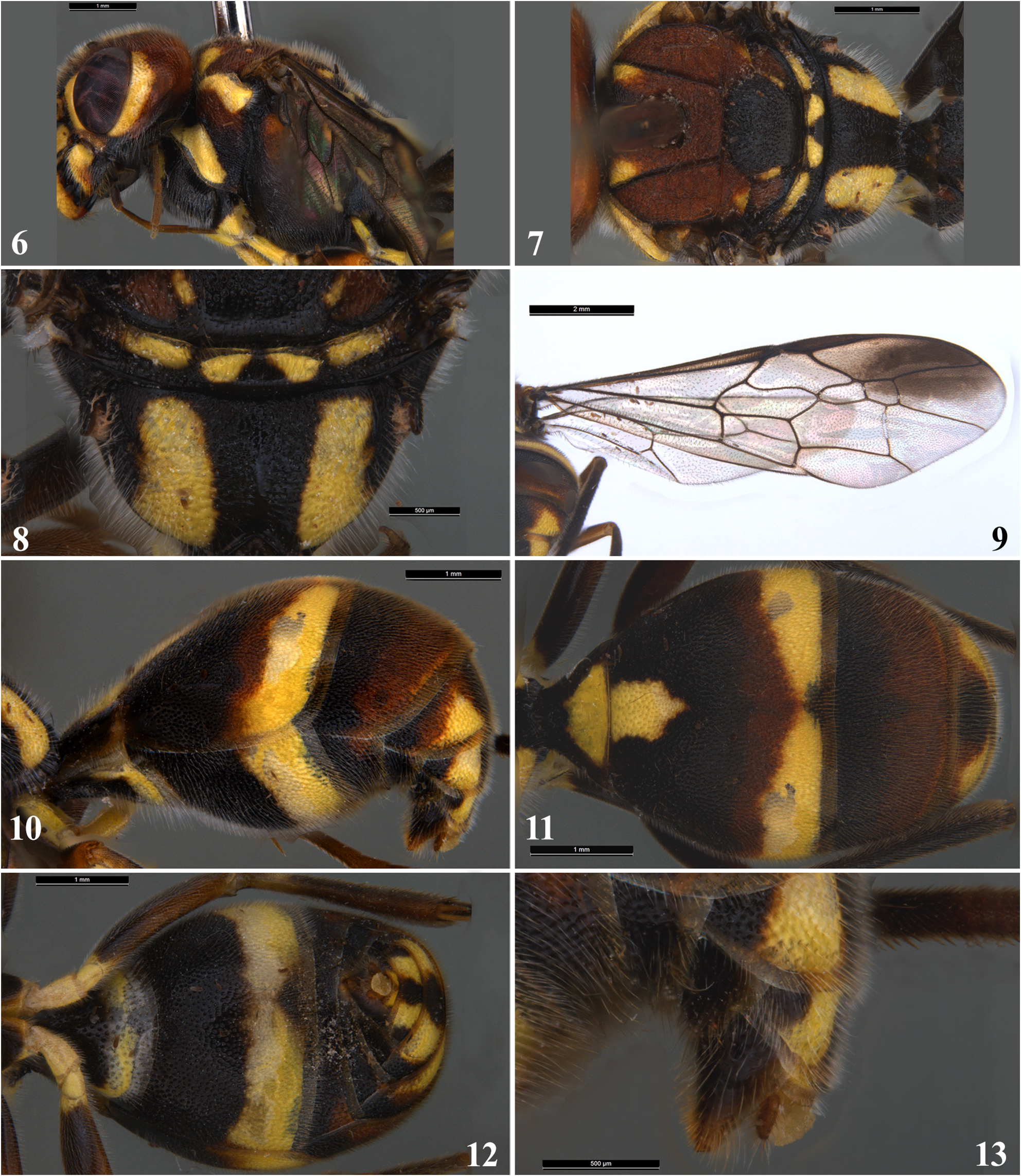
1. Second metasomal sternite produced medio-posteriorly ( Figures 5d and 5f View FIGURES 1–5 of Binoy et al., 2022); scutellum unicoloured, yellowish orange or yellow ( Figure 5b View FIGURES 1–5 of Binoy et al., 2022).......................... T. fulvoscutellata (Ayyar, 1919)
- Second sternite truncate medio-posteriorly ( Fig. 12 View FIGURES 6–13 & Figures 2e, 2h View FIGURES 1–5 , 7d, 10d, 10f, 12h View FIGURES 6–13 and 15e of Binoy et al., 2022); scutellum bicoloured or tricoloured ( Fig. 7 View FIGURES 6–13 & Figures 2b View FIGURES 1–5 , 7b and 10b View FIGURES 6–13 of Binoy et al., 2022)................................... 2
2. Head, mesosoma and metasoma predominantly bicoloured (black/blackish brown and yellow) ( Figures 6a and 11a View FIGURES 6–13 of Binoy et al., 2022)............................................................................................ 3
- Head, mesosoma and metasoma predominantly tricoloured (red/orange, black/blackish brown and yellow/yellowish white) ( Fig. 1 View FIGURES 1–5 & Figures 1a View FIGURES 1–5 , 9a View FIGURES 6–13 and 14a View FIGURE 14 of Binoy et al., 2022)........................................................ 4
3. Second metasomal tergite densely punctured, interspaces distinctly less than the diameter of punctures, matt ( Figure 7e View FIGURES 6–13 of Binoy et al., 2022); antenna black with yellow fleck on scape ( Figures 6b and 6e View FIGURES 6–13 of Binoy et al., 2022); frons and vertex with extensive yellow areas ( Figures 6d and 6e View FIGURES 6–13 of Binoy et al., 2022)............................. T. gestroi (Schulz, 1908)
- Second tergite not densely punctured, interspaces distinctly more than the diameter of punctures, glossy ( Figure 12g View FIGURES 6–13 of Binoy et al., 2022); antenna entirely brown ( Figure 11b and 6e View FIGURES 6–13 of Binoy et al., 2022); frons and vertex entirely black ( Figures 11d and 11e View FIGURES 6–13 of Binoy et al., 2022)...................................................... T. latae Polaszek & Binoy, 2022
4. Second submarginal cell of fore wing petiolate anteriorly which is connecting with discal cell ( Figure 10c View FIGURES 6–13 of Binoy et al., 2022)............................................................................. T. kerala (Ayyar, 1919)
- Second submarginal cell sessile anteriorly which is either connecting with discal cell at a point ( Figure 14a View FIGURE 14 of Binoy et al., 2022) or distinctly forming a vertical vein ( Fig. 9 View FIGURES 6–13 & Figure 2d View FIGURES 1–5 of Binoy et al., 2022)................................ 5
5. Second submarginal cell of fore wing sessile anteriorly which is connecting with discal cell at a point ( Figure 14a View FIGURE 14 of Binoy et al., 2022); middle of scutellum reddish ( Figure 14d View FIGURE 14 of Binoy et al., 2022)................................................................................................ T. thwaitesii (Westwood, 1874) [Extralimital: Sri Lanka]
- Second submarginal cell sessile anteriorly which is connecting with discal cell by forming a vertical vein ( Fig. 9 View FIGURES 6–13 & Figure 2d View FIGURES 1–5 of Binoy et al., 2022); middle of scutellum black ( Fig. 7 View FIGURES 6–13 & Figure 2b View FIGURES 1–5 of Binoy et al., 2022).......................... 6
6. Second metasomal tergite with basal yellow spot ( Fig. 11 View FIGURES 6–13 ); vertex dorsally reddish ( Fig. 4 View FIGURES 1–5 ); 21 flagellomeres ( Fig. 2 View FIGURES 1–5 ); yellow apical band of second metasomal tergite laterally widened and narrowed medially ( Fig. 11 View FIGURES 6–13 ); yellow apical band of second metasomal sternite comparatively wide medially ( Fig. 12 View FIGURES 6–13 )................... T. dhritiae Girish Kumar & Hegde sp. nov.
- Second tergite without basal yellow spot ( Figure 2g View FIGURES 1–5 of Binoy et al., 2022); vertex dorsally black with reddish markings ( Figure 1d View FIGURES 1–5 of Binoy et al., 2022); 22 flagellomeres ( Figure 1b View FIGURES 1–5 of Binoy et al., 2022); yellowish white apical band of second tergite with almost uniform thickness ( Figure 2g View FIGURES 1–5 of Binoy et al., 2022); yellowish white apical band of second sternite narrow medially ( Figure 2h View FIGURES 1–5 of Binoy et al., 2022)............................. T. ayyari Binoy, van Achterberg & Girish Kumar, 2022

No known copyright restrictions apply. See Agosti, D., Egloff, W., 2009. Taxonomic information exchange and copyright: the Plazi approach. BMC Research Notes 2009, 2:53 for further explanation.
|
Kingdom |
|
|
Phylum |
|
|
Class |
|
|
Order |
|
|
Family |