
Pteropagurus spina McLaughlin & Rahayu, 2006
|
publication ID |
https://doi.org/10.5281/zenodo.4525838 |
|
persistent identifier |
https://treatment.plazi.org/id/C22CA730-D403-0C2A-6F43-FF7CB2E329DD |
|
treatment provided by |
Felipe |
|
scientific name |
Pteropagurus spina McLaughlin & Rahayu, 2006 |
| status |
|
Pteropagurus spina McLaughlin & Rahayu, 2006 View in CoL
( Fig. 1 View FIG )
Pteropagurus spina McLaughlin & Rahayu, 2006: 64 View in CoL View Cited Treatment , fig. 5. — McLaughlin 2007: 508.
HOLOTYPE. — New Caledonia. MUSORSTOM 5, stn DW 274 , 24°44.8’S, 159°41.0’E, 285 m, 9.X.1986, ovig. ♀ ( 1.2 mm) (MNHN-Pg 7637). GoogleMaps
NEW MATERIAL EXAMINED. — New Caledonia. EBISCO, stn DW 2492, 24°44.0’S, 159°41.0’E, 285 m, 6.X.2005, 1 ♂ ( 1.3 mm) (MNHN-Pg 7763).
DISTRIBUTION. — Presently known only from the type locality, Chesterfield Plateau , and vicinity, New Caledonia ; 285 m.
DESCRIPTION OF MALE
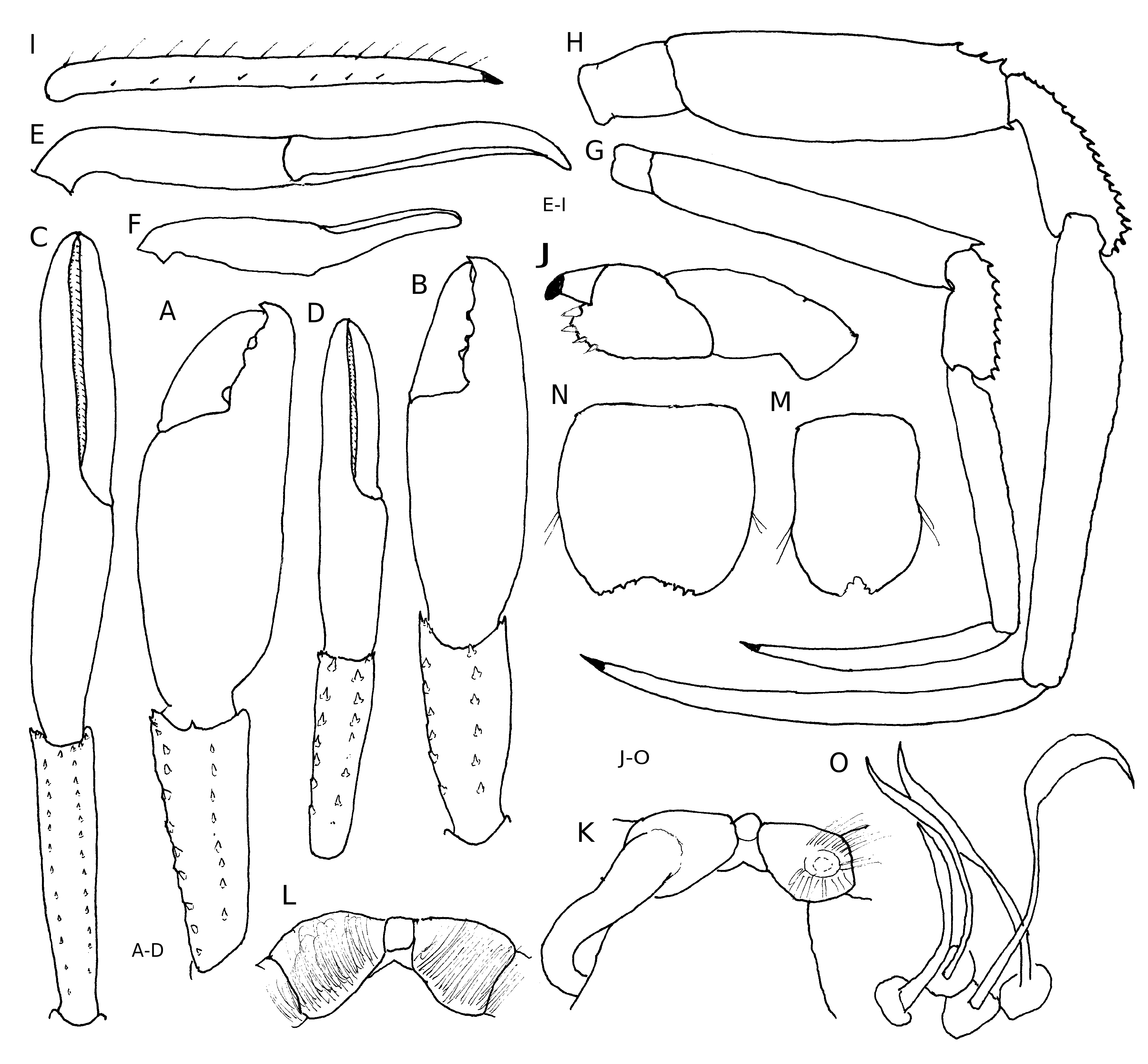
Shield ( Fig. 1A View FIG ) slightly broader than long, weakly calcified; anterior margin between rostrum and lateral projections concave; anterolateral margins sloping; posterior margin roundly truncate. Triangular rostrum produced slightly beyond midlength of ocular acicles; lateral projections each with acute marginal spine. Carapace lateral lobes reaching 0.4 of shield.Posterior carapace with moderately narrow median plate; cardiac sulci reaching to posterior margin. Branchiostegites membranous, unarmed.
Ocular peduncles moderately stout but not noticeably broadened distally, 0.7 length of shield; corneal diameter 0.5 of total peduncular length; ocular acicles narrowly triangular, reaching to or beyond proximal margin of ultimate peduncular segment, each with moderately large submarginal spine.
Antennular peduncles overreaching distal margins of corneas by slightly less than lengths of ultimate peduncular segments; ultimate segment with 1 long, stiff seta on dorsodistal margin; penultimate segment with very few scattered setae; basal segment with acute spine on dorsolateral margin, ventral margin not produced distally.
Antennal peduncles overreaching distal corneal margins by 0.5 lengths of ultimate segments. Fifth and fourth segments with very few scattered setae; third segment with small spine at ventrodistal angle; second segment with dorsolateral distal angle produced, terminating in small spine, dorsomesial angle with prominent spine; first segment with spine at dorsolateral distal margin, ventrolateral margin unarmed. Antennal acicle reaching beyond distal margin of fourth peduncular segment, moderately slender, terminating in simple spine. Antennal flagellum with 1 or 2 very short to moderately long setae every article.
Chelipeds subequal in length, but right slightly stouter; dactyls and fixed fingers each with small hiatus. Right cheliped ( Fig. 1B View FIG ) with dactyl equal to length of palm; dorsomesial margin smooth, dorsal surface somewhat convex, unarmed, but with few scattered, short setae mesially and ventrally; cutting edge with 2 low, broad, calcareous teeth, terminating in tiny corneous claw, slightly overlapped by fixed finger. Palm longer than carpus, dorsomesial margin delimited by 3 widely-spaced, acute spines and sparse tufts of setae, dorsal surface weakly convex, unarmed; dorsal surface of fixed finger similarly unarmed, dorsolateral margin smooth and with few scattered short setae; cutting edge with 3 broad, low calcareous teeth, terminating in tiny corneous claw. Carpus approximately equal to length of merus; dorsomesial distal angle with acute spine and 1 additional small spine on dorsomesial margin, dorsal surface with 2 small spines in lateral 0.3, dorsolateral margin not distinctly delimited; remaining surfaces unarmed. Merus with all surfaces unarmed; ventromesial and ventrolateral margins each with small spine at distal angle. Ischium unarmed.
Left cheliped ( Fig. 1C View FIG ) approximately equal to length of right, slightly slenderer. Dactyl equal to length of palm; surfaces rounded and unarmed, but with sparsely scattered, moderately long setae ventrally; cutting edge with row of minute calcareous teeth, terminating in small corneous claw. Palm with convex dorsal surface unarmed but with few scattered setae; dorsomesial margin with 3 small spines, dorsolateral margin not delimited; fixed finger rounded and unarmed but with several moderately long setae laterally; cutting edge with row of minute calcareous teeth, terminating in small corneous claw. Carpus equal to length of palm; dorsomesial margin not distinctly delimited but with acute spine at distal angle, without small spine at midlength, dorsal surface with 2 small spines laterad of midline, dorsolateral margin not delimited;remaining surfaces unarmed but with few scattered setae, ventromesial distal angle with small spine. Merus with surfaces unarmed but with few scattered short setae, ventromesial and ventrolateral distal angles each with small spine. Ischium unarmed.
Second and third pereopods ( Fig. 1D, E View FIG ) similar in armature but unequal in length, second shorter. Dactyls relatively straight in dorsal and lateral views, slightly shorter to slightly longer than propodi, dorsal surfaces each with row of widely-spaced setae; ventral surfaces each with 4 or 5 corneous spines and few setae. Propodi slightly to considerably longer than carpi; surfaces all unarmed but with sparse setae dorsally and ventrally. Carpi of second pereopods each with dorsodistal spine and 1 additional spine on dorsal surface in proximal 0.5; carpi of third each without minute spinule at dorsodistal margin, other surfaces unarmed but all with sparse setae. Meri unarmed, but each with few widely-spaced setae dorsally. Ischia unarmed. Sternite of third pereopods ( Fig. 1F View FIG ) not produced posteriorly. Fourth pereopods semichelate; propodal rasp ( Fig. 1G View FIG ) with 1 row of scales.
Extremely long sexual tube developed from coxa of left fifth pereopod ( Fig. 1H View FIG ), directed toward exterior, curved up and over dorsal surface of pleon and ventrally to level of coxa of right fifth pereopod; right coxa with gonopore circumscribed by moderately long, stiff setae.
Uropods symmetrical, each with 2 rows of scales on endopod and exopod. Telson ( Fig. 1I View FIG ) with posterior lobes separated by shallow median cleft, oblique terminal margins each with 3 or 4 very small spines, largest at apex.
Coloration
Not known.
Habitat
Empty shells of Cuvierina columnella (Rang, 1827) .
REMARKS
Because of the paucity of material, the mouthparts of P. spina have not been removed for description and illustration, but appear to be in general agreement with those illustrated by McLaughlin & Rahayu (2006) for P. inermis , including the several teeth on the crista dentata of the third maxilliped. The general morphology of the male of P. spina differs only slightly from the female in proportions and spination of the chelipeds and ambulatory legs. However, the scales of the propodal rasp of the fourth pereopod form only a single row in this male. Having only one specimen of each sex, it is not possible to determine whether the two rows of spines in the female reflect a sexually dimorphic character or simply variation.
Pteropagurus spinulocarpus McLaughlin, 2007 View in CoL ( Figs 2 View FIG ; 3 View FIG )
Pteropagurus spinulocarpus McLaughlin, 2007: 505 View in CoL , fig. 1.
HOLOTYPE. — New Caledonia. BIOCAL, stn DW 44 , 22°47.30’S, 167°14.30’E, 440-450 m, 30.VIII.1985, ♂ ( 1.6 mm, posterior portion of pleon, uropods and telson missing) (MNHN-Pg 7738). GoogleMaps
PARATYPES. — New Caledonia. MUSORSTOM 4, stn CC 212, 22°47.40’S, 167°10.50’E, 375-380 m, 28.IX.1985, 1 ovig. ♀ ( 1.5 mm, right cheliped and right second pereopod missing, left third pereopod regenerating) (MNHN-Pg 7737). — Stn DW 222, 22°57.60’S, 167°33’E, 410-440 m, 30.IX.1985, 1 ♀ ( 1.5 mm, with rhizocephalan, missing both chelipeds, left second and third right pereopods) (MNHN-Pg 7701).
MUSORSTOM 5, stn DW 274 , 24°44.84’S, 159°41.00’E, 285 m, 9.X.1986, 1 ♀ ( 1.2 mm, missing right cheliped and both third pereopods) (MNHN-Pg 7702). — Stn DC 361 , 19°52.50’S, 158°38.10’E, 400 m, 19.X.1986, 1 ♂ ( 1.5 mm, right cheliped regenerating; left cheliped with fixed finger broken off; left third pereopod missing) (MNHN-Pg 7700) GoogleMaps .
NEW MATERIAL EXAMINED. — New Caledonia. EBISCO, stn DW 2492, 24°44.0’S, 159°41.0’E, 285 m, 6.X.2005, 12 ♂♂ ( 1.2-1.7 mm, 1 with rhizocephalan), 7 ♀♀ ( 1.4-1.7 mm), 15 ovig. ♀♀ ( 1.4-1.6 mm) (MNHN-Pg 7764).
DISTRIBUTION. — New Caledonia from the Chesterfield Plateau to south of the Isle of Pines ; 285-440, possibly 450 m.
SUPPLEMENTAL DESCRIPTION
Shield ( Fig. 2A View FIG ) with rostral lobe slightly to moderately produced, very faintly rounded to broadly and bluntly triangular, unarmed; lateral projections obsolete to roundly triangular, unarmed or with tiny marginal spinule. Ocular peduncles short and stout, appreciably broadened distally; ocular acicles narrowly to ovately triangular, slightly less to slightly more than 0.5 peduncular lengths; each terminating acutely or with prominent terminal spine. Antennular peduncles overreaching distal margins of corneas by 0.2-0.5 lengths of penultimate peduncular segments. Ultimate segment with 1-5 long setae at dorsodistal margin. Antennal peduncles overreaching distal corneal margins by 0.3-0.5 lengths of ultimate segments; second segment with dorsolateral distal angle produced, with acute terminal spine and often 1, occasionally 2, smaller spines on lateral margin subterminally; first segment with small to prominent spine on laterodistal margin; antennal acicle moderately slender, reaching to or beyond distal margin of fourth peduncular segment.
Mandible ( Fig. 2B View FIG ) with 2-segmented palp. Maxillule ( Fig. 2C View FIG ) with external lobe of endopod obsolete, internal lobe with 1 seta; basial endite with very slender tooth-like marginal processes. Maxilla ( Fig. 2D View FIG ) with endopod slightly overreaching distal margin of scaphognathite. First maxilliped ( Fig. 2E View FIG ) with endopod overreaching basial endite; slender exopod with few marginal setae. Second maxilliped ( Fig. 2F View FIG ) with very long exopod. Third maxilliped ( Fig. 2G View FIG ) with basis-ischium almost entirely fused; crista dentata ( Fig. 2H View FIG ) reduced to few very tiny teeth.
Right cheliped ( Fig. 3A, B View FIG ) slightly longer to appreciably shorter than left, but considerably stouter; dactyl and fixed finger with or without slight hiatus. Dactyl 0.6-0.7 length of palm; dorsomesial margin rounded, all surfaces unarmed, but with few scattered setae, particularly ventrally; cutting edge with 2 or 3 moderate to large calcareous teeth. Palm slightly shorter to slightly longer than carpus; dorsomesial and dorsolateral margins rounded, unarmed, convex dorsal and ventral surfaces also unarmed. Carpus slightly longer than merus; dorsomesial and dorsolateral margins each with row of 4-9 small spines, number increasing with increased animal size, other surfaces unarmed, but distomesial margin often with 2 or 3 smaller spines. Merus glabrous or with row of widely-spaced, individual bristles on dorsal surface; other surfaces unarmed but with few scattered setae; ventromesial and ventrolateral distal angles each usually with small spine.
Left cheliped ( Fig. 3 View FIG C-F) long, slender, considerably longer than right with tips prominently curved in larger male specimens; dactyl and fixed finger upwardly flexed ( Fig. 3F View FIG ) and with slight hiatus; dactyl shorter to approximately 1.2 length of palm; surfaces all unarmed but with sparse scattering of short setae particularly ventrally. Palm long, slen- der, surfaces unarmed but with few scattered setae. Carpus approximately equal to length of merus; dorsomesial and dorsolateral margins each with row of small spines increasing in number with increasing body size, other surfaces unarmed but with few scattered setae.
Second ( Fig.3G View FIG ) and third ( Fig. 3H, I View FIG ) pereopods similar in armature, but dissimilar in length and proportions, third appreciably longer and stouter. Dactyls slightly shorter to approximately equal to lengths of propodi; terminating in small corneous claws; mesial faces each with row of widely-spaced small bristles or corneous spinules adjacent to ventral margin ( Fig. 3I View FIG ). Propodi approximately twice length of carpi, unarmed or with very low protuberances and sparse row of setae on each dorsal surface. Carpi each with row of small spines on dorsal surface. Meri of second pereopods each usually with dorsodistal spine, occasionally unarmed; third each with 1 dorsodistal and 1-3 subdistal spinulose protuberances or small spines and 1 or 2 setae, ventrolateral distal angles each with tiny spinule or small spine. Fourth pereopods ( Fig. 3J View FIG ) semichelate; propodal rasp with 1 row of 3-5 spiniform corneous scales.
Sternites of second and third pereopods ( Fig. 2I, J View FIG ) broad; third broadly subrectangular anteriorly and drawn out posteriorly into broad, terminally rounded plate in both sexes. Sternite of fifth pereopods ( Fig. 3K, L View FIG ) with simple or faintly bilobed median projection in both sexes.
Male with coxae of fifth pereopods approximately equal, right with long, stout sexual tube developed as posterior coxal extension ( Fig. 3K View FIG ) directed posteriorly and exteriorly and curving over
anterior portion of dorsal pleon; coxa of left with gonopore and sometimes papilla. Coxae of fifth pereopods ( Fig. 3L View FIG ) in females each with dense setae on ventral surface.
Telson ( Fig. 3M, N View FIG ) with V- or U-shaped median cleft, or only slight median concavity; terminal margins oblique, each with 2-4 tiny blunt or acute spines; lateral margins each with 1 or 2 moderately long setae.
Coloration
Unknown.
Habitat
Empty shells of Cuvierina columnella (Rang, 1827) .
Larval development
Only limited information available from prematurely hatched zoeae. Carapace with no rostral spine apparent; posterolateral angles rounded. Appendages typical of first stage zoeae, but none with setae extruded. Pleon with 5 somites and telson; posterior telsonal margin ( Fig. 2K View FIG ) with small U-shaped median cleft and 7+7 processes: outermost – small spine, second – anomuran hair, third through seventh – plumodenticulate setae, fourth longest.
REMARKS
The mouthparts of P. spinulocarpus are in general agreement with those described for P. inermis by McLaughlin & Rahayu (2006) as representative of the genus. However, the crista dentata of P. spinulocarpus consists of considerably fewer teeth than were illustrated but not specified for P. inermis . Although the right chelipeds of males of P. spinulocarpus are generally a little stouter than females, both are very similar morphologically and do not change with growth. McLaughlin’s (2007) description of the left chela being ventrally curved in both sexes is misleading. In lateral or mesial view, the dactyl and fixed finger are flexed upward in relation to the ventral margin of the palm ( Fig. 3F View FIG ) with the ventral margins of the former slightly curved. This flexion readily distinguishes the left cheliped of P. spinulocarpus from the otherwise structurally similar and sexually dimorphic left cheliped of C. spinicarpus . Nonetheless, the left chelipeds of P. spinulocarpus exhibit comparable sexual dimorphism to that seen in C. spinicarpus , such as an increase in length with increased animal size and the prominent ventral curvature of the distal portions of the dactyl and fixed finger, which accompanies the increase in overall length in males ( Fig. 3C, E View FIG ), but not in females ( Fig. 3D, F View FIG ). Sexual dimorphism is exhibited by females of P. spinulocarpus in the presence of moderately dense setation on the ventral surfaces of the coxae of the fifth pereopods ( Fig. 3L View FIG ).
The increases in the number of spines on the dorsal surfaces of the carpi of the chelipeds and ambulatory legs occur with increased body size in both sexes and appear to be entirely functions of growth. The variations seen in the breadth, median cleft, or armature of the telson ( Fig. 3M, N View FIG ) do not appear to be dimorphic or growth related.
McLaughlin & Rahayu (2006) speculated that the longer third pereopods of Pteropagurus species enabled the animals to elevate the shells above the substrate when walking, as none of the four shells examined in their original sample had any marks on the exterior surfaces. The numerous carcinoecia of P. spinulocarpus in the recent sample support their supposition. The external shell surfaces are clear and smooth, showing no signs of scuffing from being dragged across the substrate. The apertures are usually smooth and unbroken. In the preserved state, the appendages of the hermits often were at least partially extended; however, the entire animals and their appendages could be retracted completely into the carcinoecia. On the internal surfaces of several of the pteropod shells occupied by P. spinulocarpus were what appeared to be clusters of a stalked protozoan ( Fig. 3O View FIG ) probably of the phylum Ciliophora. Although Williams & McDermott (2004) indicated that protozoan attachments included shell surfaces and other symbionts as well as the exoskeletons of the crabs, Fernandez-Leborans & Tato-Porto (2000a, b) and Fernandez-Leborans (2001) reported such attachments only on the gills, integuments, and setae of several hermit crabs. Clusters similar to those on the shell walls occasionally also were observed on the bodies of P. spinulocarpus .


| CC |
CSIRO Canberra Rhizobium Collection |
No known copyright restrictions apply. See Agosti, D., Egloff, W., 2009. Taxonomic information exchange and copyright: the Plazi approach. BMC Research Notes 2009, 2:53 for further explanation.
|
Kingdom |
|
|
Phylum |
|
|
Class |
|
|
Order |
|
|
Family |
|
|
Genus |
Pteropagurus spina McLaughlin & Rahayu, 2006
| McLAUGHLIN, Patsy A. & Rahayu, Dwi Listyo 2008 |
Pteropagurus spinulocarpus
| MCLAUGHLIN P. A. 2007: 505 |