
Synapturanus latebrosus, Osorno-Muñoz & Gutiérrez-Lamus & Lynch & Keeffe & Caicedo-Portilla & Chan & Tonini & De Sá, 2023
|
publication ID |
https://doi.org/10.11646/zootaxa.5258.2.1 |
|
publication LSID |
lsid:zoobank.org:pub:33F922CD-6372-427A-912E-DC3CF4641F8D |
|
DOI |
https://doi.org/10.5281/zenodo.7777056 |
|
persistent identifier |
https://treatment.plazi.org/id/C23387E2-FFC8-FF82-C9F4-FAC36009F88A |
|
treatment provided by |
Plazi |
|
scientific name |
Synapturanus latebrosus |
| status |
sp. nov. |
Synapturanus latebrosus sp. nov.
Synapturanus sp. “ Caquetá ”
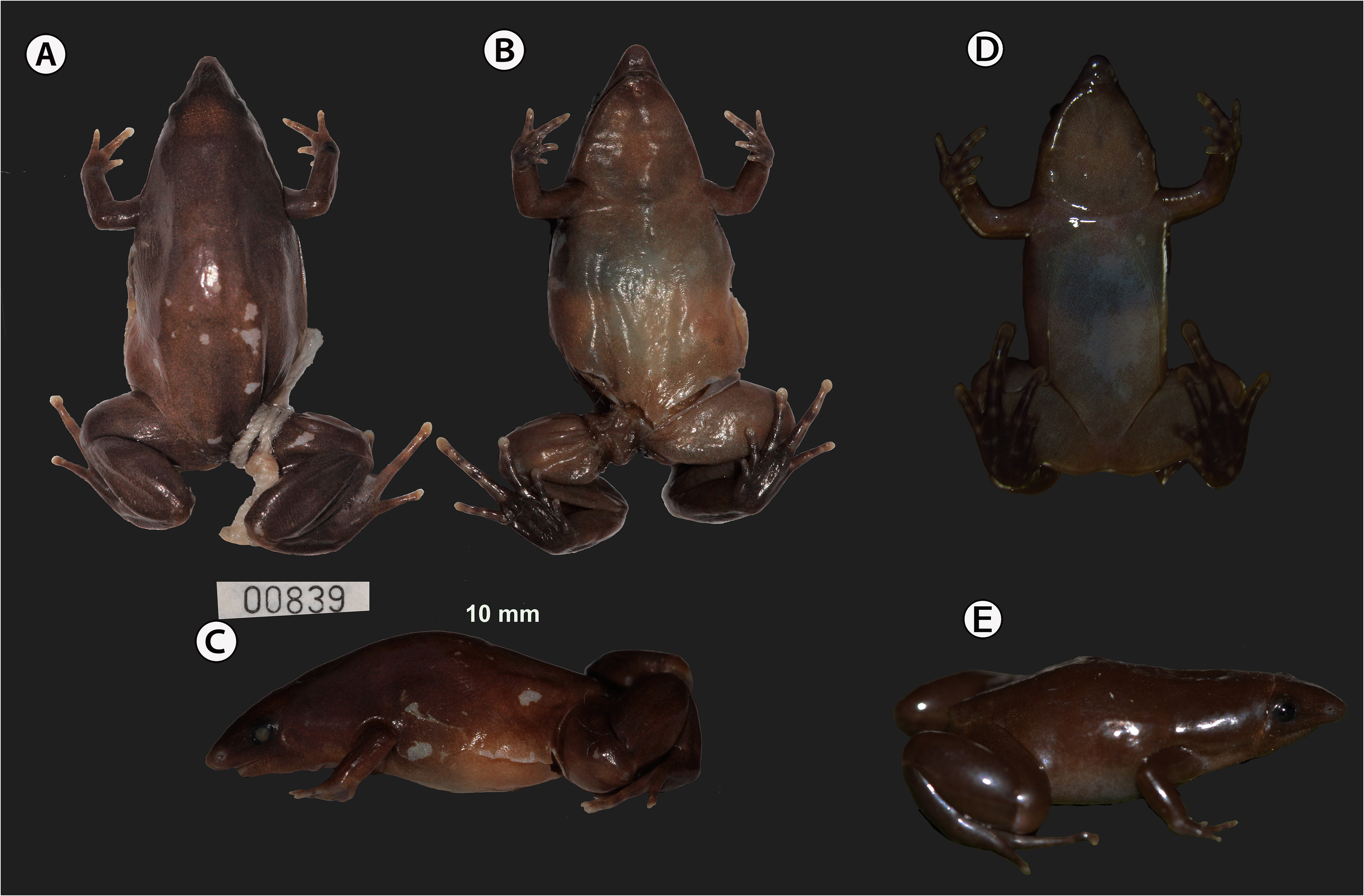
Holotype ( Fig. 3 View FIGURE 3 ). SINCHI-A 839 (MOM 2517), adult female, collected at finca El Cairo, vereda Sinaí, Municipio Morelia, Departamento Caquetá, Colombia, ( 01°23′31.7″ N, 75°45′06.2″ W), ca. 278 m., on August 4, 2011 by Yunner Fabian González, Diego Huseth Ruiz, Doris Laurinette Gutiérrez, and Mariela Osorno-Muñoz. GoogleMaps
Paratopotypes. SINCHI-A 840-841 (MOM 2518-2519), adult females, and SINCHI-A 842 (MOM 2520), cleared and double-stained specimen, SINCHI-A 843-845 (MOM 2521-2523) adult males, all collected with the holotype.
Paratypes. Female SINCHI-A 2678 (JARM 139) ( Fig. 3B View FIGURE 3 ) and male SINCHI-A 2679 (JARM 140) a cleared and double-stained specimen, collected at hacienda Villa Mery , vereda Sinaí , Municipio Morelia, Departamento Caquetá, Colombia ( 01°24′00.1″ N, 75°43′52.3″ W), 218 m., on December 16, 2015 GoogleMaps ; female SINCHI-A 2703 (JARM 172), collected at vereda La Mono, Municipio Belén de los Andaquíes, Departamento Caquetá, Colombia, ( 01°18′27.8″ N, 75°48′13.9″ W), ca. 262 m., on February 18, 2016 GoogleMaps ; females: SINCHI-A 5642–5643 (JARM 220- 221) collected at vereda Alto Caldas, Municipio Florencia, Departamento Caquetá, Colombia ( 01°39′N, 75°37′W), ca. 560 m., on 01 April, 2016 GoogleMaps ; female SINCHI-A 5671 (JARM 254) collected at finca Alsacia , vereda la Primavera, Municipio Florencia, Departamento Caquetá, Colombia ( 01°39′ 4.87″ N 75°37′4.7″ W), ca. 530 m., on April 23, 2016 GoogleMaps , by Julián Andrés Rojas Morales , and Fabián Andrés Cabrera Vargas. Females : SINCHI-A 258–259 (MOM 1840 -1841) ( Fig.3A View FIGURE 3 ), collected at vereda La Recreo, Municipio Solita, Departamento Caquetá, Colombia ( 0°55′39.2″ N, 75°40′35″ W), ca. 220 m., collected by Hernándo Trujillo on October 09, 2009 GoogleMaps .
Referred specimens. Males SINCHI-A 846–847 (MOM 2524-2525), female SINCHI-A 848 (MOM 2526), all juveniles collected along with the type series, male SINCHI-A 5665 (JARM 248) collected at finca Alsacia, vereda la Primavera, Municipio Florencia, Departamento Caquetá, Colombia ( 01°39′ 4.87″ N 75°37′4.7″ W), ca. 530 m. on April 23, 2016, by Julián Andrés Rojas Morales, and Fabián Andrés Cabrera Vargas GoogleMaps .
Diagnosis. A species of Synapturanus diagnosed by the following combination of characters: 1) SVL median size: adult females 20.0–22.0 mm (χ = 20.9 ± 0.3, n = 8), adult males 18.1–19.0 (χ = 18.7 ± 0.3, n = 3 mm), 2) stout and elongated body, 3) head narrower than body, snout pointed in dorsal view, rounded in lateral view, and ventrally distinctly projecting beyond the anterior edge of the upper jaw, 4) symphysis of lower jaw with an unpigmented notch and external nares bear a wide and unpigmented rim, 5) tympanum indistinct, tympanic annulus visible below the skin, particularly its anteroventral edge, 6) vocal slits absent, 7) choanae rounded, larger in diameter than the unpigmented rim of the external nares, 8) vomerine teeth absent, 9) hand formula III>IV>II>I, digits becoming thinner towards their distal ends, rounded or slightly pointed finger tips, fingers bordered by a thin fringe, interdigital membrane absent, 10) subarticular tubercles absent; thenar tubercle elongated, palmar tubercle rounded with undefined edges, 11) adult males bear an elongated gland on the internal surface of the anterior forearm extending half of its length and broader at the wrist, 12) toe lengths IV>III>V>II>I, toes are thin and subcylindrical with a slight distal rounded or lanceolate widening, except toe I which is pointed, toes narrowly fringed, fringes more distinct distally and around the distal expansion, toes without webs, 13) inner metatarsal tubercle small and elongated, outer metatarsal tubercle absent, subarticular tubercles absent, subarticular spots unpigmented on toes, 14) skin folds on knee, heel, and wrist, 15) cephalic groove distinct, extending over the tympanum reaching and slightly extending beyond the lower jaw, 16) in life, upper surfaces of body uniformly brown, lighter brown on the area of the cephalic groove, tympanum brownish, and body flanks orange with a greyish brown ventral area, 17) light canthal stripe present or absent; if present, dorsal to the nares and the eyes, canthal stripe continuous or broken into a series of variable size spots; confluent or not on the distal tip of snout; posteriorly, the canthal stripe could reach the area over the tympanum or above the shoulder, 18) ventral surfaces overall brown, darker on edges of mandible, snout, arms, and hidden surfaces of legs and feet; lighter on thighs, chest, and edges of abdominal region; medially the belly has a narrow and irregular shaped whitish and translucent area, hands and feet dark brown; thenar, internal metatarsal tubercles, subarticular surfaces and distal digits without coloration, 19) elongated forearm gland cream-colored in preserved specimens extending from the wrist to half the length of the forearm.
Synapturanus latebrosus sp. nov. differs from S. rabus (traits in parenthesis) by its larger size, adult females SVL 20.0-22.0 mm (vs. 17.2-19.0 mm), adult males SVL 18.1-19.0 mm (vs. 16.2-16.6 mm), a shorter tibia length TBL/SVL 38% in females and 37% in males (vs. 41% in both sexes). Eyes are smaller in S. latebrosus sp. nov. ED/ SVL is 54% (vs. 73%) and the ratio of the eye diameter to the eye-nare distance is also smaller, ED/END females χ = 0.5 (vs. 0.8), ED/END males χ = 0.6 (vs. 0.9) ( Tables 2 View TABLE 2 and 3 View TABLE 3 ). In life, S. latebrosus sp. nov. has greyish ventral flanks (vs. overall dark brown); canthal stripe, if present, extending to the shoulder area (vs. canthal stripe may extend onto the body); posterior limbs without spotting (vs. most specimens have irregular light spots on one or both legs), tympanum partially hidden (visible tympanum).
Synapturanus latebrosus sp. nov. males are smaller (SVL 18.1-19.0 mm) than S. mirandariberoi (SVL males 27.0- 31.7 mm), S. salseri , (SVL males 23.7-26.4 mm), S. zombie (SVL males 37.0- 40.6 mm), S. mesomorphus (SVL males 22.9-26.0 mm), and S. ajuricaba (SVL males 29.3-33.2 mm); S. latebrosus is larger than S. danta (SVL males 17.6-17.9). Adults of Synapturanus danta and S. latebrosus sp. nov. have a uniform dark brown dorsal coloration in life whereas the dorsum of S. mirandariberoi , S. salseri , S. zombie , S. mesomorphus , and S. ajuricaba have noticeable mottled patterns made up of speckles, spots, or blotches.
The forearm gland is not conspicuous in life in Synapturanus latebrosus sp. nov. and S. rabus (i.e., gray-brown, similar to the rest of the arm), whereas in S. salseri and S. mirandariberoi it was described as a pale wrist gland contrasting with the darker coloration of the rest of the arm (Pyburn, 1976). Furthermore, the gland of S. salseri is protuberant on the dorsal and inner area of the distal forearm and becoming slightly triangular with its apex towards the posterior forearm; in S. latebrosus the gland is slightly wider on the wrists and narrowing posteriorly, in some specimens reaching the midpoint of forearm.
Description of the Holotype. An adult female with two large, unpigmented ovarian eggs (3.6 and 4.0 mm diameter), body smooth, slightly ovoid in dorsal view, SVL = 21.5 mm; head triangular in shape, broader than longer (HW = 5.9 mm, HL = 5.5 mm); snout tip acuminate, snout projects beyond the anterior edge of the upper jaw (SL/SW = 0.6), nostrils with a distinct light rim, directed laterally, the distance from the eye to the nostril is 2.0 mm, being twice the diameter of the eye; canthus rostralis defined, slightly concave, loreal region marked by a distinct groove that extends from the anteroventral edge of eye to the posteroventral edge of nostril, eyes small and slightly protruding, interorbital area concave, IOD = 2.5 mm; occipital groove indistinct across the head and tympanum and visible just beyond the jaw; tympanum mostly concealed, anteroventral edge of tympanic ring barely visible, upper jaw distinctly projecting beyond lower one, with an unpigmented median notch in the anterior end of the lower jaw; the tongue is as wide as the oral cavity, its posterior edges are thin and wide; vomerine teeth absent; choanae round and widely separated. Anterior and posterior limbs short and robust, hands without interdigital membranes, finger relative lengths III>IV>II>I, fingers narrowing distally with distal tips pointed or rounded, slightly fringed, subarticular tubercles absent, subarticular area light colored, thenar tubercle elongated to oval, light colored and located at the base of finger I; distinct fold on knee and heel, less distinct folds also on wrist and metatarsal area; toes overall subcylindrical and slightly broader and rounded distally, fringes noticeable in toes I, II and distally in toes III, IV, V, interdigital membrane absent, in lateral view the distal tip of the digits are slightly flattened, subarticular areas light colored without subarticular tubercles, inner metatarsal tubercle very small, elongated, placed at base of toe I, outer metatarsal tubercle absent, toe formula IV>III>V>II>I; tibia length 8.3 mm, about 39% of snout–vent length.
Live coloration. Dorsal surfaces uniformly brown, except the cephalic groove that is lighter; ventral body flank greyish brown; lateral head anterior to the arm light brown, rim of nares and tip of snout grey; tympanum brown/ slightly orange; iris dark brown, canthal stripe formed by very small and discontinuous cream spots that do not reach the shoulder, ventral surface brown, belly with a narrow, medial, and unpigmented area; dorsal surfaces of hands and feet brown, articulation and distal tips of digits unpigmented, ventrally dark brown with unpigmented subarticular areas; a black spot on the external region of the right wrist.
Coloration of Preserved Specimens. Dorsal surfaces brown, light brown around the occipital groove on top head, anterior body flank brown, medial and posterior body flank brown-cream, ventral surfaces of throat, chest, arms and legs light brown cream, central belly light cream, ventral surfaces of hands and feet dark brown, unpigmented subarticular surfaces, and tips of digits cream-colored ( Fig. 4 View FIGURE 4 ).
Measurements of Holotype (mm). SLV 21.5, HL 5.5, HW 5.9, HL/ESD 1.6, SL/SW 0.6, ESD 3.5, END 2.0, ED 1.0, ED/END 0.5, IOD 2.5, TBL 8.3, TBL/SVL 0.4
Variation in the type series. Measurement data of the type series are given in Table 2 View TABLE 2 and Table S2 View TABLE 2 . Overall, the type series agrees with the holotype coloration. Two adult females (SINCHI-A 840 and SINCHI-A 5643) lack canthal stripes, three adult males (SINCHI-A 843-845) and five females (SINCHI-A 841, SINCHI-A 2678, SINCHI-A 2703, SINCHI-A 5642 and SINCHI-A 5671) have a canthal stripe being less distinct in the holotype. The medial unpigmented area of belly is variable, in two females (SINCHI-A 840 and SINCHI-A 841) it extends toward the ventral flanks, in males the unpigmented medial area bears a few small brownish dots.
Etymology. Latin adjective, meaning ignored, alluding to the ecological and behavioral habits that make the species imperceptible and probably even allow it to live in forest fragments, as small as those found in the type locality.
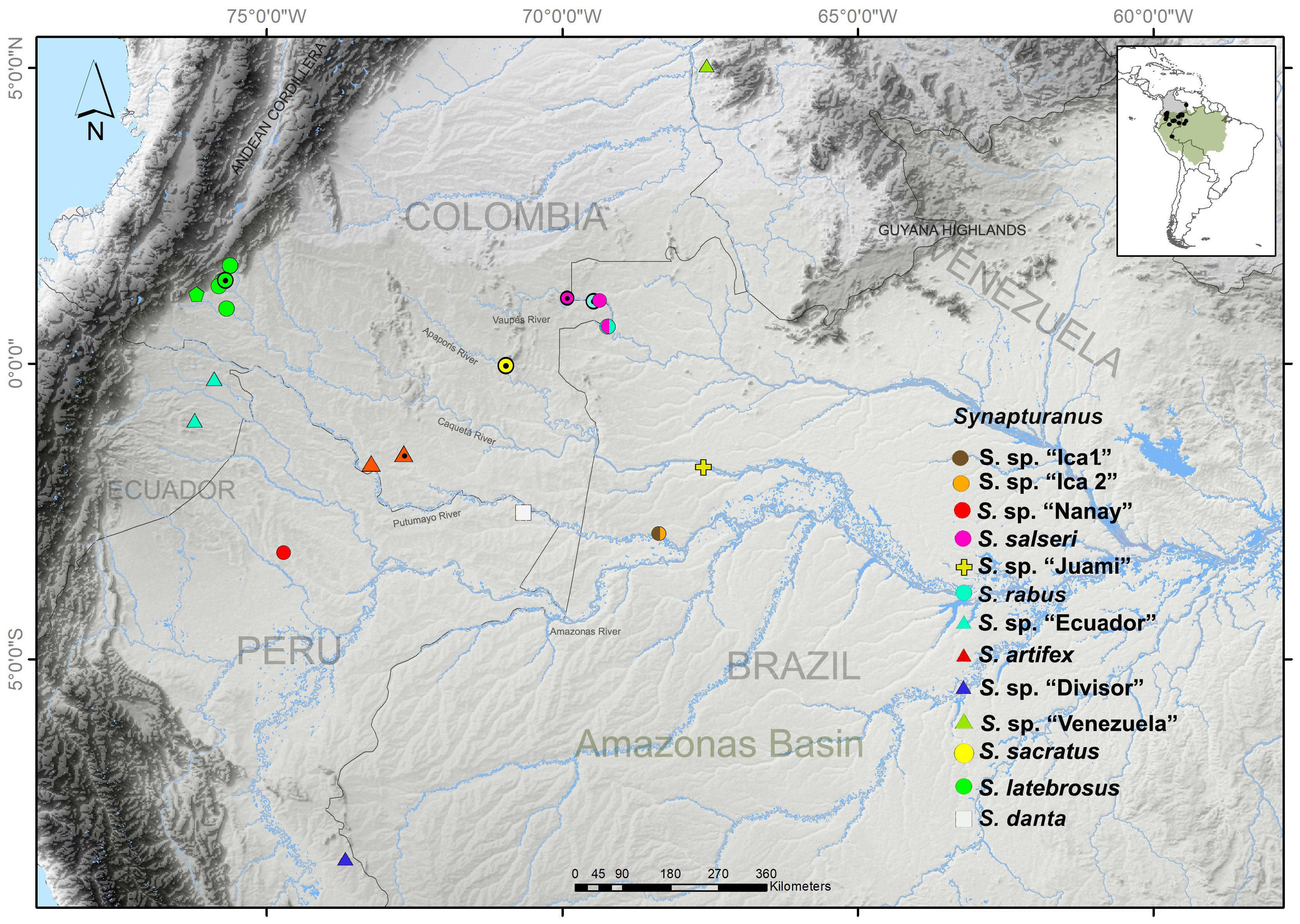
Distribution ( Fig. 4 View FIGURE 4 ). The northernmost locality currently known for Synapturanus latebrosus sp. nov. is the Municipio Florencia, Departamento Caquetá; likely the species has a continuous distribution from the Municipio Morelia and Belén de los Andaquíes to Municipio Solita, north of the Caquetá River.
Tadpole and Advertisement Call. Unknown
Natural History. The holotype and paratopotypes of Synapturanus latebrosus sp. nov. were collected in a fragment of primary forest (<10 hectares), surrounded by meadows for livestock and located on the top of a low hill. A few specimens were found in the afternoon moving over the leaf litter. At night, in the same place, we selected an area of about 1x2 mts, removed the fallen leaves and cut the network of fine roots into a rectangle; we carefully rolled the rectangle and lifted it exposing the clay substrate where we found and collected additional specimens. At the time of collection, all the specimens were placed together; consequently, we do not know if the specimens moving around in the afternoon were males or females. Of the series of paratypes and referred material, two males, SINCHI-A 5665 and SINCHI-A 2679, were found under leaf litter and among a decomposing trunk, the females were found both under and above the leaf litter.
Osteology of Synapturanus latebrosus sp. nov.
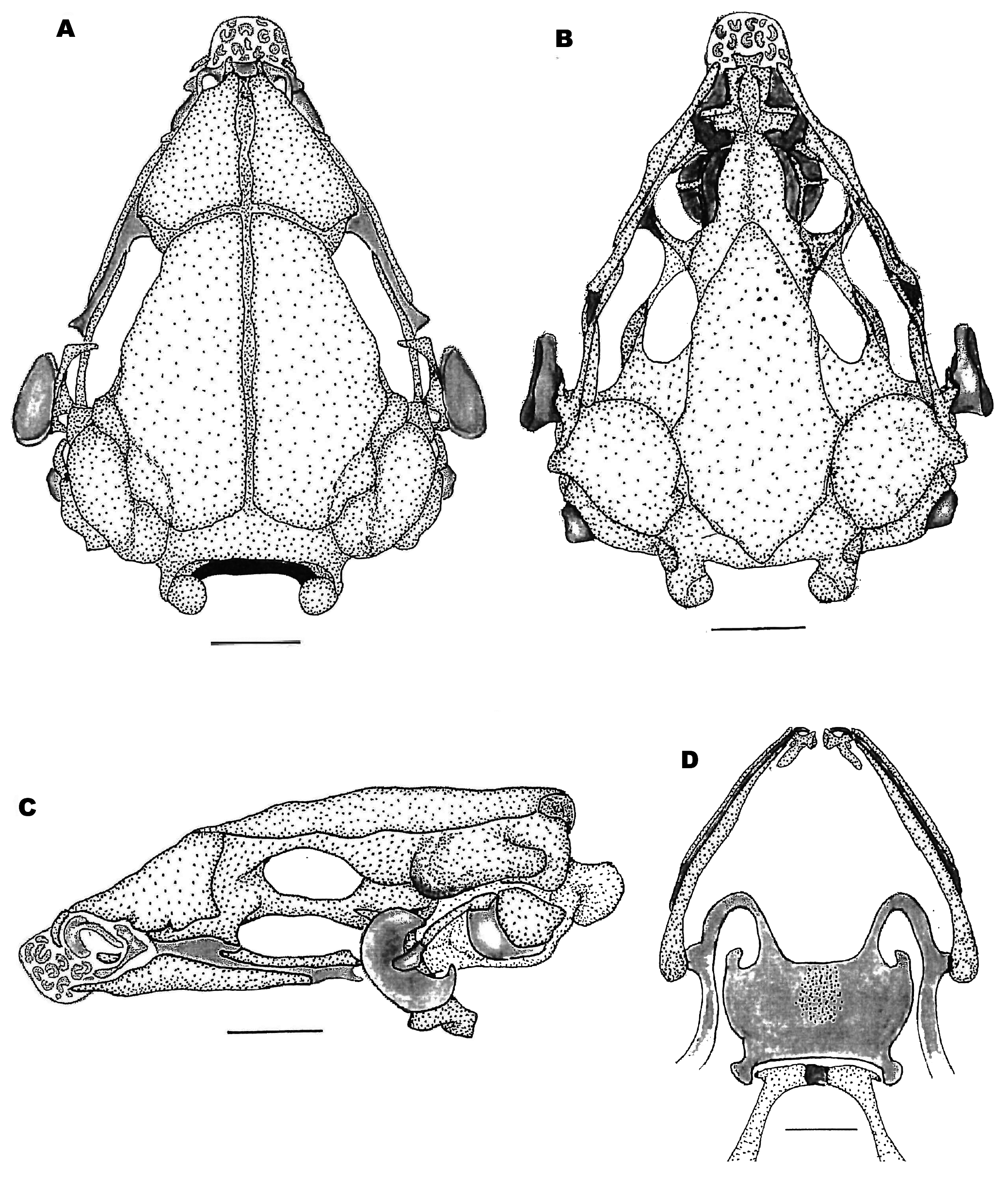
Overall, the skull of the genus Synapturanus is well ossified, with very little cartilaginous areas, mostly restricted to the nasal capsule and tympanic ring. Osteological description of Synapturanus latebrosus sp. nov. is based on male specimen SINCHI-A 2679, variation with male SINCHI-A 842 is noted and compared with an adult female of S. sacratus sp. nov. (ICN 56893). The skull of S. latebrosus sp. nov. is overall triangular, slightly longer than wide ( Fig. 9A, B View FIGURE 9 , and C) and widest at the level of the jaw articulation. The jaw articulation lies at the most anterior edge to the otic capsule. The planum anterorbitalis is ossified and oriented anterolaterally forming the posterolateral walls of the nasal capsules and the anterior wall of the orbits. The auditory capsules, except its most anteroventral edge that is mostly cartilaginous, and the crista parotica are fully ossified. Between the anteroventral cartilaginous edge of the otic capsule and the crista parotica, lies a large and cartilaginous operculum; by transparency a large fenestra ovalis is clearly visible. From the most anterior edge of the operculum and running horizontally, between the crista parotica and the cartilaginous anterior edge of the otic capsule, lies the ossified columella. The columella then bends slightly ventrally towards the squamosal, before reaching the posterior edge of the squamosal, it articulates with a cartilaginous externa plectri that connects with the cartilaginous tympanic ring. The tympanic ring is not complete, but lack its dorsoposterior third.
Endocranium. Sphenethmoid. The paired sphenethmoid are well developed, fused into a single bone, ossified, and form the anterolateral wall of the neurocranium and the anterior margin of the optic fenestra. Dorsally, the sphenethmoid is mostly covered by the nasals and frontoparietals and only visible between the nasals and the frontoparietals. Ventrally, it is visible anteriorly and posteriorly on both sides of the cultriform process of the parasphenoid and forming the ventral border of a very large optic fenestra. Anteriorly it does not reach the posterior border of the choanae. Posterolaterally, the sphenethmoid indistinguishable fuses with prootic and surrounds the optic fenestra.
Prootics and exoccipitals. The prootics are fused with the exoccipitals contributing to the posterior part of the braincase, both are well ossified. The prootic ossifies on the posterolateral wall of the neurocranium, forming the dorsal and posterior margin of the optic foramen and entirely enclose the prootic foramen. Dorsally, their anteromedial margins are overlapped by the frontoparietals. Ventrally, the medial and posterior margins of the prootic are overlapped by the parasphenoid. The prootics also form the anterior and ventrolateral walls of the otic capsules; dorsally they form the epiotic eminences. The epiotic eminences are ossified, large, and visible; medially they fuse gradually to the skull. Most of the ossified crista parotica does not extend beyond the level of the otic capsule; however, a small distinct extension is found on the anterolateral corner of the crista parotica. The otic capsule is mostly ossified except in its most anteroventral area. A relatively large prootic foramen lies on the anteroventral surface of the otic capsule. In anurans, usually smaller oculomotor and trochlear foramina are found between the optic fenestra and prootic fenestra; these two foramina are not visible in S. latebrosus , and likely were integrated to the large optic fenestra. The exoccipitals form the posterior part of the otic capsules, the margins of the foramen magnum, and the occipital condyles. Dorsally, the exoccipitals are largely overlapped by the frontoparietals and ventrally by the alae and posteromedial process of the parasphenoid. The exoccipitals are only visible forming the dorsal and ventral edge of the foramen magnum and the occipital condyles. The occipital condyles bear oval-shaped articular surfaces.
Plectral apparatus. The plectral apparatus is found ventral to the crista parotica. The columella ( pars media plectri) is a long, cylindrical bone, that proximal to the operculum has an expanded base, and runs almost horizontally and then it bends slightly and gradually ventrally towards the squamosal to about half the height of the squamosal. Distally, it connects with the cartilaginous pars externa plectri which is surrounded by a dorsally incomplete and wide ring, the cartilaginous tympanic annuli. The proximal end of the columella contacts with the anterodorsal edge of the cartilaginous operculum. The operculum is well developed, cartilaginous, and occludes the large fenestra ovalis.
Exocranium. Frontoparietals. The broad, large, and paired frontoparietal bones are narrowly separated medially along their length and completely roofing the frontoparietal fenestra. The frontoparietals do not contact with the nasal bones and overlap the sphenethmoid. The anterior tip of the frontoparietals is close to the midline and from there they slant outwardly and posteriorly to the anterodorsal edge of the orbit. Posteriorly the frontoparietals extend over the prootic, partially overlap the epiotic eminences, and the exoccipitals. In specimen SINCHI 842, the separation of the frontoparietals is much narrower on its posterior 1/3.
Nasal. The paired nasals are extensive and well-ossified bones, they cover the nasal capsule and the sphenethmoid, they are medially separated, and do not contact with the frontoparietals. From the midline, they extend posteriorly slanted in a 45-degree angle to reaching the dorso-anterior edge of the orbit. The nasals cover the olfactory capsules and curve ventrolateral to about half the diameter of the external nostril (they do not reach the maxillae) and then turn upward over and dorsally surrounding the nasal capsule. Dorsally, between the anterior tip of the nasals and between the anterior tip of the cartilaginous nasal capsule, a thin ossification of the septum nasi is visible between the nasals with its apex directed posteriorly.
Parasphenoid. The large parasphenoid lacks ornamentation. The cultriform process is broad, occupying most of the floor of the brain and laterally reaching the ventral edges of the optic foramina. Overall, it is rectangular shaped, with its anterior and broad rounded tip passing and between the anterior edge of the neopalatines; medially the anterior tip of the parasphenoid is notched. The parasphenoid alae are broad, rectangular shaped, oriented posterolateral and underlying the otic capsules in about ½ their ventral width and they are widely separated from the medial ramus of the pterygoid. The posteromedial process of the parasphenoid is distinct, overall rounded, and its posterior medial edge bears a medial notch. The parasphenoid does not reach the edge of the foramen magnum.
Vomer. The small, paired anterior vomers lack articulation with other bony elements; posterior vomer are absent. Ventrally, the vomers are seen as thin ossifications on the margins of the choanae and visible, in ventral view, as a triradiate bone over the wall of the choana. The pre- and post-choanal processes support the choana’s anterior and anteromedial margins. Vomerine teeth and odontoids are absent. In specimen SINCHI-A 2679, there is a thin, lateral, projection, at the mid length of the vomer, towards and close to the anterior edge of the internal choana.
Neopalatines. The neopalatines ossify and partially cover the planum anterorbitalis; laterally, the planum anterorbitalis remains cartilaginous in the adult. The medial tips of the neopalatines are rounded, placed slightly below the anterior tip of the parasphenoid, whereas the distal ones are bifurcated. The anterior edge of the neopalatines borders the posterior edge of internal choana.
Premaxillae. The edentate premaxillae are narrowly separated and embedded within the snout of the species ( Fig. 9 View FIGURE 9 ). The premaxillae are not found as the most anterior bones of the upper jaw. They are placed slightly back in the palate (i.e., the maxillae extend beyond the premaxillae) and between the anterior tips of the maxillae; laterally they do not articulate with the maxillae ( Fig. 9 View FIGURE 9 ). The alary processes of the premaxillae are well developed, inclined anteriorly, and their tip end slightly before the anterior tip of the maxillae. The pars palatina are broad, almost rectangular but their outer edge is anteriorly inclined towards the maxillae; they do not contact with the pars palatina of the maxillae. In specimens SINCHI-A 842, the pars palatina are overall broad and triangular and anteriorly, on their outer edge have a variable size projection.
Septomaxillae. The septomaxillae are small, thin, and triradiate bones that are not visible from outside; ventrally they can be seen within the nasal capsule. The anterior end of the nasal capsule is imbedded within the genus characteristic projecting ‘snout’. The snout has multiple, small, and incomplete cartilaginous rings (i.e., Alcian blue positive).
Maxillae. The edentate maxillae lack pre and postorbital processes; the pars facialis is moderately developed, overall triangular at the level of the posterior half of the nasals, but remains widely separated from the nasals. The maxilla is relatively short with its posterior end found at about half the length of the optic foramen; over its posterior third, the maxillae articulates with the anterior ramus of the pterygoid. The anterior end of the maxillae lacks pars palatina, which is visible posterior to and does not contact with the pars palatina of the premaxillae; it is anteriorly expanded and tapes gradually to the posterior tip of the maxillae.
Quadratojugals. The quadratojugals are lost and the maxillary arch is incomplete.
Suspensorium. Pterygoid. The pterygoid is triradiate. The anterior ramus is long and its anterior 1/3 length overlaps with the posterior part of the maxillae and its tip reaches planum anterorbitalis. The medial ramus is short, seen only as a short, blunt, and ossified triangular blunt project which does not contact the otic capsule; the connections remain cartilaginous. The posterior ramus is short and overlaps the ventral ramus of the squamosal posteriorly; sometimes remains of the cartilaginous palatoquadrate are found between these two elements. The ventral ramus of the squamosal and the posterior ramus of the pterygoid form the articular surface for the lower jaw.
Squamosal. The squamosal is “T”-shaped consisting of ventral, otic, and zygomatic rami. The ventral ramus is well developed, broad, and robust, whereas otic and zygomatic rami are poorly developed, the zygomatic is slightly longer. The ventral ramus descends almost straight, forming a 60º–80º angle with the maxilla. The otic ramus articulates with the cartilaginous anterolateral margin of the crista parotica; the crista parotica is variably calcified.
Mandible. The dentaries are smooth, lacking odontoids or ridges. The dentaries are thin and elongated bones and overlap about half of the length of the angulosplenials; anteriorly, they articulate with the main body of the mentomeckelian bones. The angulosplenial are long bones extending from almost the level of the mentomeckelian to the articulation with the upper jaw. The dentaries overlap the anterolateral outer surfaces of Meckel’s cartilage and the angulosplenials the its inner and ventral surfaces. Overall, the angulosplenial are wider and longer than the dentaries. The mentomeckelian bones form the mandibular symphysis, they are well ossified but remain cartilaginous medially. The posterior tip of the mentomeckelian bones have a short, visible, posterior projection. Medially the mentomeckelian are connected by cartilage. The external margin has a distinct and long cartilaginous projection that curves and extends towards, but not contact with, the angulosplenial. These posterior projections are mostly well-ossified although their distal tips remain slightly cartilaginous
Hyobranchial skeleton. Hyoid ( Fig. 9D View FIGURE 9 ). The hyoglossal sinus is a slightly narrow U-shaped. The hyoid is cartilaginous with an oval mineralized area located medially and anteriorly. The anterolateral processes of the hyoid plate are wide lateral expansions with small anterior projection and the posterolateral processes are slender and posteriorly curved. The hyale project anteriorly from the hyod plate and then they curve posterolateral; they are homogenous in width and at their anterior end, where they curve, they have poorly chondrified semicircular cartilaginous expansions. A narrow slit separates the hyoid plate from the posteromedial bones. The posteromedial processes are anteriorly expanded (medially connected by a small cartilaginous area) and posteriorly long, slender, narrow, and fully ossified.
Postcranial osteology
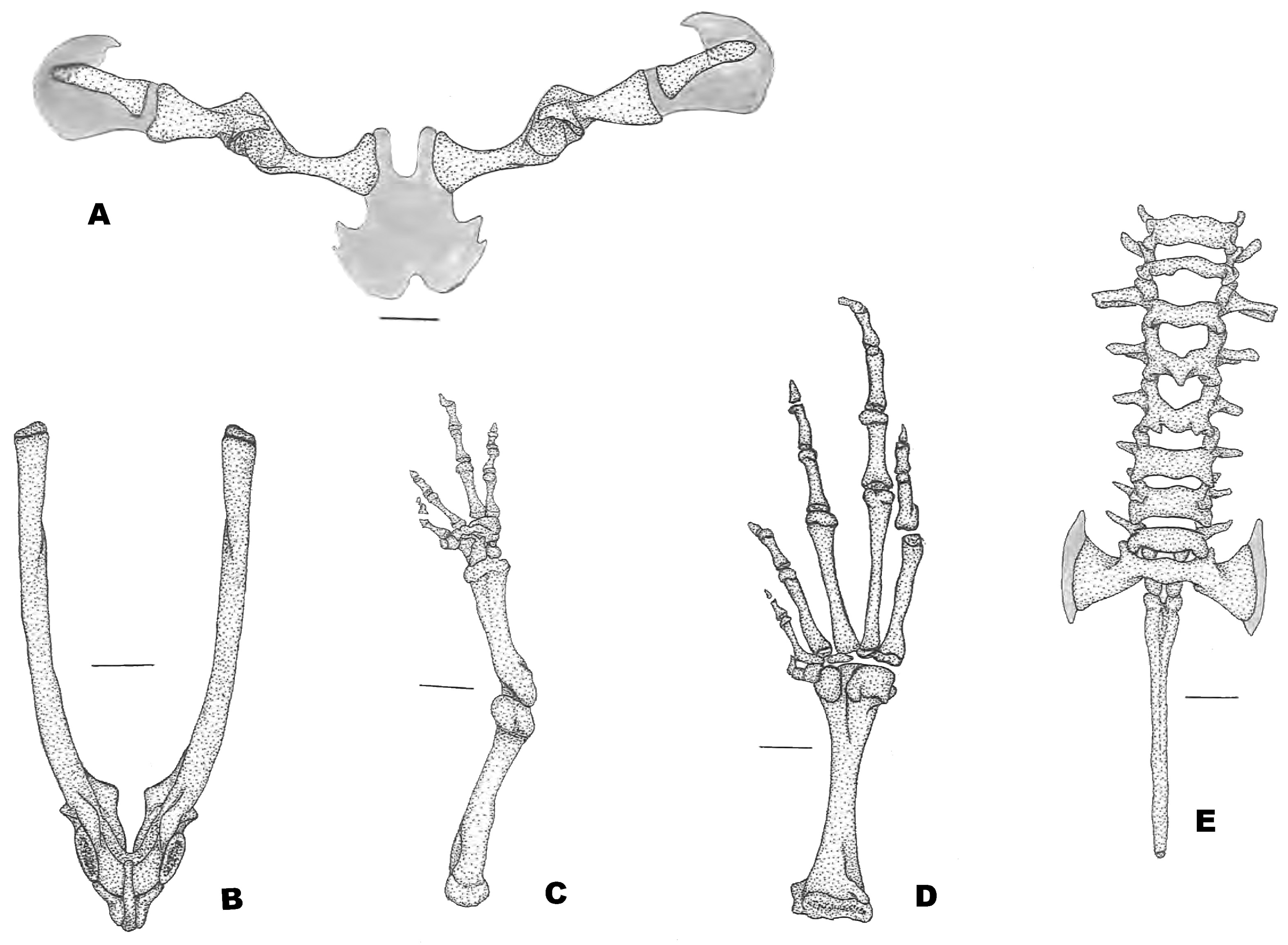
Axial Skeleton. Vertebral column ( Fig. 10E View FIGURE 10 ). The vertebral column consists of 8 presacral vertebrae (I–VII procoelous and VIII is diplasiocoelous. The atlas bears distinct and widely separated cervical cotyles. All vertebrae are non-imbricate and lack neural spines, centrum of vertebrae I–VI are wider than long, whereas that last two are more equal. Width of presacral and sacral vertebrae, including transverse processes, are: III> sacrum (including cartilaginous expansion)> IV> II> VIII> V = VI = VII> I. The transverse processes of presacral vertebrae are overall narrow with those of presacral V–VII being thinner (about than half the width of the anterior ones). The transverse processes of presacral III and V are oriented nearly perpendicular, whereas those of presacral IV are oriented slightly posteriorly, presacral II, VI–VIII are oriented anteriorly (more distinctly in II and VIII). Sacral diapophyses are perpendicular to the midline, broadly and symmetrically expanded. The lateral margins of the sacral diapophyses are continuous with a narrow cartilage that in its axial length reaches or passes the anterior tip of the transverse process of presacral VIII. This cartilage articulates with the anterior tip of the ilial shaft and shows some mineralization. The urostyle is rounded, lacking any lateral extension, and smooth; it is broader anteriorly just behind the point of articulation with the sacral vertebra.
Pectoral girdle ( Fig. 10A View FIGURE 10 ). The firmisternal pectoral girdle lacks clavicles and procoracoid cartilages. The sternum is a broad cartilaginous plate, lacking any mineralization and continuous with the epicoracoid cartilage; the latter is restricted between the coracoids. The coracoids are in a slight angle with the body’s midline; they are medially narrow. The coracoid and the scapula are robust, fully ossified, and fused with each other forming the glenoid fossa. The suprascapula is entirely cartilaginous, distally it is widely expanded and bears a distinct “hook-like” projecting posteriorly. The thin cleithrun lies on the anterior edge of the suprascapula, it is elongated and narrow, its proximal end is slightly broader.
Pelvic girdle ( Fig. 10B View FIGURE 10 ). In dorsal view, the space between the ilial shafts is U-shaped. Anteriorly and posterodorsally, the acetabulum is formed by the ilium and ischium, ventrally the acetabulum is bounded by a calcified pubis. A small crest is visible dorsal to the acetabulum.
Limbs. Manus ( Fig. 10C View FIGURE 10 ). The phalangeal formula is 3-3-2-2, all phalanges well ossified; length of digits is III> IV> II> I. The terminal phalanges are overall triangular with pointed distal tips. Proximally, the carpus consists of a large radiale and medium size ulnare, no intermedium visible. Carpal elements 3–5 are fused into a single element, which lies at the base of the metacarpals III–V. A single distal carpal 2 lies at the base of metacarpal II and also articulates with metacarpal III. Element Y lies between and articulating with the radiale proximally and distally with carpal 2 and the posterior edge of the proximal element of the prepollex. The prepollex is formed by the ossified proximal and a smaller distal element.
Pes ( Fig. 10D View FIGURE 10 ). The phalangeal formula is 2-2-3-4-3, terminal phalanges are overall triangular with pointed distal tips. Digit lengths are IV> III> V> II> I. The tarsus bears three tarsal elements: tarsal 3 is elongated and articulates mostly with digit 3 but also laterally with digit 2; tarsal 2 is minute and located between digits II, I, and tarsal 1; and tarsal 2 is large, rounded, and articulates with digit 1 and the proximal element of the prehallux, both proximal and distal elements of the prehallux are completely ossified.
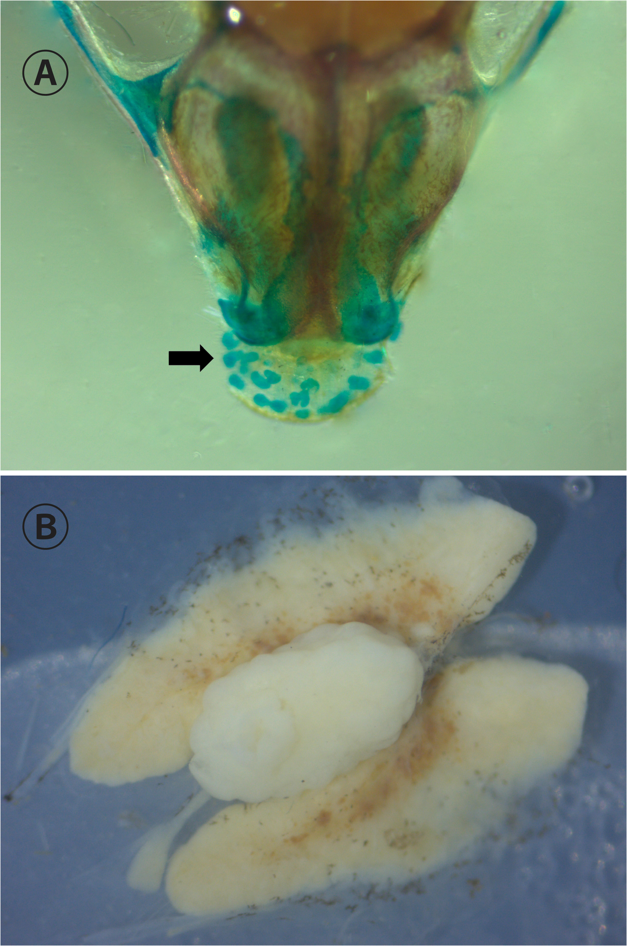
Diagnosis of Synapturanus . Based on the revision of S. mirandariberoi , S. salseri , S. rabus , S. latebrosus sp. nov., S. sacratus sp. nov., S. artifex sp. nov. (osteology and morphology) and the publication of further recently described species (Fouquet et al.., 2021b), we provide the following updated diagnosis for the genus: protruding snout supported by incomplete cartilaginous rings ( Fig. 11A View FIGURE 11 ); vertebral column diplasiocoelous, eight presacral vertebrae, sacral diapophyses expanded; maxillary arch incomplete, prevomers and quadratojugal absent; pars palatina of premaxillae posteriorly notched; pars palatina of maxillae anteriorly expanded; internasal bone present; palatines present (sometimes partially or completely fused with underlying bones); procoracoid and clavicles absent; terminal phalanges pointed; distal fingers and toes rounded, lacking disk-like expansion; transverse processes of urostyle as wide as those of the third vertebrae; adult males with a distinct wrist gland; males have a single and medial testicle ( Fig. 11B View FIGURE 11 ); W-shaped lower jaw; a small, medial, and unpigmented small area on tip of the lower jaw.
No known copyright restrictions apply. See Agosti, D., Egloff, W., 2009. Taxonomic information exchange and copyright: the Plazi approach. BMC Research Notes 2009, 2:53 for further explanation.
|
Kingdom |
|
|
Phylum |
|
|
Class |
|
|
Order |
|
|
Family |
|
|
SubFamily |
Otophryninae |
|
Genus |