
Microlia meticola ( Casey, 1911 )
|
publication ID |
https://doi.org/ 10.5281/zenodo.155781 |
|
DOI |
https://doi.org/10.5281/zenodo.6277381 |
|
persistent identifier |
https://treatment.plazi.org/id/C419FA02-FFB0-6279-CB19-A646B58DFD8A |
|
treatment provided by |
Plazi |
|
scientific name |
Microlia meticola ( Casey, 1911 ) |
| status |
|
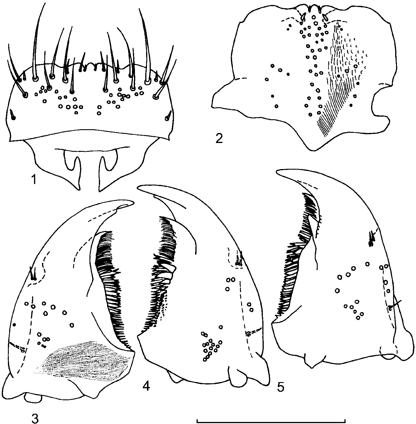
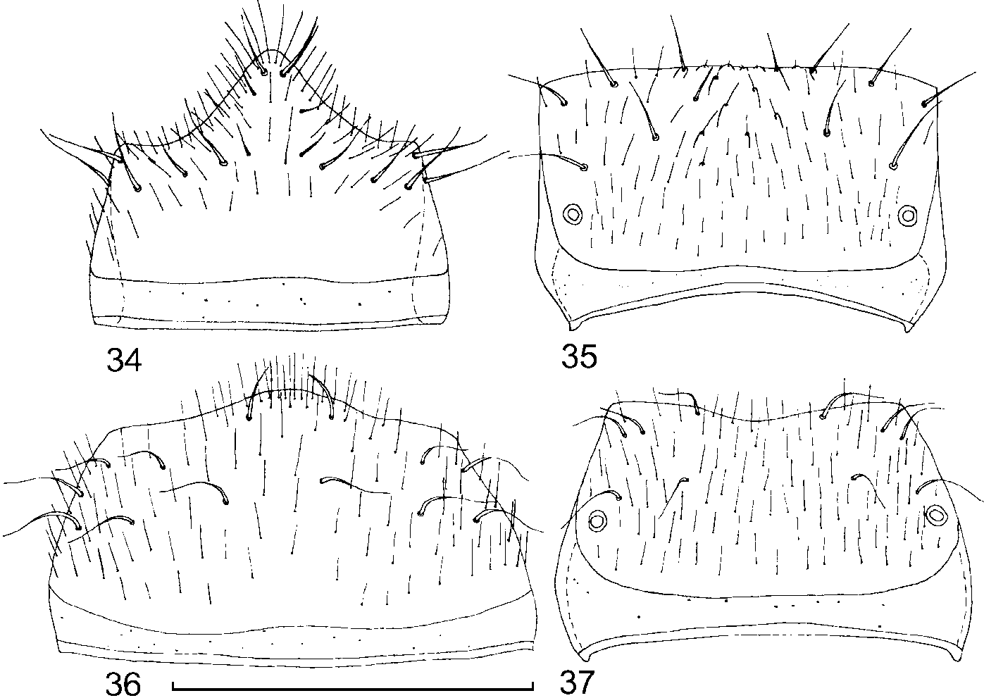
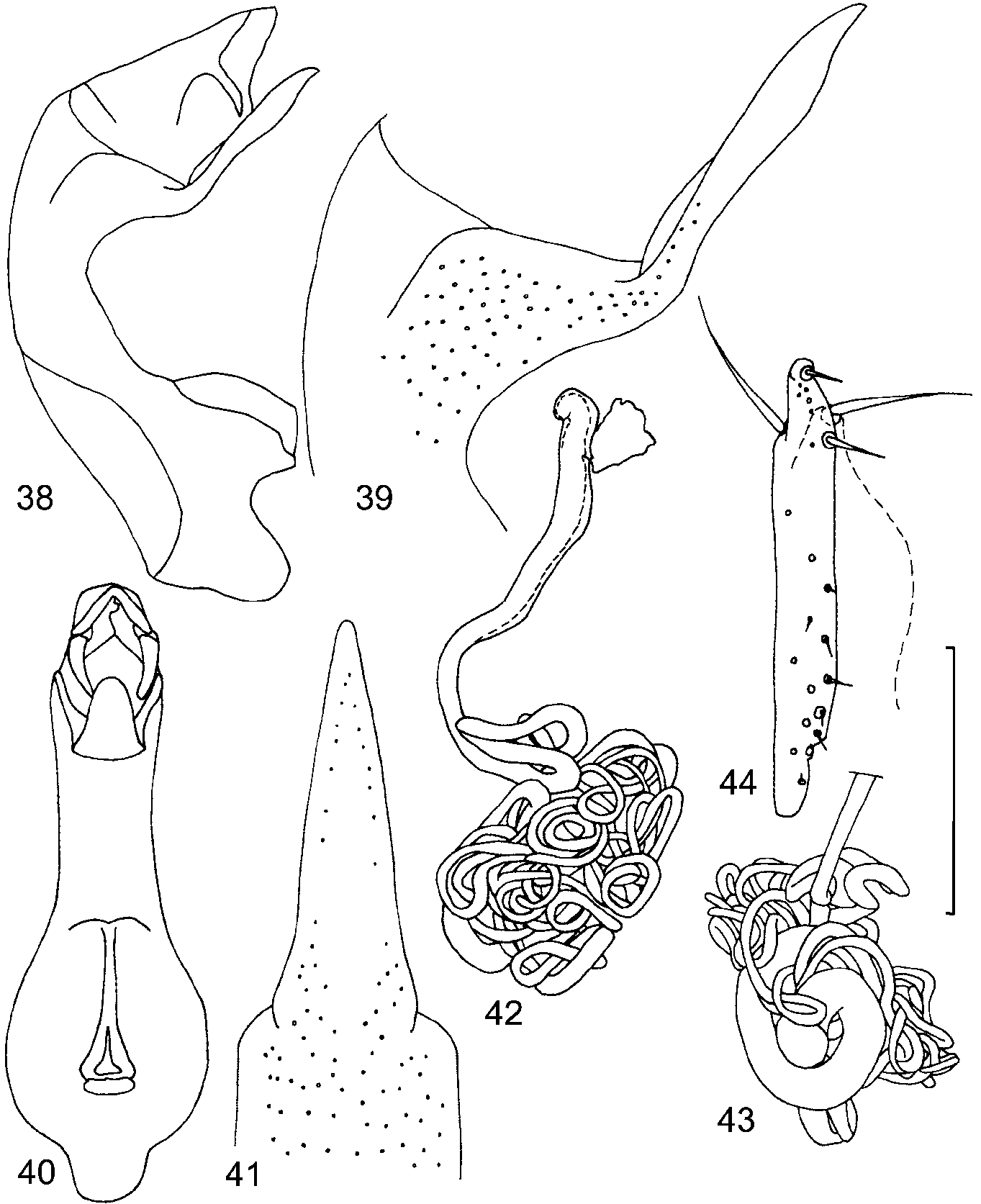
2. Microlia meticola ( Casey, 1911) View in CoL ( Figs. 115 View FIGURES 1 5 , 3444 View FIGURES 34 37 View FIGURES 38 44 )
Nosora meticola Casey, 1911: 146 View in CoL .
Nosora meticola: Leng, 1920: 122 View in CoL .
Nosora meticola: Fenyes, 1920: 308 View in CoL .
Nosora meticola: Bernhauer & Scheerpeltz, 1926: 717 View in CoL . Nosora meticola: Moore & Legner, 1975: 456 . Nosora meticola: Seevers, 1978: 143 .
Type material. Lectotype:, UNITED STATES: Arizona: Nogales (Wickham) ( NMNH). This reference to the lectotype is not to be considered as lectotype designation. The lectotype will be designated by Hanley (in press, c).
Additional material: UNITED STATES: Arizona: Cochise Co.: 23 specimens, Portal, Soutwest Research Station, dead Cucurbita flowers (H. & A. Howden), 22.vi.1956;, ditto but beating oak, 24.vi.1956;, ditto but 28.vi.1956 (all in CNC); 67 specimens, Southwest Research Station, Datura flowers (M.Weiser), 29.vii.1993 ( KSEM); Texas:, 2 miles W of Fort Davis (H. & A. Howden), 14.vii.1956 ( CNC).
Diagnosis: Microlia meticola can be distinguished from other species of Microlia by the shape of the aedeagus, particularly the apical process of median lobe ( Figs. 3841 View FIGURES 38 44 ) and spermatheca with numerous irregular loops ( Fig. 4243 View FIGURES 38 44 ).
Description: Length 1.92.1 mm. Head from brownish yellow to brown; pronotum from brownish yellow to brown, with lighter borders; elytra from yellowish brown with lighter humeral angles to brown; abdominal segments 34 yellow to brown, segments 57 brown, segment 8 brownish yellow to brown; 4 or 5 basal antennal articles brownish yellow, 6 or 7 apical articles brown; legs and mouthparts yellow; in most specimens pronotum and abdominal segments 34 and 8 lighter than head, elytra and abdominal segments 57.
Head surface glossy, on disk with weak isodiametric microsculpture, puncturation fine, distance between punctures 12 times their diameter. Eyes 2.53.5 times longer than temples.
Pronotum strongly transverse, 1.3 times wider than head, width 0.440.56 mm, length 0.290.39 mm, width to length ratio 1.5, surface glossy, with weak and poorly visible (at x70) transverse microsculpture; puncturation as on head or stronger, distance between punctures 12 times their diameter. Elytra wider (0.510.66 mm) and longer (0.460.54 mm, measured from humeral angle) than pronotum (pronotal length to elytral length ratio 0.68), 1.2 times wider than long, surface glossy, with weak transverse microsculpture, puncturation slightly asperate, distance between punctures 12 times their diameter. Mesotarsus with 4 segments.
Abdominal terga glossy, with fine and poorly visible (at x70) microsculpture consisting of meshes, with fine puncturation, distance between punctures 13 times their diameter.
Male tergum 7 in front of posterior margin and posterior half of male tergum 8 with numerous longitudinal tubercles, posterior margin of male tergum 8 straight ( Fig. 35 View FIGURES 34 37 ). Posterior margin of male sternum 8 with big triangular lobe ( Fig. 34 View FIGURES 34 37 ). Aedeagus as in Figs. 3841, 44 View FIGURES 38 44 . Median lobe with long and narrow apical process ( Figs. 41 View FIGURES 38 44 ).
Female tergum 8 with concave posterior margin ( Fig. 37 View FIGURES 34 37 ), sternum 8 with convex posterior margin ( Fig. 36 View FIGURES 34 37 ). Spermatheca forming numerous irregular loops ( Fig. 4243 View FIGURES 38 44 ). No female accessory sclerites.
Variability: Body coloration varies from yellowish brown (as in M. silacea ) in light specimens to entirely brown in dark specimens. The strength of pronotal puncturation is variable.
Distribution: Known from Arizona and Texas ( Fig. 80 View FIGURE 80 ).
Natural History: Long series of M. meticola were collected in flowers of Cucurbita and Datura .

No known copyright restrictions apply. See Agosti, D., Egloff, W., 2009. Taxonomic information exchange and copyright: the Plazi approach. BMC Research Notes 2009, 2:53 for further explanation.
|
Kingdom |
|
|
Phylum |
|
|
Class |
|
|
Order |
|
|
Family |
|
|
SubFamily |
Aleocharinae |
|
Tribe |
Hoplandriini |
|
Genus |
|
Kingdom |
|
|
Phylum |
|
|
Class |
|
|
Order |
|
|
Family |
|
|
SubFamily |
Aleocharinae |
|
Tribe |
Hoplandriini |
|
Genus |
|
Kingdom |
|
|
Phylum |
|
|
Class |
|
|
Order |
|
|
Family |
|
|
SubFamily |
Aleocharinae |
|
Tribe |
Hoplandriini |
|
Genus |
|
Kingdom |
|
|
Phylum |
|
|
Class |
|
|
Order |
|
|
Family |
|
|
SubFamily |
Aleocharinae |
|
Tribe |
Hoplandriini |
|
Genus |
|
Kingdom |
|
|
Phylum |
|
|
Class |
|
|
Order |
|
|
Family |
|
|
SubFamily |
Aleocharinae |
|
Tribe |
Hoplandriini |
|
Genus |
|
Kingdom |
|
|
Phylum |
|
|
Class |
|
|
Order |
|
|
Family |
|
|
SubFamily |
Aleocharinae |
|
Tribe |
Hoplandriini |
|
Genus |
|
Kingdom |
|
|
Phylum |
|
|
Class |
|
|
Order |
|
|
Family |
|
|
SubFamily |
Aleocharinae |
|
Tribe |
Hoplandriini |
|
Genus |
|
Kingdom |
|
|
Phylum |
|
|
Class |
|
|
Order |
|
|
Family |
|
|
SubFamily |
Aleocharinae |
|
Tribe |
Hoplandriini |
|
Genus |
|
Kingdom |
|
|
Phylum |
|
|
Class |
|
|
Order |
|
|
Family |
|
|
SubFamily |
Aleocharinae |
|
Tribe |
Hoplandriini |
|
Genus |
|
Kingdom |
|
|
Phylum |
|
|
Class |
|
|
Order |
|
|
Family |
|
|
SubFamily |
Aleocharinae |
|
Tribe |
Hoplandriini |
|
Genus |
|
Kingdom |
|
|
Phylum |
|
|
Class |
|
|
Order |
|
|
Family |
|
|
SubFamily |
Aleocharinae |
|
Tribe |
Hoplandriini |
|
Genus |
Microlia meticola ( Casey, 1911 )
| Gusarov, Vladimir I. 2002 |
Nosora meticola:
| Seevers 1978: 143 |
| Moore 1975: 456 |
| Bernhauer 1926: 717 |
Nosora meticola:
| Leng 1920: 122 |
Nosora meticola:
| Fenyes 1920: 308 |
Nosora meticola
| Casey 1911: 146 |