
Ronzotherium elongatum Heissig, 1969
|
publication ID |
https://doi.org/ 10.5852/ejt.2021.753.1389 |
|
publication LSID |
lsid:zoobank.org:pub:8009DD3B-53B0-45C9-921E-58D04C9C0B48 |
|
DOI |
https://doi.org/10.5281/zenodo.4959524 |
|
persistent identifier |
https://treatment.plazi.org/id/C53FFD4D-D711-FFBD-FDBB-D9221E00A413 |
|
treatment provided by |
Felipe |
|
scientific name |
Ronzotherium elongatum Heissig, 1969 |
| status |
|
Ronzotherium elongatum Heissig, 1969
Figs 8–10 View Fig View Fig View Fig
Ronzotherium filholi elongatum Heissig, 1969: 46–55 , 68, 71, 116, 119, fig. 18d (from Pernes and Kleinblauen).
Rhinoceros filholi – Jenny 1905: 125.
Aceratherium filholi – Jenny 1905: 125. — Roman 1910: 1559 (from Pernes and Kleinblauen); 1912a: 17, 27, 45–50, 57–58, figs 14.1, 15, 18, pl. V figs 1–2 (from Pernes and Kleinblauen); 1912b: 360– 364, fig. 2. — Stehlin 1914: 185 (from Kleinblauen). — Gignoux 1928: 148, 151, fig. 3 (from Pernes and Kleinblauen).
Praeaceratherium filholi – Spillmann 1969: figs 11, 13, 16.
Ronzotherium filholi – Brunet 1979: 105, table 2 (from Pernes and Kleinblauen). — Becker 2003: 212–213, 230–231, 234, 256, pl. II fig. a–d (from Kleinblauen); 2009: 490, 493–495, fig. 4h–l, table 1 (from Kleinblauen). — Ménouret & Guérin 2009: 296 (from Pernes and Kleinblauen).
Non Ronzotherium filholi elongatum – Heissig 1969: 46–55, figs 16–17, 18a–c, 19 (from Villebramar, Bumbach, Montans, Cournon).
Historical diagnosis
From Heissig (1969), translated by the authors: “A subspecies of Ronzotherium filholi with the following characteristics: corpus mandibulae low, very slender, fossa masseterica deeply concave, foramen mandibulae at about the level of the teeth neck, strongly enlarged, symphysis long, flat forward; i2 still shearing towards I1, i1 present; angle of jaw branches very pointed; upper molars elongated with very broad medisinus, extremely short post-fossette and strong lingual cingulum; upper P3 and P4 premolariform to semimolariform, P2 molariform, protocone and hypocone widely separated, all upper premolars strongly widened, inside slightly rounded, metaloph curved and S-shaped, often with complicated folds, hypostyle missing; lower molars with strong labial cingulum and relatively long anterolingual cingulum, relatively long, narrow and conspicuously low, talonid pit unclear or notched; lower premolars, especially p3 often lengthened to the front, protoconid fold strong, metalophid strongly backwards, labial cingulum strong, p2 strongly narrowed, p1 single-rooted.”
However, this diagnosis is not only based on the type material, but also on referred material from other localities, such as Villebramar or Bumbach that we refer to other species. We thus propose an emended diagnosis.
Emended diagnosis
The paraoccipital process is poorly developed. The roots of the upper cheek teeth are lingually fused, P2 is molariform with a lingual bridge connecting the protocone and hypocone, the protocone and hypocone form a lingual wall on P3 and P4, with a well-marked lingual groove above the cingulum, especially on P4. Upper premolars usually bear a simple crochet, the protocone is slightly constricted, the metaloph curved and S-shaped and the hypostyle missing. The protocone is usually constricted on upper molars and the lingual cingulum is strong and continuous, except under the hypocone of M1–2 and the protocone of M2. The labial cingulum of the lower molars is always present and continuous.
Differs from Ronzotherium filholi by the presence of a processus postorbitalis on the zygomatic arch and by its poorly developed processus paraoccipitalis.
Type material
Holotype
FRANCE • two-parts well preserved skull with almost complete cheek teeth rows, the two parts are joined together by plaster, which does not reflect the original morphology; Vaucluse, Pernes-les- Fontaines; probably MP23; FSL-9601 .
Additional material
No other material is known from this locality.
Type horizon and locality
Pernes (= Pernes-les-Fontaines, Vaucluse, France), probably dated from MP23. The ‘sands and green sandstones of the Valette-de-Pernes’ in which this skull was found, have been dated from MP 23 in Murs, another locality 20 km from Pernes.
Stratigraphical distribution
Early Oligocene.
Geographical distribution
France: Pernes. Switzerland: Kleinblauen.
Description
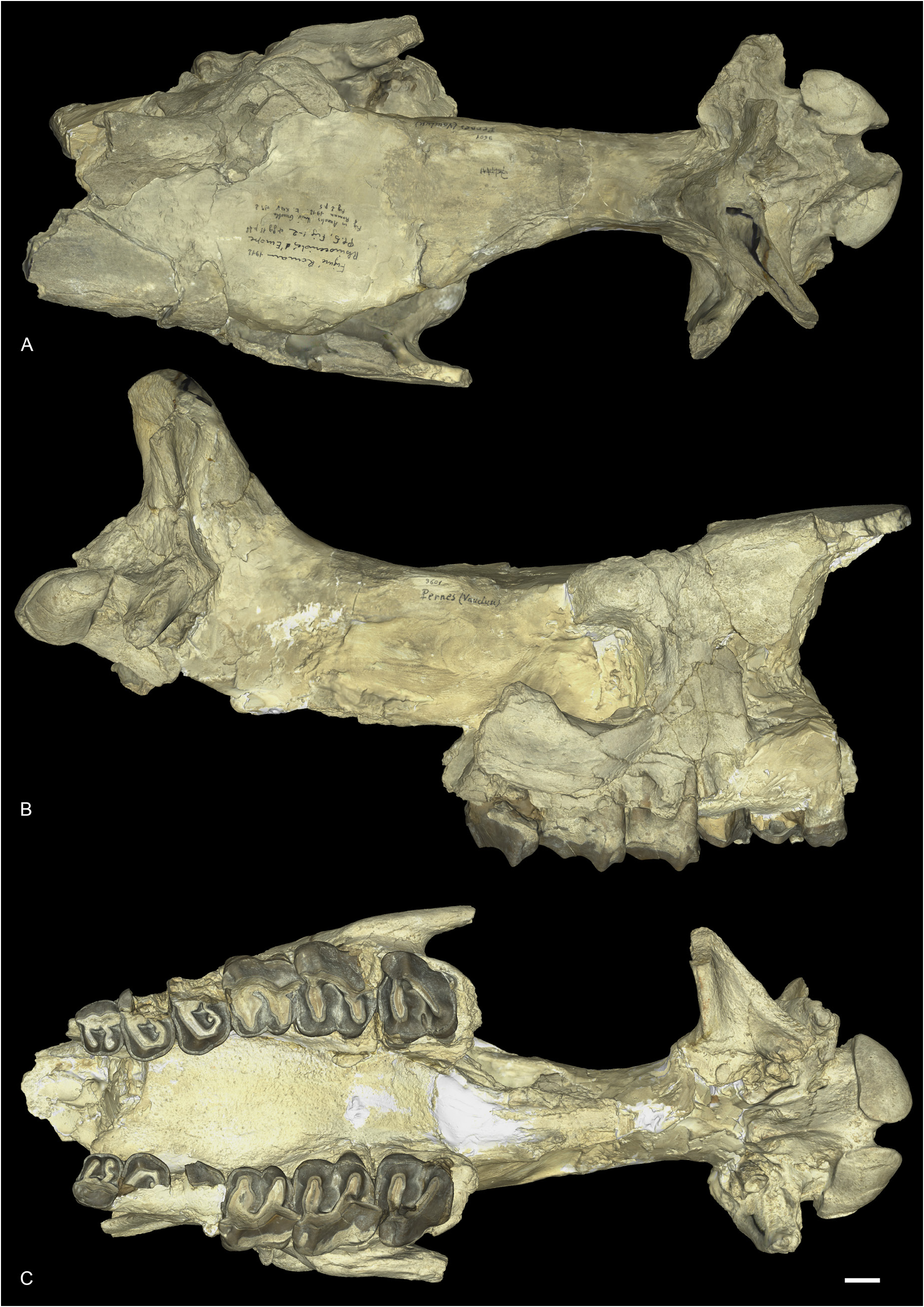
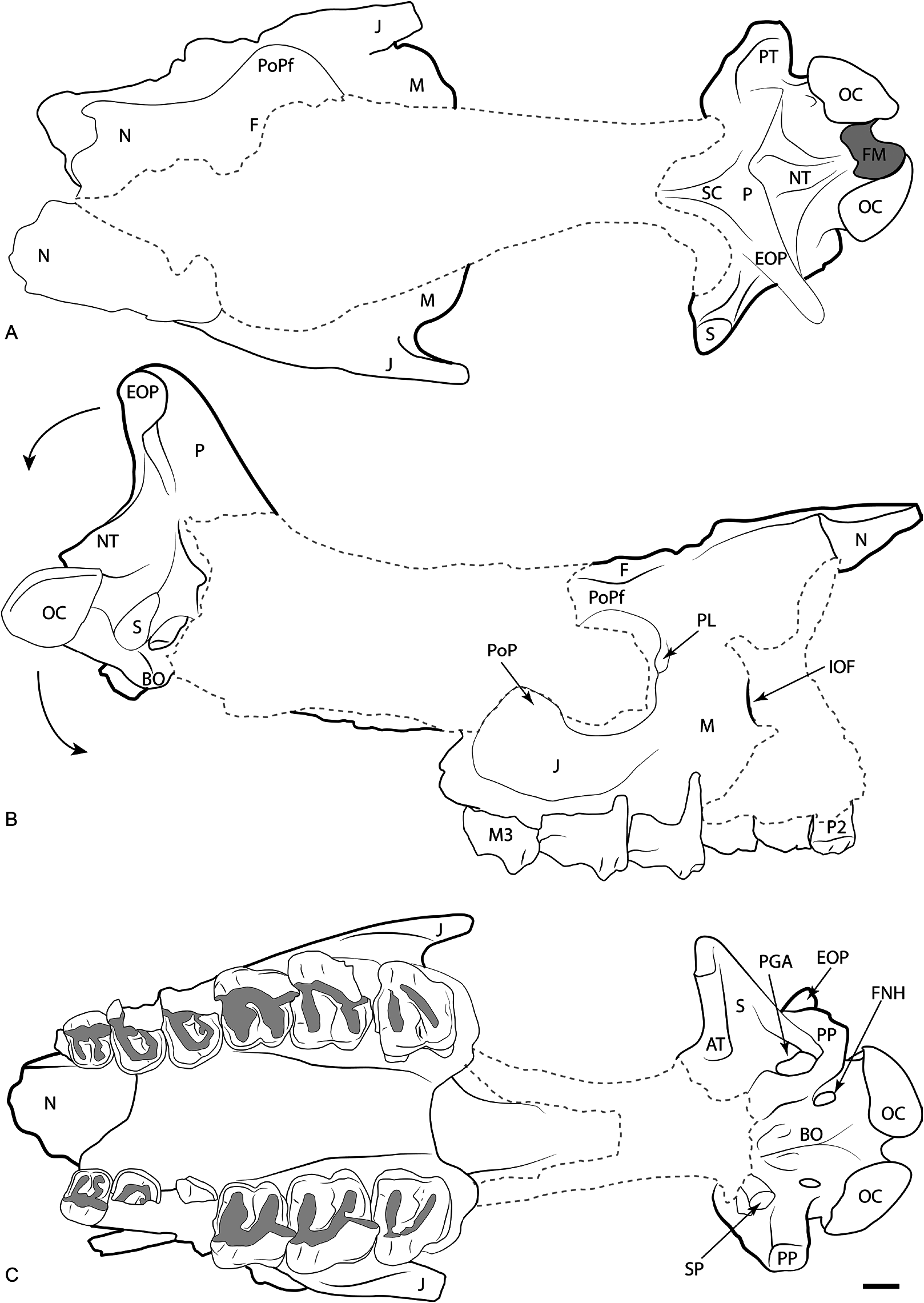
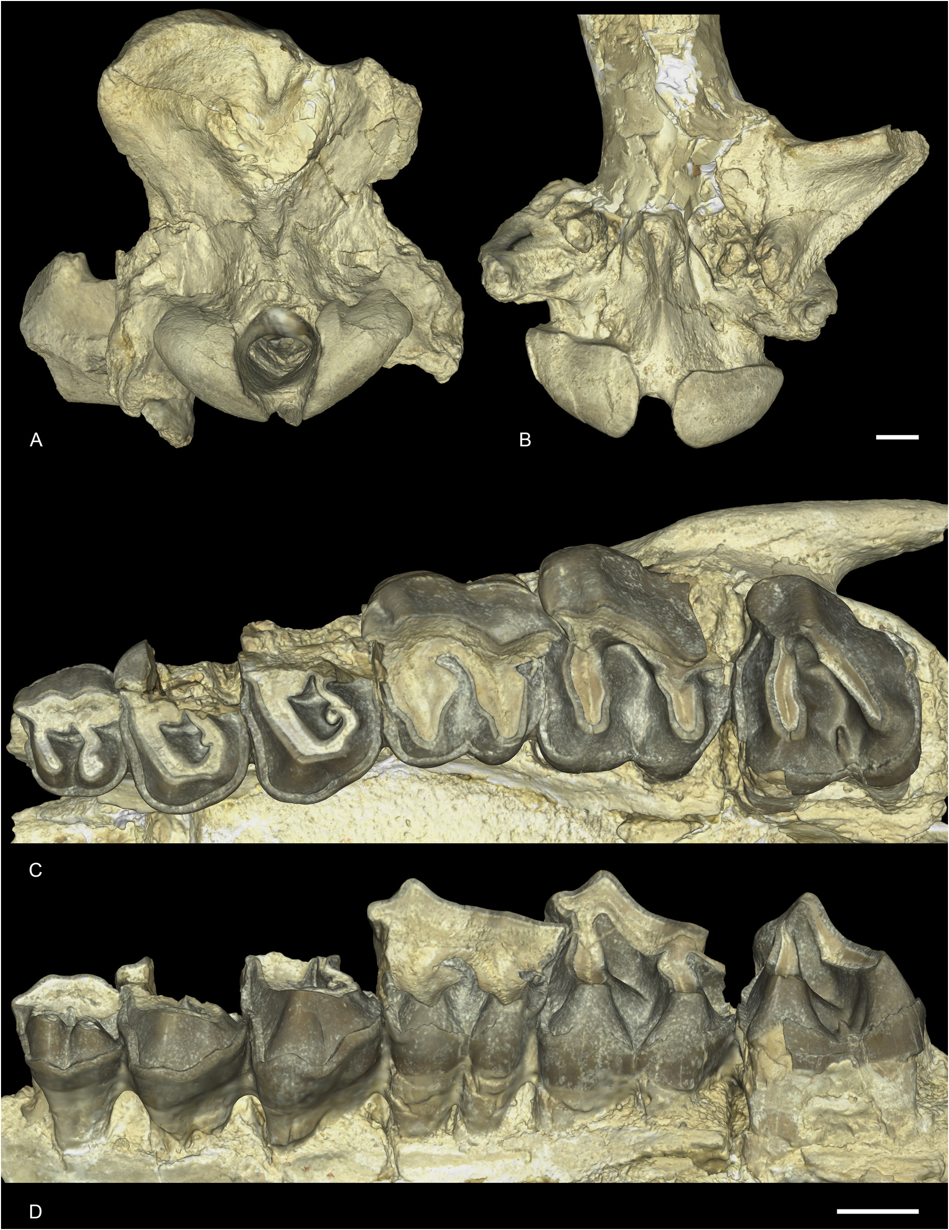
SKULL. The skull was originally described by Roman (1912a, 1912b), who attributed it to Ronzotherium filholi . It is heavily reconstructed in plaster, especially the frontals and parietals, but it is nonetheless possible to identify the original bony material ( Figs 8–9 View Fig View Fig ). The nasals are very fragmentary, the anterior part is broken. The lateral apophysis is not preserved. The infraorbital foramen opens above P4. The posterior border of the nasal incision is above P3 and the anterior border of the orbit is above the middle of M1. The lachrymal process is well developed and there is a large postorbital process of the frontals above the orbit. Only the anterior parts of the jugal bones are preserved, and the anterior base of the zygomatic arch is high above the teeth neck. The postorbital process of the zygomatic arch is large and on the jugal. The squamosals are not preserved. The dorsal profile of the skull is difficult to interpret, because of the heavy reconstruction, yet it was probably concave, though not as much as suggested by the reconstruction. The area between the temporal and nuchal crests is very concave. The external auditory pseudomeatus is ventrally open, between the postglenoid and posttympanic apophyses. The nuchal tubercle is well-developed. From the preserved part of the parietal bone, we can observe a wide parietal crest. The occipital crest is concave. In ventral view, the anterior part of the zygomatic arch does not strongly diverge from the maxilla. The vomer is badly preserved. The articular tubercle of the squamosal is smooth and tranversally straight. The postglenoid apophysis is rounded and convex anteriorly, and anteroposteriorly elongated. The foramen nervi hypoglossi is in the middle of the condylar fossa. There is a strong and high sagittal crest on the basilar process of the basioccipital. In occipital view, the paraoccipital and posttympanic processes are fused. The posttympanic process is well-developed and the paraoccipital process is partly broken. The foramen magnum is circular. There is neither a median crest nor a medial truncation on the occipital condyles.
UPPER CHEEK TEETH. No anterior teeth are preserved on the skull, only the cheek teeth ( Figs 8B–C View Fig , 9B–C View Fig , 10C–D View Fig ). The three molars are well preserved on both sides, but the ectolophs of P3–4 are missing, whereas P2 is well preserved and P1 is absent on both sides. There is, however, a single broken root still preserved on the left side which means that this tooth was present in the juvenile at least. The premolar series is short compared to the molar series (LP3–4/LM1–3 = 0.48). There are no enamel folds and the cement is absent. The crown of the cheek teeth is low.
The labial cingulum is strong and continuous on P2, but the ectolophs are broken on P3–4 so we cannot determine whether it was present or absent. The lingual cingulum is very strong and continuous on P2–4 and is rippled in lingual view, especially on P4. There is a short but well-defined crochet on P3–4. It is simple, directed towards the protocone and completely missing on P2. The metaloph is not constricted and the postfossette is narrow. The antecrochet is always absent. The protocone and hypocone of P2 are connected by a low bridge and are rather equal in size. The protoloph of P2 is directed slightly postero-lingually while the metaloph is S-shaped and transverse. They are both joining the ectoloph. The paracone and metacone folds of P2 are present and wide. The medifossette is always absent on premolars and the protocone is never constricted. The protocone and hypocone of P3–4 form a lingual wall, and a lingual groove is present. The metaloph of P3–4 is S-shaped and directed postero-lingually. The protoloph and metaloph of P3–4 are connected to the ectoloph.
The labial cingulum is strong under the metastyle of M1–2 and the parastyle of M1 but is absent otherwise. The lingual cingulum is also strong and almost completely continuous on all upper molars. It is only fainted under the hypocone of M1 and the protocone of M2. The anterior and posterior cingulum are continuous. The antecrochet is present, but poorly defined and only appears effectively on the protoloph with very strong wear. The crochet, crista and medifossette are always absent on upper molars. The protocone is always weakly constricted. The paracone fold is strong and there is neither a metacone fold nor a mesostyle. The metastyle and metaloph are long and the posterior part of the ectoloph is straight. The hypocone is never constricted and the anterior groove of the metaloph is very shallow or absent. The postfossette is short, but deep, below the posterior cingulum. The ectoloph and metaloph of M3 are completely fused, and the posterior groove is very shallow. It is quadrangular in occlusal view. The protoloph is transverse. There is a small crest in the median valley of the left M3, that seem to have been broken on the right one. It may be caused by individual variation and is completely absent on other molars.
Remark
This species is the most recently one erected, though it was originally considered a subspecies of R. filholi . Brunet (1979) and subsequent authors considered it as a junior synonym of R. filholi . Based on our comparative work and our phylogeny, we consider it as a valid species.
No known copyright restrictions apply. See Agosti, D., Egloff, W., 2009. Taxonomic information exchange and copyright: the Plazi approach. BMC Research Notes 2009, 2:53 for further explanation.
|
Kingdom |
|
|
Phylum |
|
|
Class |
|
|
Order |
|
|
Family |
|
|
Genus |
Ronzotherium elongatum Heissig, 1969
| Tissier, Jérémy, Antoine, Pierre-Olivier & Becker, Damien 2021 |
Ronzotherium filholi elongatum
| Heissig K. 1969: 55 |
Ronzotherium filholi elongatum
| Heissig K. 1969: 46 |
Rhinoceros filholi
| Jenny F. 1905: 125 |
Aceratherium filholi
| Gignoux M. 1928: 148 |
| Stehlin H. G. 1914: 185 |
| Roman F. 1910: 1559 |
| Jenny F. 1905: 125 |