
Ronzotherium heissigi, Tissier & Antoine & Becker, 2021
|
publication ID |
https://doi.org/ 10.5852/ejt.2021.753.1389 |
|
publication LSID |
lsid:zoobank.org:pub:8009DD3B-53B0-45C9-921E-58D04C9C0B48 |
|
DOI |
https://doi.org/10.5281/zenodo.4959444 |
|
persistent identifier |
https://treatment.plazi.org/id/D1CFA5AE-6BC9-479B-AE43-F46EB9D86A49 |
|
taxon LSID |
lsid:zoobank.org:act:D1CFA5AE-6BC9-479B-AE43-F46EB9D86A49 |
|
treatment provided by |
Felipe |
|
scientific name |
Ronzotherium heissigi |
| status |
sp. nov. |
Ronzotherium heissigi sp. nov.
urn:lsid:zoobank.org:act:D1CFA5AE-6BC9-479B-AE43-F46EB9D86A49
Figs 22–26 View Fig View Fig View Fig View Fig View Fig
Acerotherium lemanense – Roman 1912a: 61–62, pl. VII (from Lamothe-Capdeville).
Aceratherium filholi – Stehlin 1914: 183, 85 (Bumbach).
Ronzotherium filholi – Lavocat 1951: 116, pl. 19 fig. 3, pl. 26 fig. 1 (from Vendèze). — Brunet 1979: 105 (from Bumbach). — Becker 2003: 213–214.
Ronzotherium velaunum – Heissig 1969: 20–36, 77, fig. 8b (from Vendèze).
Ronzotherium filholi elongatum – Heissig 1969: 46–55, 71, 75–77, 82–83 (from Bumbach).
Ronzotherium filholi romani – Heissig 1969: 63 (from Lamothe-Capdeville).
Ronzotherium romani – Brunet 1979: 135–136, fig. 15, pls XVII–XVIII (from Vendèze).
Diaceratherium lemanense – Antoine & Becker 2013: 140 (from Lamothe-Capdeville).
Diagnosis
Differs from R. romani by the mandibular ramus inclined forward, the P1 sometimes absent and without anterolingual cingulum, the angular and V-shaped external groove of the lower cheek teeth, the lower premolars without lingual cingulum, the d/p1 always absent in the adult, the deep and wide gutter for the m. extensor carpi on the radius, the concave proximal border of the anterior side of the magnum, the salient insertion of the m. extensor carpalis of the metacarpals and the oval proximal side of the cuboid.
Differs from R. velaunum by the presence of a lingual groove on the corpus mandibulae, the curved and not constricted paralophid on p2 and the deep median constriction of the distal humeral articulation.
Differs from R. filholi by a foramen infraorbitalis above P3, a zygomatic width/frontal width ratio above 1.5, a concave occipital crest, the reduced paraconid on p2, the high posterior expansion of the scaphoid facet on the radius, the open angle between the diaphysis of the ulna and the olecranon and the curved magnum facet on the McII.
Differs from R. elongatum by the absence of processus lacrymalis, the reduction of the postorbital process on the zygomatic arch, its poorly developed processus posttympanicus and by the metaloph of P2 directed postero-lingually.
Further differs from R. filholi and R. elongatum by a convex processus postglenoidalis of the squamosal and by a narrow and V-shaped lingual opening of the lower premolars.
Etymology
The specific epithet honours Prof. Dr Kurt Heissig for his major and imperishable contributions on the study of the Rhinocerotidae , and for providing the first systematic revision on Ronzotherium more than 40 years ago.
Type material
Holotype
FRANCE • complete skull and associated mandible; Auvergne-Rhône-Alpes, Cantal , Vendèze near St- Flour; 45°02′36.8″ N, 3°06′13.1″ E; MNHN.F.LIM181. GoogleMaps
According to the MNHN registry, it was discovered by M. Lauby (possibly Antoine Lauby), but sold to the MNHN by M. Hugon, from St-Flour on the 19 th of June 1909. It bears the old MNHN inventory number MNHN.F.1909-25.
Additional material
SWITZERLAND – Bumbach (MP25) • 1 very poorly preserved and incomplete skull; NMB-UM-200 • 1 fragment of parietal bone with occipital crest; NMBE-5035820 • 1 P 1; NMB-UM-463 • 1 P 2; NMB-UM-126a • 1 P 2; NMBE-5014494 • 1 P 3; NMBE-5035822 • 1 P 3; MGL-4264 • 2 P 4; MGL-5265 , MGL-5266 • 1 M 2; NMBE-5014495 • 1 subcomplete mandible; NMB-UM-6132 • 1 d2; MGL-5275 • 1 d4; NMB-UM-13 • 1 p 2; NMBE-5035824 • 1 p 3; MGL-5274 • 1 fragment of mandible with p3–4; NMBE-5035825 • 2 p 4; NMBE-5035826 , NMBE-5035827 • 1 p 4; NMB-UM-6133 • 1 m 1; NMB-UM-806 • 1 m 1; NMBE-5035828 • 1 m 2; NMB-6278 • 3 m 2; NMBE-5035829 , NMBE-5035830 , NMBE-5035831 • 1 m 3; NMBE-5035832 • 2 incomplete humeri; NMB-UM-132 , NMB-UM-129a • 1 radius; NMB-UM127a • 1 ulna; NMB-UM-131b , NMB-UM-131c • 1 lunate; NMBE-5035833 • 2 trapezoids; NMB-UM-6136b , NMB-UM-6 • 1 magnum; NMB-UM-6136c • 1 McII; NMB-UM-6136a • 1 McII; NMB-UM-121 • 1 proximal fragment of femur; NMBE-5035835 • 1 cuboid; NMBE-5035834 .
Type horizon and locality
Vendèze near St-Flour, Cantal, Auvergne-Rhône-Alpes, France ( MP 24, late early Oligocene), approximative coordinates: 45°02′36.8″ N, 3°06′13.1″ E.
Stratigraphical distribution
MP24–MP25.
Geographical distribution
France: ‘Auvergne’ (without precision, which could possibly correspond to Vendèze), Lamothe- Capdeville, Vendèze. Switzerland: Bumbach.
Description
Holotype
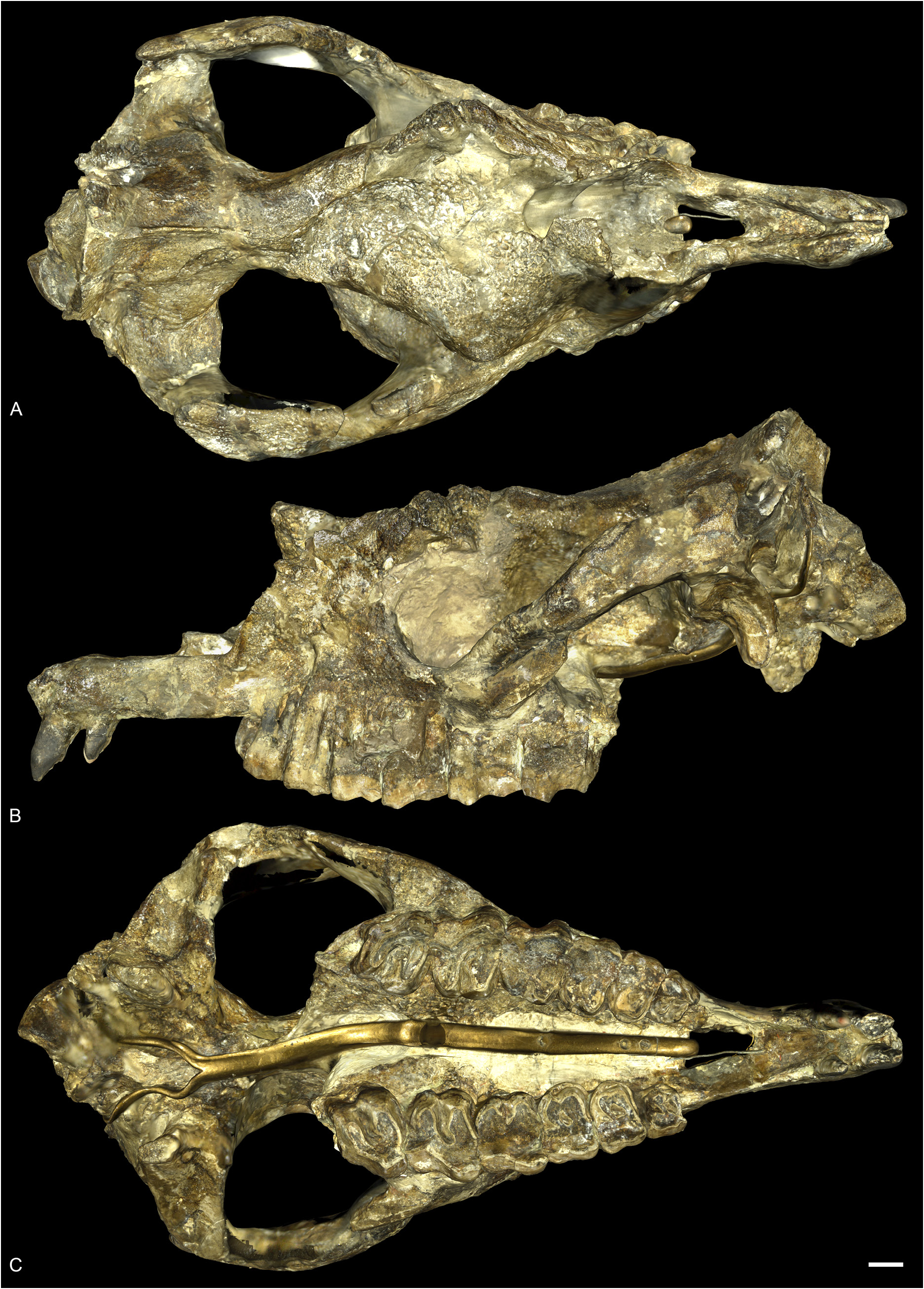
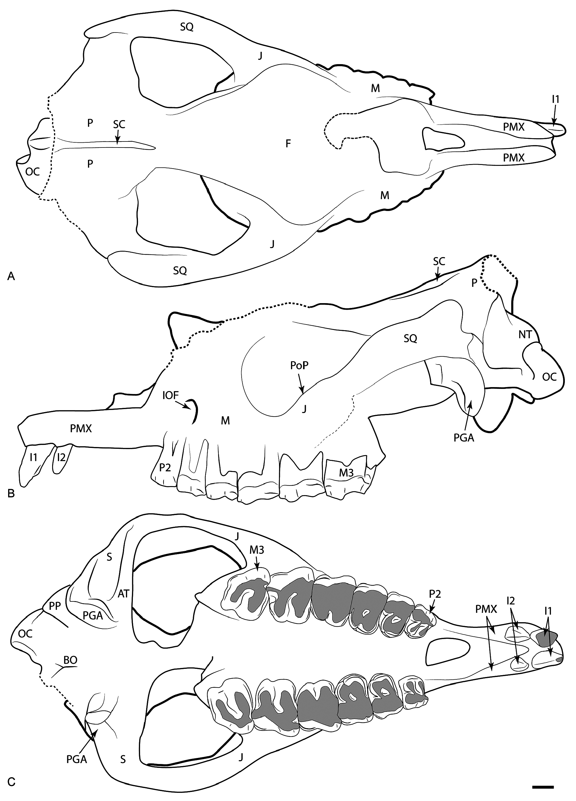
SKULL. The skull MNHN.F.LIM181 is quite well preserved but the nasals are broken ( Figs 22–23 View Fig View Fig ). The premaxillae are long and contact each other only at their anterior extremity. The nasal notch extends up to P2 and the foramen infraorbitalis is located above P3. The nasal septum is not ossified. The suture between the nasals and the lacrimal is not visible and the lacrimal process is absent. The orbit is large and its anterior border is above the anterior side of M1. The processus postrobitalis of the frontal is large. The anterior base of the zygomatic process is high above the teeth neck. The zygomatic arch is high in lateral view, it is almost reaching the dorsal border of the skull. The postorbital process of the zygomatic arch is almost absent and very poorly distinguishable. The dorsal profile of the skull is overall concave in lateral view. The external auditory pseudomeatus is partially closed and the occipital side is inclined forward. The nuchal tubercle is developed. The back of the teeth row is in the posterior half of the skull in lateral view. In dorsal view, the skull is brachycephalic and it is hornless. The orbit is not laterally projected. The zygomatic width/frontal width ratio is above 1.5. A very thin sagittal crest is present, and the occipital crest is not preserved. In ventral view, the anterior tip of the zygomatic arch diverges progressively from the maxilla, without a sharp angle. The vomer and most of the basicranium are not preserved. The articular tubercle of the squamosal is rather smooth and its transverse profile is straight. The anterolateral sides of the processus postglenoidalis form a right dihedron. There is no posterior groove on the zygomatic process. The processus posttympanicus and paraoccipitalis are fused at their base. The processus post-tympanicus is poorly developed while the paraoccipitalis is developed. The foramen magnum is circular and there are no median ridges on the condyles, neither medial truncation.
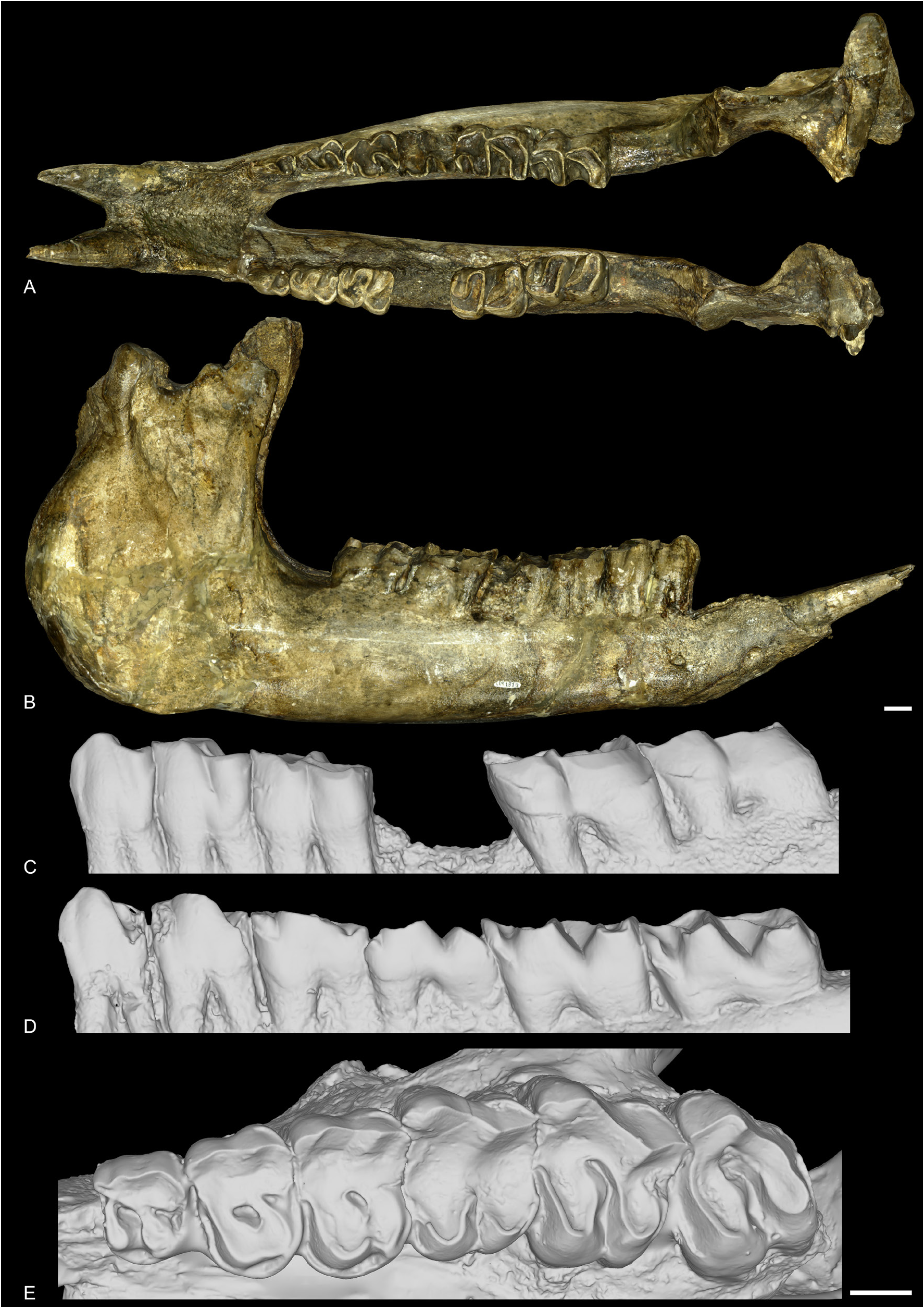
MANDIBLE. The mandibular symphysis ( Fig. 24A–B View Fig ) is slightly upraised and it is long and massive in dorsal view. Its posterior margin is at the level of p2. There are two mental foramen, one bellow p2 and one below the root of i2. The lingual groove of the sulcus mylohyoideus is slightly marked on the lingual
border of the corpus. The ventral base of the corpus is completely straight. The ramus is inclined forward in lateral view, and the coronoid process is well developed. In medial view, the foramen mandibulare is located below the teeth neck.
DENTITION. The complete dental formula is I1–2, P2–M3 / i2, p2–m3. The Length(P3–4)/Length(M1–3) ratio is between 0.42 and 0.5. The cement is absent and the crowns are very low ( Figs 22–24 View Fig View Fig View Fig ).
I1 is oval in cross-section, pointed and not chisel-shaped. It is directed downwards, and it bears two crests, one anterior and one posterior. There was no contact with the lower incisor (no visible wear on any of them). It is separated from I2 by a very short diastema. I2 overall has the same shape as I1 but is smaller and less pointed. The diastema between I2 and P2 is very long. The i1 is absent, and the space between the two i2 is very short. The i2 are large, tusk-shaped and parallel.
The labial cingulum of the upper premolars is always present, posteriorly, below the metastyle. The lingual one is always present, continuous and undulating in lingual view: it is high below the protocone, very low at the level of the median valley, and very high at the level of the hypocone. The crochet is always absent. The hypocone is connected to the ectoloph. The postfossette is narrow and the antecrochet is always absent. The P1 is absent. The protocone and hypocone of P2 are separated and the hypocone is posterior to the metacone. The metacone fold is strong. The protocone is same sized as the hypocone. The protoloph of P2 is joined to the ectoloph. The medifossette is always absent on the premolars and the protocone is never constricted. On P3–4, the protocone and hypocone are connected by a lingual wall. The metacone fold is strong. The hypocone is posterior to the metacone as on P2. The protoloph of P3 is joined to the ectoloph and the crista and pseudometaloph are absent. The antecrochet is always absent on P3–4. The metaloph of P4 is continuous.
The labial cingulum of the upper molars is always present, as on the premolars, below the metastyle. The antecrochet is present on M2, absent on M3 and not visible on M1. The crochet, crista, cristella and medifossette are always absent. The lingual cingulum is always absent. The protocone is never constricted. The paracone fold is strong and the metacone fold absent. The metastyle is long, but the parastyle is rather short. The metaloph is long. The posterior part of the ectoloph is straight. The posterior cingulum is continuous. There is no lingual groove on the protocone of M2 and the mesostyle is absent. The antecrochet and hypocone are well separated. The ectoloph and metaloph of M3 are fused into an ectometaloph, and it is quadrangular. The protocone is not constricted and the protoloph is transverse. The posterior groove of the ectometaloph is present.
The lower p2–3 do not bear vertical external rugosities. The external groove of the lower cheek teeth is angular and is developed until the neck. The trigonid is angular and forms an acute dihedron. The metaconid and entoconid are always joined to the hypolophid. The lingual opening of the lower premolars is V-shaped. The lingual cingulum is always absent on all lower cheek teeth and the labial one is basically absent, except anteriorly below the paralophid. The d/p1 is always absent. The paralophid of p2 is curved, without constriction and the paraconid is quite reduced. The posterior valley is lingually open. The lingual branch of the paralophid of p3 is long and developed. The anterolingual cingulum does not reach the metaconid. The hypolophid of the lower molar is transverse and there is no lingual groove of the entoconid.
Material from Bumbach
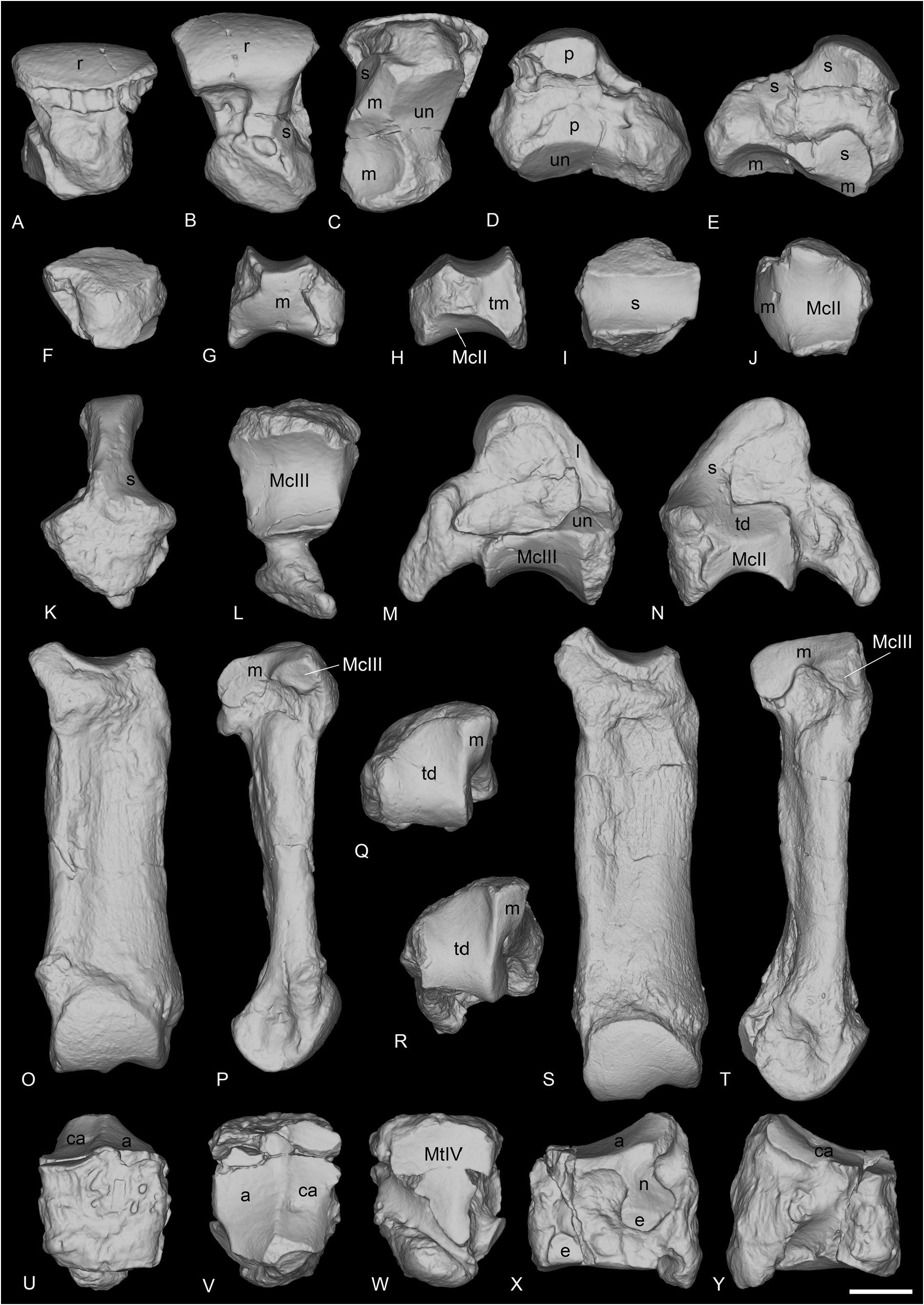
Newly prepared specimens are referred to as Ronzotherium heissigi sp. nov. and they document the only known postcranial remains of this species.
HUMERUS. The two humeri from Bumbach are proximally broken ( Fig. 25E–H View Fig ). In posterior view, the fossa olecrani is high on NMB-UM-132, but lower on the other humerus, NMB-UM-6132, possibly due to taphonomical deformation. The lateral epicondyle is very prominent, and distally elongated. The distal articulation is hourglass-shaped in anterior view, with a deep proximal incision between the two lips of the trochlea and the articulation is oblique. The epicondylar crest is high. The distal gutter on the epicondyle is strong in posterior view.
RADIUS. The radius from Bumbach (NMB-UM-127a, Fig. 25I–J View Fig ) belongs to the same individual as the humerus NMB-UM-132. The anterior border of the proximal articulation is straight. The medial border of the diaphysis is straight. The insertion of the m. biceps brachii is deep and the gutter for the m. extensor carpi is deep and wide. The posterior expansion of the scaphoid facet is high and there is no secondary distal articulation for the ulna.
ULNA. The ulna NMB-UM-131b-c ( Fig. 25K–M View Fig ) belongs to the same individual as the radius (NMB-UM-127a) and the humerus (NMB-UM-132). It is very poorly preserved. The olecranon of the ulna is rather long and makes an open angle with the diaphysis. The distal end is large, the anterior tubercle and the lunate facet are absent, and the pyramidal facet is concave.
LUNATE. The lunate NMBE-5035833 is complete and very well preserved ( Fig. 26A–E View Fig ). The proximal articulation for the radius is very large and convex anteroposteriorly. There is no articulation with the ulna. In proximal view, there is a long drop-like posterior extension of the radius facet on the medial side. The anterior side is deeply keeled, and the distal border is acute in anterior view. Medially, the two proximal articulations for the scaphoid are fused in a single facet. It is separated from the distal scaphoid facet by a deep groove. The distal facet for the scaphoid is large. In lateral view, the proximal and distal articulations for the pyramidal are large and the proximal one is medially displaced. In distal view, there are three articulation facets: a large anterior one for the unciform, and two for the magnum, one of which is distal and very concave and the other thin, flat and elongated is anterior, and located between the scaphoid and unciform facets.
TRAPEZOID. Two trapezoids are preserved from Bumbach (NMB-UM-6 and NMB-UM-6136b, Fig. 26F–J View Fig ). The latter is slightly different from the former, and it articulates with the magnum NMB-UM-6136c and the McII NMB-UM-6136a. In anterior view, they are both wider than high. The proximal border is sigmoid on NMB-UM-6136b, whereas it is symmetric on NMB-UM-6. The magnum facet occupies most of the lateral side, while the medial side is occupied by the medio-distal articulation
for the trapezium. The proximal side is fully occupied by the anteroposteriorly concave scaphoid facet. The distal articulation for the McII is also anteroposteriorly concave.
MAGNUM. The magnum NMB-UM-6136c is complete ( Fig. 26K–N View Fig ). In anterior view, the proximal border of the anterior side is concave and the anterior face is wider than high. The proximal apophysis is very high, very convex and narrow. The lunate facet on this apophysis is very long and contacts the small unciform facet. On the other side of the proximal apophysis, the facet for the scaphoid is also long and is very poorly distinguished from the medial facet for the trapezoid. This latter facet is longer than high. The medio-distal McII facet is flat and its distal border is very concave in medial view. On the distal side, the McIII facet is very large, quadrate, as long as wide and very concave anteroposteriorly. The posterior tuberosity of the magnum is short and curved.
MCII. There are two McII preserved from Bumbach (NMB-UM-121 and McII NMB-6136a, Fig. 26R– T View Fig ). The outline of the proximal side is trapezoidal. The articulation for the trapezoid is concave lateromedially and slightly convex anteroposteriorly. On the lateral side, the magnum facet is curved and band-shaped. The anterior facet for the McIII is present but the posterior is absent. There is no trapezium facet. The insertion for the m. extensor carpalis is salient.
FEMUR. Only a proximal fragment is preserved in Bumbach (NMBE-5035835). The head is hemispheric and the fovea capitis is high and narrow. The major trochanter is lower than the head and separated from it by a short neck. It is nearly triangular in proximal view and in lateral view the posterior part is higher than the anterior. The trochanteric fossa is deep on the posterior side, with a well-marked intertrochanteric crest. A fossa is also present on the anterior side, overhung by the major trochanter.
CUBOID. The cuboid NMBE-5035834 is complete ( Fig. 26U–Y View Fig ). The anterior side is approximately as high as wide. In lateral view, the posterior side is higher than the anterior and the posterior apophysis of the cuboid is slightly lower than the distal articulation. In proximal view, the posterior apophysis is almost not visible. The proximal side is oval and the astragalus and calcaneal facets are almost equalsized. On the medial side, the postero-proximal and elongated facet for the navicular is concave and contacts the small, square and postero-mesial facet for the ectocuneiform. The navicular facet bears a thin extension up to the anterior border, bordering the astragalus facet. The small anterodistal facet for the ectocuneiform is separated from the posterior one by a wide groove. On the lateral side, the groove for the tendon of the m. fibularis longus is very large and deep. The distal articulation for the MtIV is flat and triangular.

No known copyright restrictions apply. See Agosti, D., Egloff, W., 2009. Taxonomic information exchange and copyright: the Plazi approach. BMC Research Notes 2009, 2:53 for further explanation.
|
Kingdom |
|
|
Phylum |
|
|
Class |
|
|
Order |
|
|
Family |
|
|
Genus |
Ronzotherium heissigi
| Tissier, Jérémy, Antoine, Pierre-Olivier & Becker, Damien 2021 |
Diaceratherium lemanense
| Antoine P. - O. & Becker D. 2013: 140 |
Ronzotherium romani
| Brunet M. 1979: 135 |
Ronzotherium velaunum
| Heissig K. 1969: 20 |
Ronzotherium filholi elongatum
| Heissig K. 1969: 46 |
Ronzotherium filholi romani
| Heissig K. 1969: 63 |
Ronzotherium filholi
| Becker D. 2003: 213 |
| Brunet M. 1979: 105 |
| Lavocat R. 1951: 116 |
Aceratherium filholi
| Stehlin H. G. 1914: 183 |
Acerotherium lemanense
| Roman F. 1912: 61 |