
Parahaploposthia longituba, Hooge & Tyler, 2007
|
publication ID |
https://doi.org/ 10.11646/zootaxa.1479.1.3 |
|
publication LSID |
lsid:zoobank.org:pub:00598FD9-9272-4511-992E-B8312A26860D |
|
DOI |
https://doi.org/10.5281/zenodo.5086399 |
|
persistent identifier |
https://treatment.plazi.org/id/C6048246-FFF2-FFA7-4EDF-853FFAE3F8F1 |
|
treatment provided by |
Felipe |
|
scientific name |
Parahaploposthia longituba |
| status |
sp. nov. |
Parahaploposthia longituba sp. nov.
( Figs. 2–4 View FIGURE 2 View FIGURE 3 View FIGURE 4 )
Type material. Holotype. USNM 1096760 About USNM , one set of 2-µm-thick serial sagittal sections of epoxy-embedded specimen stained with toluidine blue . Paratype. USNM 1096761 About USNM , epoxy-embedded whole mount .
Type locality. South of Carrie Bow Cay at 3– 8 m depth from sand collected at a sand bore (1645’53” N, 88°07’09” W), and south of Carrie Bow Cay (16°48’09” N, 88°04’55” W), from medium-grained in a sand trough at 5 m water depth GoogleMaps .
Other material examined. Living specimens in squeeze preparations; three sets of 2-µm-thick serial sections of epoxy-embedded specimens; whole mount for fluorescence imaging of musculature.
Etymology. Species name refers to the long tube-like nature of the male antrum.
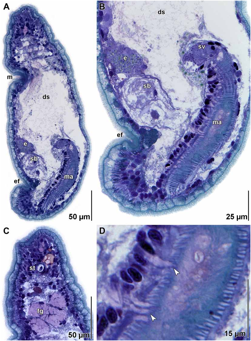
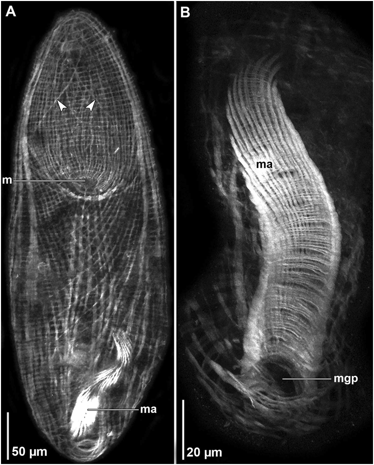
Description. Unsqueezed, mature specimens ~ 400 µm long and ~ 100 µm wide ( Figs. 2A View FIGURE 2 , 3A View FIGURE 3 , 4A View FIGURE 4 ). Body cylindrical. Anterior and posterior ends rounded. Body color yellow by transmitted light. Epidermis completely ciliated. Without rhabdoid glands. Frontal organ well developed ( Fig. 3C View FIGURE 3 ). Mouth opening on ventral surface, anterior half of body ( Fig. 3A View FIGURE 3 , 4A View FIGURE 4 ).
Body-wall musculature with circular muscles that encircle the body along entire length of animal; straight longitudinal muscles present between frontal pore and anterior edge of mouth; longitudinal-cross-over muscles (fibers with a longitudinal orientation anteriorly, but bend medially to cross diagonally) present in both dorsal and ventral body wall; longitudinal muscles in anterior half of body that wrap around posterior rim of mouth (U-shaped muscles) present in ventral body wall; without anterior ventral diagonal muscles ( Fig. 4A View FIGURE 4 ).
Ovary unpaired, ventral. Testes paired, lateral to ovary, compact.
Female gonopore and vagina absent. Seminal bursa, with sponge-like tissue wall, positioned in caudal portion of body, ventral to seminal vesicle ( Figs. 2B View FIGURE 2 , 3A, B View FIGURE 3 ).
Male gonopore terminal at posterior end of body opens to long (~ 140 µm), ciliated male antrum ( Figs. 2A, B View FIGURE 2 , 3A, B, D View FIGURE 3 ). Male antrum wall composed of outer longitudinal and inner circular muscle fibers ( Fig. 4B View FIGURE 4 ). Epithelium of antrum penetrated by necks of gland cells that empty into lumen of antrum ( Fig. 3D View FIGURE 3 ). Proximal end of male antrum capped with small, sperm-filled, seminal vesicle with thin tissue wall lacking musculature ( Fig. 3B View FIGURE 3 ).
Remarks. As is the case with Parahaploposthia longituba , the five previously described species of Parahaploposthia have an unpaired ovary, paired testes, and a ciliated male antrum (see Tyler et al. 2006). Parahaploposthia longituba stands distinct from all the others in having well-developed frontal glands and an exceptionally long male antrum. The antrum of P. thiophilus Fegley , Smith, & Rieger, 1984, is similarly elongated but only about half the length of that of P. longituba (~ 80 µm vs 140 µm). Like P. velvetum Hooge & Tyler, 2001 , and P. thiophilus , P. longitubus has a seminal bursa, but it lacks the vagina that connects the bursa to the posterior end of the body in those two species ( Fegley et al. 1984, Hooge & Tyler 2001). P. longituba is further distinguished from P. thiophilus in having pale coloration (brown in P. thiophilus ), a more anteriorly positioned mouth (mid-body in P. thiophilus ) and frontal glands extending posterior to the statocyst.

No known copyright restrictions apply. See Agosti, D., Egloff, W., 2009. Taxonomic information exchange and copyright: the Plazi approach. BMC Research Notes 2009, 2:53 for further explanation.
|
Kingdom |
|
|
Phylum |
|
|
Class |
|
|
Order |
|
|
Family |
|
|
Genus |