
Pseudohaplogonaria rodmani, Hooge & Tyler, 2007
|
publication ID |
https://doi.org/ 10.11646/zootaxa.1479.1.3 |
|
publication LSID |
lsid:zoobank.org:pub:00598FD9-9272-4511-992E-B8312A26860D |
|
persistent identifier |
https://treatment.plazi.org/id/C6048246-FFF6-FFA5-4EDF-82DAFF2BFC0E |
|
treatment provided by |
Felipe |
|
scientific name |
Pseudohaplogonaria rodmani |
| status |
sp. nov. |
Pseudohaplogonaria rodmani sp. nov.
( Fig. 5 View FIGURE 5 )
Type material. Holotype. USNM 1096762 About USNM , one set of 2-µm-thick serial sagittal sections of epoxy-embedded specimen stained with toluidine blue.
Type locality. East of Carrie Bow Cay (16°48’09” N, 88°04’55” W), from fine-grained sand in a trough at 30 m depth, south of Carrie Bow Cay at 3–8 m depth from sand collected at a sand bore (16°45’53” N, 88°07’09” W), and from Twin Cays near West Bay (16°49’56” N, 88°06’29” W), from subtidal sand among mangrove roots and Thalassia sp GoogleMaps .
Other material examined. Living specimens in squeeze preparations; one set of 2-µm-thick serial sections of epoxy-embedded specimen.
Etymology. Named in honor of James Rodman of the U.S. National Science Foundation, a champion of systematics and taxonomy.
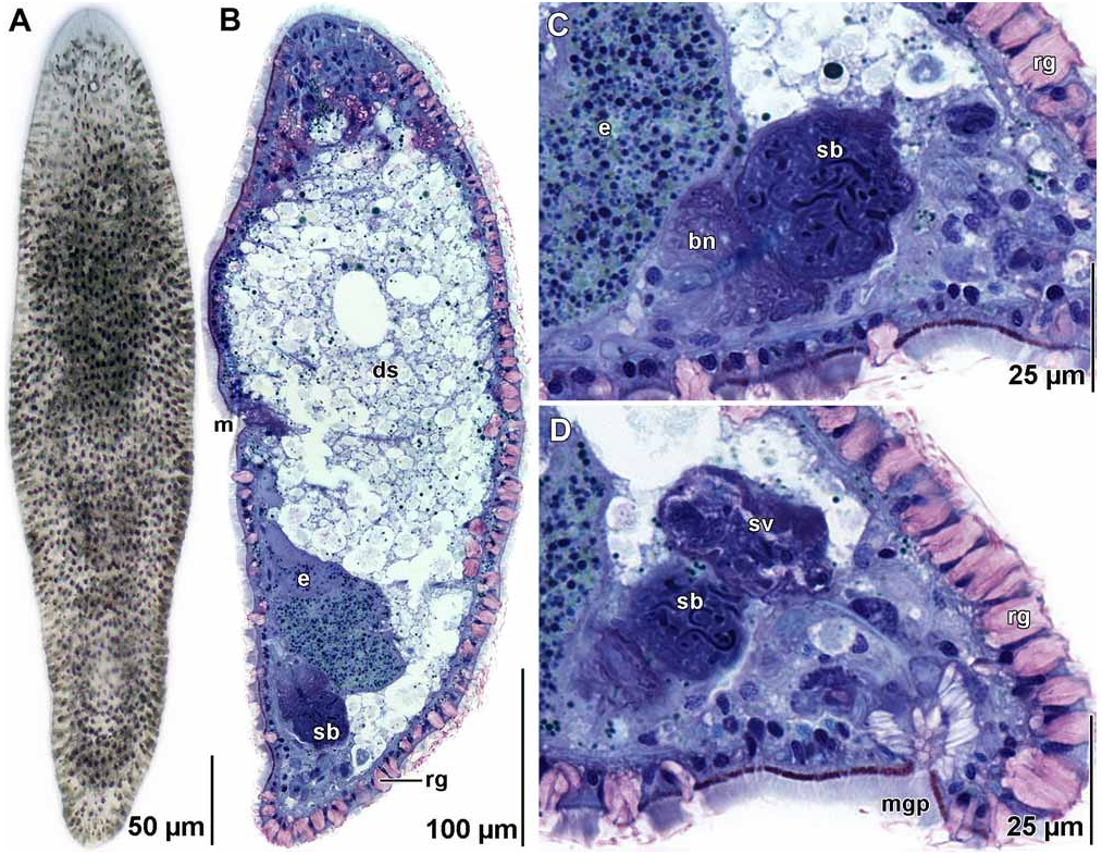
Description. Mature specimens ~ 450 µm long and ~ 100 µm wide ( Figs. 5A, B View FIGURE 5 ). Anterior and posterior ends rounded. Epidermis uncolored by transmitted light. Epidermis completely ciliated. Numerous red rhab- doids present in body wall, especially concentrated on dorsal side ( Figs. 5A, B View FIGURE 5 ). Frontal organ well developed. Mouth opening on ventral surface, middle of body ( Fig. 5B View FIGURE 5 ).
Ovary unpaired, ventral, extends from mouth posteriorly to seminal bursa. Testes paired, lateral to ovary, compact, separate from ovary, extend from level of statocyst posteriorly to seminal vesicle.
Female gonopore and vagina absent. Seminal bursa positioned caudally, ventral to seminal vesicle. Bursa with ventro-anteriorly directed bursal nozzle ( Fig. 5C View FIGURE 5 ).
Male gonopore ventral, at posterior end of body ( Figs. 5D View FIGURE 5 ). Gonopore surrounded by large gland cells that do not stain distinctly in toluidine blue ( Fig. 5D View FIGURE 5 ). Seminal vesicle positioned slightly anterior to gonopore gland cells ( Fig. 5D View FIGURE 5 ).
Remarks. This species was easily distinguished from other acoels in our samples by its numerous red rhabdoid glands. While most specimens we collected were of a similar size, we encountered one specimen that was much larger and seemed to have a significantly larger copulatory organ; we were unable to confirm if this specimen was conspecific.
Pseudohaplogonaria rodmani is united with the five previously described species in the genus in having a seminal bursa with a sclerotized bursal nozzle and a weakly developed or absent seminal vesicle (see Tyler et al. 2006). The relative simplicity of the male and female copulatory organs of this species is similar to that of the much smaller (~ 200 µm long) P. minima Ehlers and Dörjes, 1979 , from the Galapagos; however, unlike P. minima , P. rodmani lacks a vagina connecting the seminal bursa to the common gonopore ( Ehlers & Dörjes 1979).
No known copyright restrictions apply. See Agosti, D., Egloff, W., 2009. Taxonomic information exchange and copyright: the Plazi approach. BMC Research Notes 2009, 2:53 for further explanation.
|
Kingdom |
|
|
Phylum |
|
|
Class |
|
|
Order |
|
|
Family |
|
|
Genus |