
Platycoelostoma rauppi Foldi
|
publication ID |
https://doi.org/10.5281/zenodo.191685 |
|
DOI |
https://doi.org/10.5281/zenodo.6217015 |
|
persistent identifier |
https://treatment.plazi.org/id/C8140C4D-FF98-FF88-FF41-F9BCFD032D1E |
|
treatment provided by |
Plazi |
|
scientific name |
Platycoelostoma rauppi Foldi |
| status |
sp. nov. |
Platycoelostoma rauppi Foldi n. sp.
Material examined. Holotype adult female. PERU: Cusco, Tambo Machay, ruins near Puca Pucara, on roots of alfalfa, Medicago sativa (Fabaceae) , 1000 m, 10 February 1980, M. J. Raupp coll., USNM.
Note. Some of the collection data given on the slide label are in error and more accurate data have been provided by the collector, M.J. Raupp (personal communication). The location “Tombo Muchi” should be “Tambo Machay”, which is an Inca ruin near Puca Pucara in the hills to the north of Cusco; also the elevation of 1000 m recorded on the label is too low since Cusco is at over 3,300 m and thus the collection site must be at higher elevation
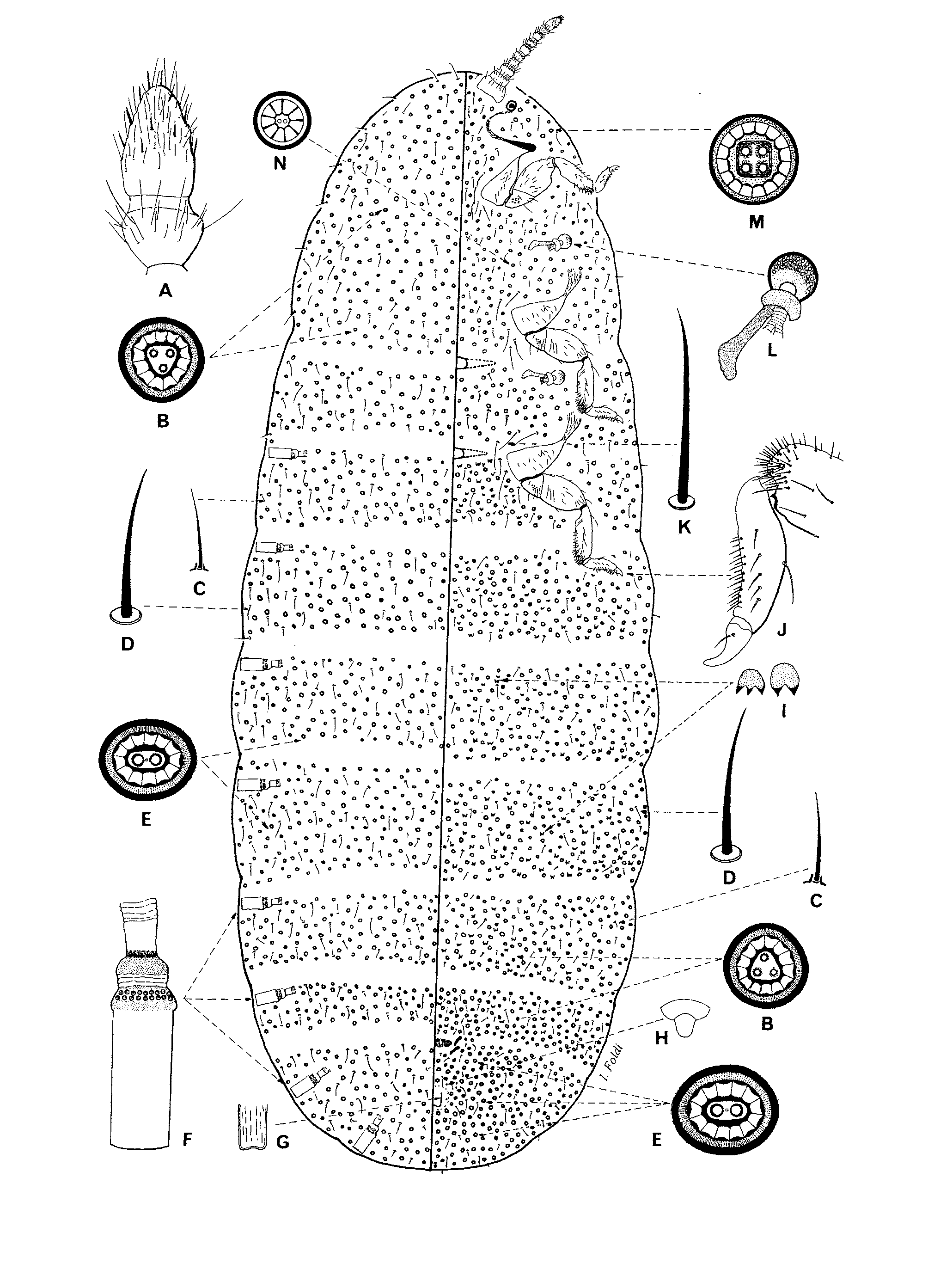
ADULT FEMALE ( Fig. 10 View FIGURE 10 )
Description based on holotype.
Mounted material. Body elongate, narrowing at both ends, approximately 14 mm long, 4.7 mm wide, with abdomen particularly long, representing nearly 2/3rds of body length. Body densely covered by multilocular pores, particularly on abdomen; setae fewer. Each abdominal spiracular opening with a plug of white wax.
Dorsum. Derm membranous, with multilocular pores ( Figs. 8 View FIGURE 8 B, 8E) either broadly ovoid or slightly triangular, both similar to those described on venter, in a quite dense transverse band 20–30 pores wide, distributed throughout body. Hair-ilke setae ( Fig. 8 View FIGURE 8 D), each about 30–45 µm long, scattered on head and thorax, but in transverse rows 2 or 3 setae wide on abdomen; longest setae, each 40–45 µm long, at anterior end; slender flagellate setae ( Fig. 8 View FIGURE 8 C), each 18–25 µm long, scattered.
Venter. Multilocular pores densely distributed throughout, most frequent on abdomen in dense transverse rows 24–32 pores wide across all segments. The following types of pores observed: (i) a broadly oval pore ( Fig. 8 View FIGURE 8 E) with a thick rim, each about 11–13 µm greatest width, each with a large bilocular centre plus a tiny central loculus, and with about 12–14 outer loculi; most common type; (ii) a smaller, slightly triangular pore ( Fig. 8 View FIGURE 8 B), each about 10 µm in greatest width, with a thick rim and a triangular-shaped centre with 3 large loculi plus a tiny central opening and with 12–14 outer loculi; less frequent, more numerous at anterior and posterior end of body; (iii) a pore about 9–10 µm greatest width, with a quadrilocular centre and 16–18 outer loculi ( Fig. 8 View FIGURE 8 M), rare; (iv) a small pore ( Fig. 8 View FIGURE 8 N) about 7–8 µm in diameter, with a thick rim and 1 or 2 tiny central openings plus about 7–9 triangular-shaped outer loculi, very rare; and (v) a small simple tubular pore ( Fig. 8 View FIGURE 8 H) about 4–5 µm long, 5–7 µm wide, restricted to between vulva and anal area. Hair-like setae ( Fig. 8 View FIGURE 8 K), each 40–80 µm long, present on head and thorax, particularly medially and submedially, and also in groups of 12–15 mesad to each pro-, meso- and metacoxa and in transverse rows 4–6 setae wide on abdomen; most numerous on submargin; also shorter and more slender flagellate setae ( Fig. 8 View FIGURE 8 K), each 20–30 µm long, scattered.
Antennae 10 segmented, length about 1280 µm; scapes placed close together, each scape wider (240 µm) than long (105 µm) with a distinct articulation with pedicel; remaining antennal segments tapering progressively to segment X, each segment with numerous long hair-like setae and a few fleshy setae; apical segment ( Fig. 8 View FIGURE 8 A) about 190 µm long, 110 µm wide, tapering to a point, with about 40–45 setae, each 30–100 µm long, plus about 6–10 slightly curved fleshy setae, each 20–30 µm long, confined on distal half. Eyespots each 150 µm in diameter, posterolateral to antennae. Thoracic spiracles ( Fig. 8 View FIGURE 8 L) each with peritreme about 90 µm wide, with a straight apodeme 160 µm long; atrium with 23–26 multilocular pores, smaller but similar to those on derm. Abdominal spiracles ( Fig. 8 View FIGURE 8 F) in 8 pairs, each with a round peritreme about 60–70 µm in diameter; each atrium divided into 3 distinct parts: outer atrium about 120–135 µm long, with approximately 2 rings of multilocular pores at inner end totalling 14–22, similar to those in thoracic spiracles; each spiracle with a smaller middle atrium with membranous folded walls, plus a sclerotised inner atrium which opens through a dense filamentous structure into a narrower sclerotised tube to which tracheae attach. Mouthparts vestigial, only represented by a non-sclerotised membranous tentorium. Legs short, robust, setose. Metathoracic legs partly visible in Fig. 8 View FIGURE 8 J: coxa ring-like, with a strong articulatory sclerosis with trochanter, about 210 µm long, 600 µm wide, with about 20 setae, each 35–70 µm long; trochanter + femur 710 µm long; trochanter with about 5–7 campaniform pores on each side and a few setae, each 25–30 µm long, one seta 70 µm long, plus a trochanteral seta on ventral surface, about 90 µm; femur with greatest width about 330 µm, with a few setae laterally, each 30–70 µm long, and with a strong articulatory sclerosis with tibia; tibia 560 µm long, 165 µm wide, distal part with a dense group of about 40–45 spur-like setae, each 40–60 µm long; tarsus 330 µm long, 100 µm widest, with about 20–24 similar spur-like setae ventrally, mostly in 2 rows, each 40–50 µm long; claw 125 Μm long, curved, narrowing gradually to a blunt apex, without a denticle; claw digitules fine and acute, each 45 µm long. Spinules ( Fig. 8 View FIGURE 8 I) present between metathoracic legs and on anterior abdominal segments medially and submedially. Vulvar opening a transverse fissure, 150–210 µm long, with 2 strong apodemes at each extremity. Anal opening 110 µm wide, surrounded by multilocular pores and a loose cluster of hair-like setae, each 30–70 µm long; anal tube ( Fig. 8 View FIGURE 8 G) simple, 180 µm long, lacking a sclerotised ring and no pores at inner end.
Remarks. Platycoelostoma rauppi n. sp. shares the following character states with other Platycoelostoma species: (i) abdominal spiracles in 8 pairs; (ii) multilocular pores with a oval, triangular, quadrate centre; (iii) thoracic spiracles with atrial pores; (iv) vulvar opening with 2 apodemes at each extremity; and (v) antennae 9–10 segmented. However, P. rauppi differs from P. c o m p re s s a and P. tasmanicum due to the following character states: (i) abdominal spiracles in 8 pairs, each well developed and each with atrial pores; in P. compressa and P. tasmanicum , the last pair of abdominal spiracles is poorly developed and without atrial pores; (ii) small circular multilocular pores with triangular-shaped outer loculi and circular centre present on thorax ventrally; and (iii) simple tubular pores present between vulvar and anal area.
Derivatio nominis. The new species rauppi is named in honour of the collector, Dr Michael Raupp, Professor at the University of Maryland, USA.

| USNM |
Smithsonian Institution, National Museum of Natural History |
No known copyright restrictions apply. See Agosti, D., Egloff, W., 2009. Taxonomic information exchange and copyright: the Plazi approach. BMC Research Notes 2009, 2:53 for further explanation.
|
Kingdom |
|
|
Phylum |
|
|
Class |
|
|
Order |
|
|
SuperFamily |
Coccoidea |
|
Family |
|
|
Genus |