
Ctenus theodorianum, Jager, 2012
|
publication ID |
https://doi.org/ 10.11646/zootaxa.3429.1.1 |
|
persistent identifier |
https://treatment.plazi.org/id/C8488786-1102-FF81-FF71-F8AC543EF950 |
|
treatment provided by |
Felipe |
|
scientific name |
Ctenus theodorianum |
| status |
sp. nov. |
Ctenus theodorianum View in CoL spec. nov.
Figs 39 View FIGURE 39 , 92–115 View FIGURES 92–95 View FIGURES 96–104 View FIGURES 105–115
Type material. Holotype male ( SMF), Laos, Oudomxai Province, Phou Hiphi provincial protected area, ca. 4 km S of Oudomxai, N 20°39'9.7'', E 102°0'10.59'', 850 m altitude, ground, vegetation, tree bark, by hand, by night, L. Nophaseud leg. 18.IV.2011 GoogleMaps . Paratypes: 1 female ( SMF) with same data as for holotype GoogleMaps . 1 female ( SMF), with same data as for holotype GoogleMaps except for: 4 km S of Oudomxai, N 20°40'4.32'', E 102°2'25.32'', 830 m altitude GoogleMaps .
Etymology. This species is named in honour of my secondary school “Gymnasium Theodorianum” in Paderborn, Germany, on the occasion of its 400th anniversary for providing an excellent education and training, therefore a good start into my life; name in apposition.
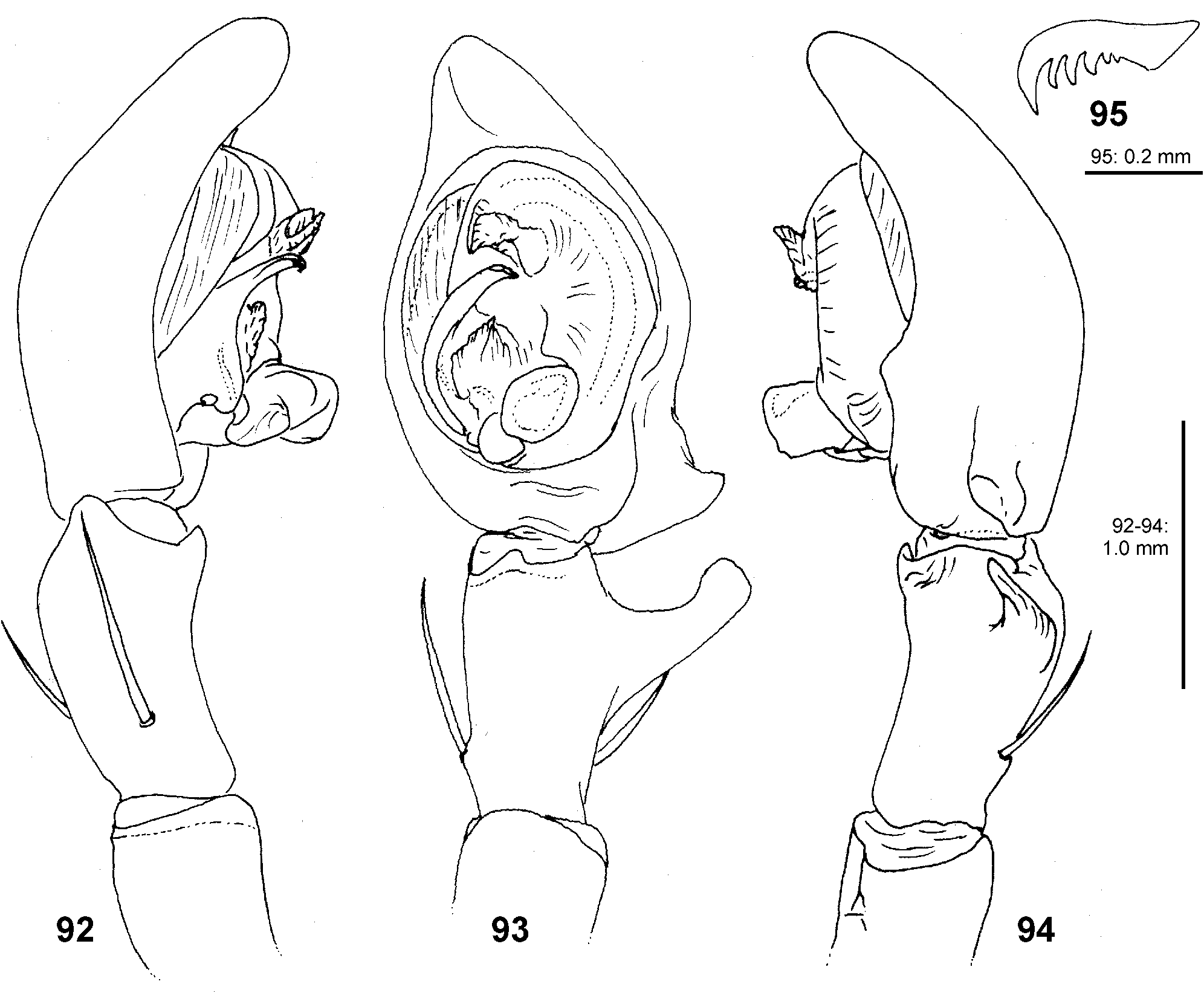
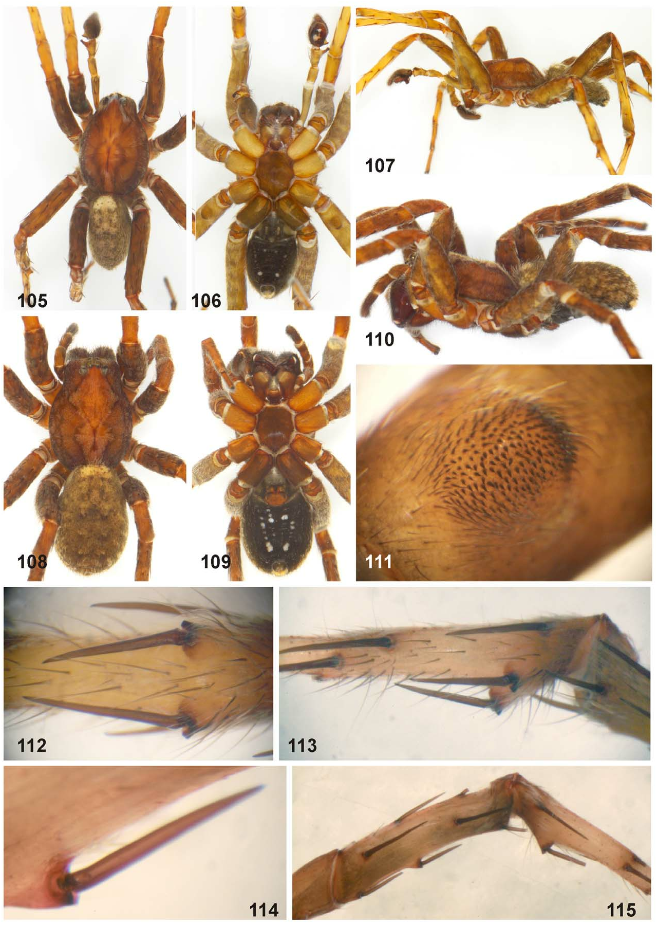
Diagnosis. Medium-sized Ctenidae (total length male 10.8, female 12.3–13.8). Distinguished from other Ctenus spp. , but not C. robustus , by the modified leg III in males with ventral hump on proximal femur covered by small stout spines ( Fig. 111 View FIGURES 105–115 ) and two ventral cone-shaped humps each with one spine on proximal metatarsus ( Figs 112–115 View FIGURES 105–115 ). For differential diagnosis see C. robustus . Females show typical characters of other Asian Ctenus spp. : broad median epigynal plate with two lateral teeth, internal duct system consisting of spherical spermathecae, short
Description. Male (holotype). PL 6.2, PW 4.8, AW 2.5, OL 4.6, OW 2.9. Eye diameters and interdistances: AME 0.32, ALE 0.21, PME 0.40, PLE 0.39, AME–AME 0.12, AME–ALE 0.33, PME–PME 0.11, PME–PLE 0.31, AME–PME 0.08, ALE–PLE 0.12, clypeus AME 0.14, clypeus ALE 0.61. Palp and leg measurements: palp 6.55 (2.4, 1.1, 1.25, -, 1.5), I 22.2 (5.8, 2.5, 6.0, 5.7, 2.2), II 19.4 (5.3, 2.3, 5.0, 5.0, 1.8), III 16.1 (4.6, 2.0, 3.4, 4.5, 1.6), IV 23.8 (6.2, 2.2, 5.7, 7.6, 2.1). Leg formula 4123. Spination of palp and legs: palp 141, 100, 1010; femora I p121,
Chelicerae with 3 promarginal, 4 retromarginal teeth, and with elongated patch of many tiny denticles along entire cheliceral furrow. Retromargin of chelicerae close to fang base with several short and thin bristles. Sparse scopula restricted almost entirely to tarsi, only metatarsi I–II with sparse scopula hairs. Right leg claw I with 5 secondary teeth ( Fig. 95 View FIGURES 92–95 ). Position of tarsal organ: I 0.90, II 0.90, III 0.80, IV 0.85.
Palp as in diagnosis (see also C. robustus ) ( Figs 92–94 View FIGURES 92–95 ). Palpal tibia with strong RTA, with broad base and blunt tip. Cymbium tip slightly conical, retro-proximally with distinct pointed extension. Embolus arising at 7.30- o’clock-position, short, its tip situated almost centrally. Conductor arising at 12-o’clock-position subdistally, partly fused with tegulum. Tegular apophysis arising at 6- to 6.30-o’clock-position from tegulum, in ventral view divided into two parts, distinctly excavated on dorsal side.
Colour ( Figs 105–107 View FIGURES 105–115 ). Brown partly with dark-brown pattern. Dorsal prosoma with broad lateral bands marginally and dark bands around distinctly marked fovea. Sternum, ventral coxae III + IV, labium and gnathocoxae dark, partly with lighter patches, coxae I + II yellowish-brown without pattern. Chelicerae as dorsal prosoma with longitudinal lines in proximal half and with dark distal half. Leg femora dark with irregular darker patches, patellae to tarsi yellowish-brown. Dorsal opisthosoma greyish-brown, mottled with small dark spots, anterior half with lighter patches. Lateral opisthosoma with spots in longitudinal rows. Ventral opisthosoma black with two pairs of white patches consisting of white hairs; epiandrium and muscle sigilla light. Anterior lateral spinnerets dark, posterior lateral and median spinnerets and anal tubercle light.
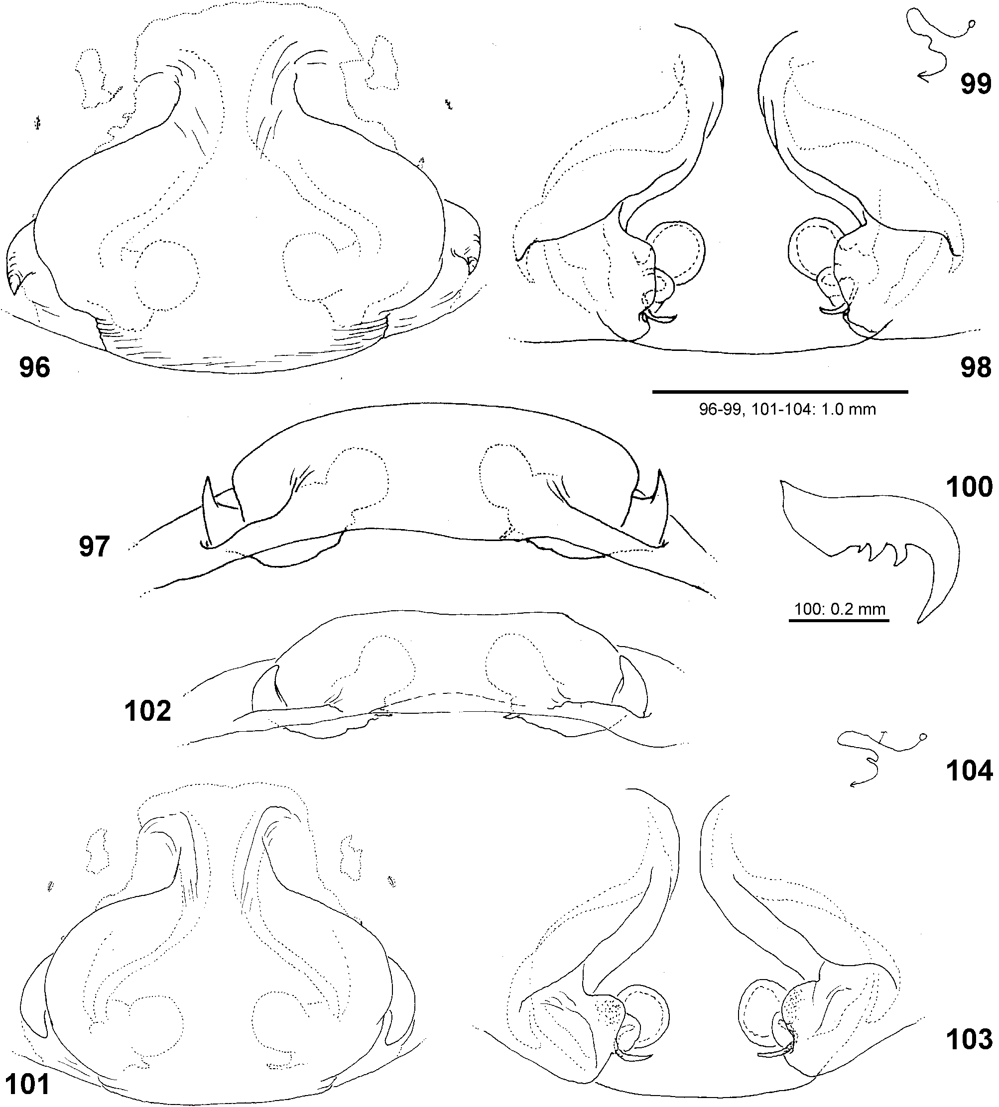
Female (paratype together with holotype). PL 6.9, PW 5.6, AW 3.6, OL 7.0, OW 4.6. Eye diameters and interdistances: AME 0.37, ALE 0.28, PME 0.47, PLE 0.39, AME–AME 0.18, AME–ALE 0.40, PME–PME 0.24, PME–PLE 0.41, AME–PME 0.11, ALE–PLE 0.15, clypeus AME 0.22, clypeus ALE 0.70. Palp and leg measurements: palp 7.0 (2.4, 1.3, 1.5, -, 1.8), I 18.15 (5.2, 2.9, 4.7, 3.9, 1.45), II 17.0 (4.9, 2.7, 4.2, 3.7, 1.5), III 14.9 (4.3, 2.2, 3.3, 3.6, 1.5), IV 20.8 (5.6, 2.5, 4.9, 6.0, 1.8). Leg formula 4123. Spination of palp and legs: palp 131, 100, 1211, 1013; femora I p021, d111, r111, II–III p112, d111, r 111, IV p111, d111, r011; patellae I–II 000, III–IV 101; tibiae I–II v22222, III–IV p11, d111, r11, v222; metatarsi I v22(1)2, II v222, III p112, d010, r112, v222, IV p112, d010, r112, v2122. Chelicerae with 3 promarginal, 4 retromarginal teeth, and with elongated patch of many tiny denticles along entire cheliceral furrow. Retromargin of chelicerae close to fang base with 13 thin bristles. Ventral tarsi and entire metatarsi with sparse scopula. Claw tufts with concave distal surface. Palpal claw with 5 secondary teeth. Right leg claw I with 3 secondary teeth ( Fig. 100 View FIGURES 96–104 ). Position of tarsal organ: I 0.90, II 0.85, III 0.76, IV 0.83.
Copulatory organ as in diagnosis (see also C. robustus ) ( Figs 96–104 View FIGURES 96–104 ). Epigynal field with two separate anterio-lateral patches, close to these patches two slit sense organs. Median plate narrow anteriorly, widening medially and narrowing posteriorly again, posteriorly with subparallel margins, lateral teeth situated at widest part. Posterior epigyne with distinct lateral furrows. Internal duct system with two large lateral folds running diagonally from medially to laterally. Spherical spermathecae separated by their diameter or more than that from each other, with a small round chamber between spermathecae and fertilisation ducts, the latter pointing medially.
Colour ( Figs 108–110 View FIGURES 105–115 ). As in male, but generally darker. Dorsal prosoma with two posteriorly diverging lines starting at PLE. Distal leg segments with indistinct patches, especially in legs III–IV. Dorsal opisthosoma with pairs of indistinct small light patches. Ventral opisthosoma with ca. 8 pairs of white patches, partly fused.
Variation. Second female paratype: PL 6.3, OL 6.3. Epigyne smaller and relatively slightly longer, with posterior parallel margins indistinct. Lateral teeth not as pointed as in syntopic paratype. In the internal duct system glandular pores could be recognised and are added in the schematic course.
Distribution. Known only from the type locality ( Fig. 39 View FIGURE 39 : 8).

| SMF |
Forschungsinstitut und Natur-Museum Senckenberg |
No known copyright restrictions apply. See Agosti, D., Egloff, W., 2009. Taxonomic information exchange and copyright: the Plazi approach. BMC Research Notes 2009, 2:53 for further explanation.