
Neuraphomimus Franz
|
publication ID |
https://doi.org/ 10.11646/zootaxa.3666.4.7 |
|
publication LSID |
lsid:zoobank.org:pub:4470BF7B-DCDB-4668-B221-600A9EDE11FA |
|
DOI |
https://doi.org/10.5281/zenodo.6146067 |
|
persistent identifier |
https://treatment.plazi.org/id/C853F731-2400-0C4B-FF34-FF0CB26FF9A1 |
|
treatment provided by |
Plazi |
|
scientific name |
Neuraphomimus Franz |
| status |
|
Subgenus Neuraphomimus Franz
Neuraphomimus Franz : 1986: 44 (replacement name for Pseudoconnus Franz, 1980 ). Pseudoconnus Franz, 1980: 216 (preoccupied, nec Leleup, 1971). Type species: Pseudoconnus simulator Franz, 1980 (original designation).
Vetusteconnus Franz, 1993: 101 (unnecessary replacement name for Pseudoconnus Franz ).
Revised diagnosis. A subgenus of Parapseudoconnus showing the following differences in relation to Parapseudoconnus s. str.: prothoracic hypomeral ridges complete; anterior margins of mesocoxal cavities distinctly demarcated by carina; mesocoxal sockets located on mesal surface of mesocoxal projections and therefore not visible in ventral view; posterior lobes of mesocoxal projections absent; and the internal armature of the aedeagus with long and slender flagellum projecting distally beyond the median lobe.
Redescription. Body of male (Fig. 45) strongly convex, elongate and relatively slender, with long appendages, vestiture distinct but unremarkable.
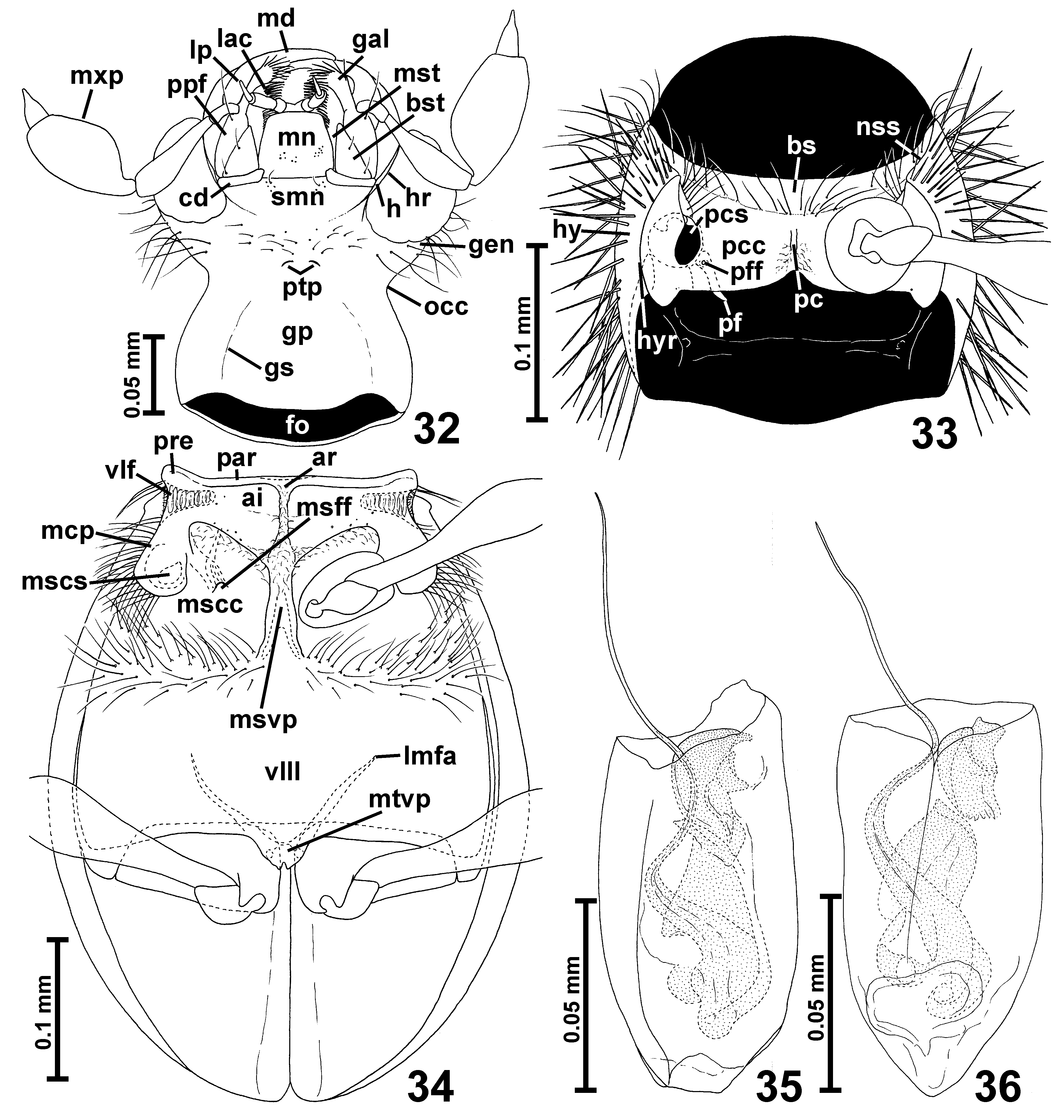
Head (Fig. 42) as in Parapseudoconnus s. str., without important differences.
Prothorax ( Fig. 33 View FIGURES 32 – 36 ) differs from that of Parapseudoconnus s. str. in ventral structures: internal parts of hypomera demarcated laterally by complete hypomeral ridges ( Fig. 33 View FIGURES 32 – 36 ; hyr), and pronotosternal sutures ( Fig. 33 View FIGURES 32 – 36 ; nss) strongly curved laterally, so that the basisternal part of prosternum is expanded laterally.
Mesoventrite ( Fig. 33 View FIGURES 32 – 36 ) as in Parapseudoconnus s. str., except for indistinctly demarcated asetose impressions ( Fig. 34 View FIGURES 32 – 36 ; ai), presence of distinct carinae demarcating antrerior margins of mesocoxal cavities ( Fig. 34 View FIGURES 32 – 36 ; mscc), and mesocoxal projections ( Fig. 34 View FIGURES 32 – 36 ; mcp), which conceal mesocoxal sockets ( Fig. 34 View FIGURES 32 – 36 ; mscs) located on their mesal margins; posterior lobes of mesocoxal projections absent.
Metaventrite ( Fig. 34 View FIGURES 32 – 36 ) as in Parapseudoconnus s. str., except for broader metaventral intercoxal process ( Fig. 34 View FIGURES 32 – 36 : mtvp).
Elytra (Fig. 45) as in Parapseudoconnus s. str.
Legs ( Figs. 21 View FIGURES 19 – 21 , 33–34 View FIGURES 32 – 36 ) long; femora clavate, tibiae slender, tarsi moderately elongate.
Abdominal sternites in the only studied specimen not preserved.
Aedeagus ( Figs. 35–36 View FIGURES 32 – 36 ) partly damaged or distorted in studied specimens, so the shape of its distal part is not possible to describe, with thin walls and complex, asymmetrical internal armature composed of a system of irregular sclerites, with long flagellum projecting distally beyond median lobe.
Distribution and composition. Three species are known to occur in the northern part of Peru.
Remarks. Specimens of Parapseudoconnus belonging to Neuraphomimus are poorly preserved in NHMW and only the male of P. simulator is nearly intact, while the type specimens of the remaining two species are partly or nearly completely damaged. The aedeagi, partly damaged or distorted during previous studies, are fragile and they were remounted only to ensure their safety and visibility in the mounting medium (previously the aedeagi were mounted in such a way that they were barely visible), as described in the Materials and Methods section. No attempts were made to obtain preparations with the aedeagi in a perfectly dorsal or ventral position, which would have required additional manipulations and might have caused further damage. Therefore, the aedeagi are here illustrated as they are embedded, not necessarily in dorsal or lateral aspects. In order to identify new specimens their aedeagi must be studied in various positions to carefully examine the shape of internal armature and compare it with that showed in Figs. 35–36 View FIGURES 32 – 36 and in Franz (1980; Fig. 205).

No known copyright restrictions apply. See Agosti, D., Egloff, W., 2009. Taxonomic information exchange and copyright: the Plazi approach. BMC Research Notes 2009, 2:53 for further explanation.