
Parapseudoconnus Franz
|
publication ID |
https://doi.org/ 10.11646/zootaxa.3666.4.7 |
|
publication LSID |
lsid:zoobank.org:pub:4470BF7B-DCDB-4668-B221-600A9EDE11FA |
|
DOI |
https://doi.org/10.5281/zenodo.6146061 |
|
persistent identifier |
https://treatment.plazi.org/id/C853F731-241A-0C57-FF34-F81AB059F9B5 |
|
treatment provided by |
Plazi |
|
scientific name |
Parapseudoconnus Franz |
| status |
|
Parapseudoconnus Franz View in CoL
Parapseudoconnus Franz, 1980 sensu Newton & Franz, 1998: 148 . Type species: Pseudoconnus aberrans Franz, 1980 (monotypy). Pseudoconnus is unavailable and the subgeneric name Parapseudoconnus has priority for the name of the genus (problem clarified by Newton & Franz, 1998).
Parapseudoconnus Franz, 1980: 218 (as subgenus of Pseudoconnus Franz, 1980 ). Type species: Pseudoconnus aberrans Franz, 1980 (monotypy).
Revised diagnosis. Male and female: head short, with vertex not expanded dorso-caudad; thick and long bristles absent on head but present on sides of prothorax; fronto-clypeal groove absent; maxillary palpomere III strongly thickened and stout; mandible without mesal sub-median tooth; antennae with club composed of antennomeres IX– XI; pronotum with rounded sides and weakly arcuate anterior and posterior margins, base of pronotum with one pair of external lateral pits and short longitudinal sub-lateral carina; basisternal part of prosternum much shorter than procoxal cavities; prosternum with fine intercoxal carina; prothoracic hypomeral ridges complete or nearly complete; postero-lateral (postcoxal) parts of prosternum fused with internal parts of prothoracic hypomera; mesoventral intercoxal process long, narrow and strongly expanding ventrally (keel-shaped); mesoventrite with asetose lateral impressions behind anterior ridge, without setose impressions; mesothorax with ventro-lateral foveae; metacoxae narrowly separated by subtrapezoidal or subtriangular metaventral intercoxal process; each elytron with single deep and setose basal fovea. Male: aedeagus without parameres, with asymmetrical internal armature.
Redescription. Body of male ( Figs. 19, 21 View FIGURES 19 – 21 ) strongly convex, elongate but moderately slender, with moderately long appendages, BL below 1 mm; cuticle glossy, brown, moderately densely setose.
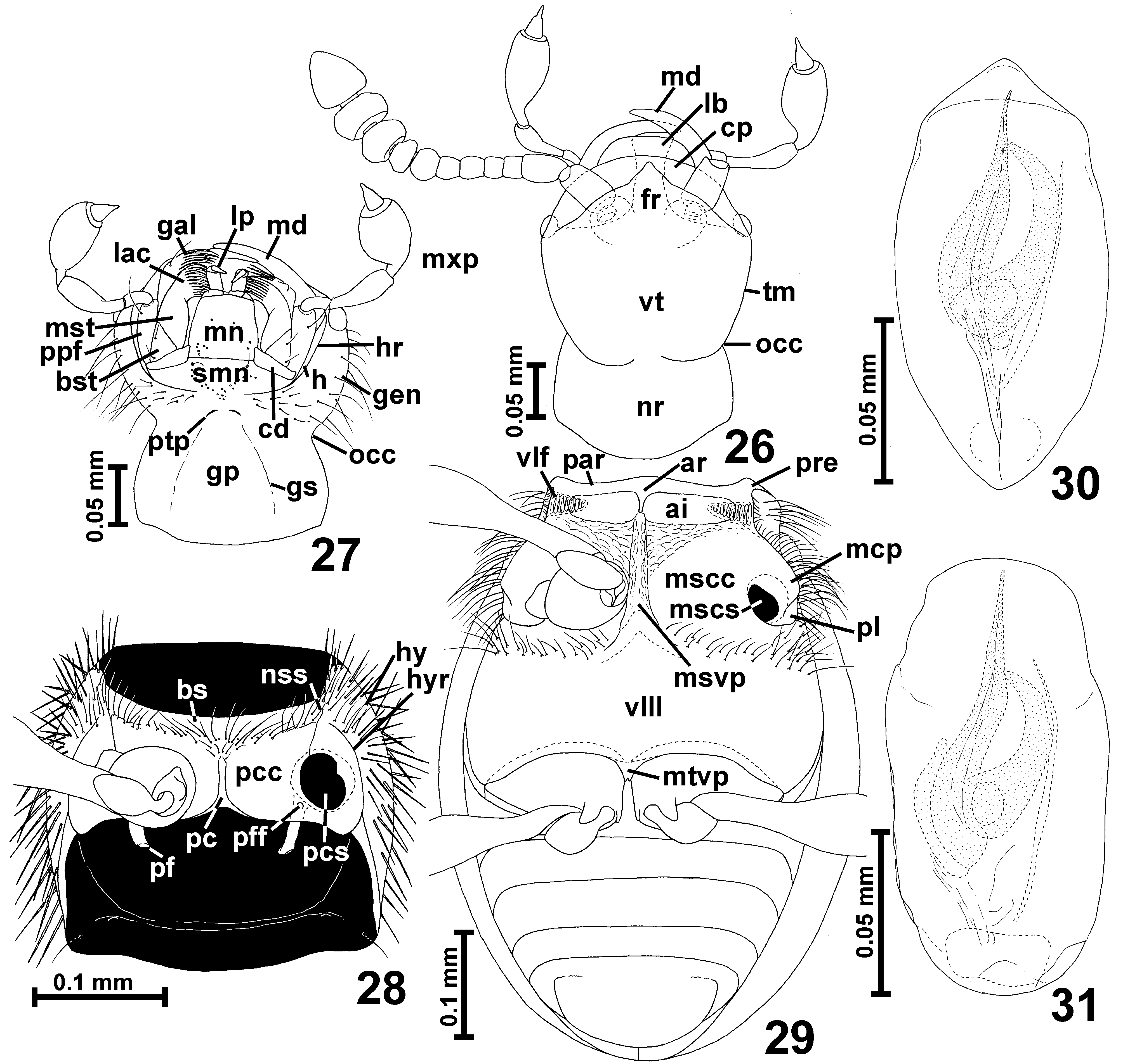
Head ( Figs. 19–21 View FIGURES 19 – 21 , 26–27 View FIGURES 26 – 31 , 32 View FIGURES 32 – 36 ) short, approximately rhomboidal or rounded, with large eyes; occipital constriction ( Figs. 27 View FIGURES 26 – 31 , 32 View FIGURES 32 – 36 ; occ) in the narrowest place much wider than half HW; tempora ( Fig. 26 View FIGURES 26 – 31 ; tm) long and convergent caudad, without bristles; vertex ( Fig. 26 View FIGURES 26 – 31 ; vt) broader than long, rounded, convex, not projecting dorsocaudad; frons ( Fig. 26 View FIGURES 26 – 31 ; fr) transverse and subtriangular; fronto-clypeal groove absent; antennal insertions broadly separated.
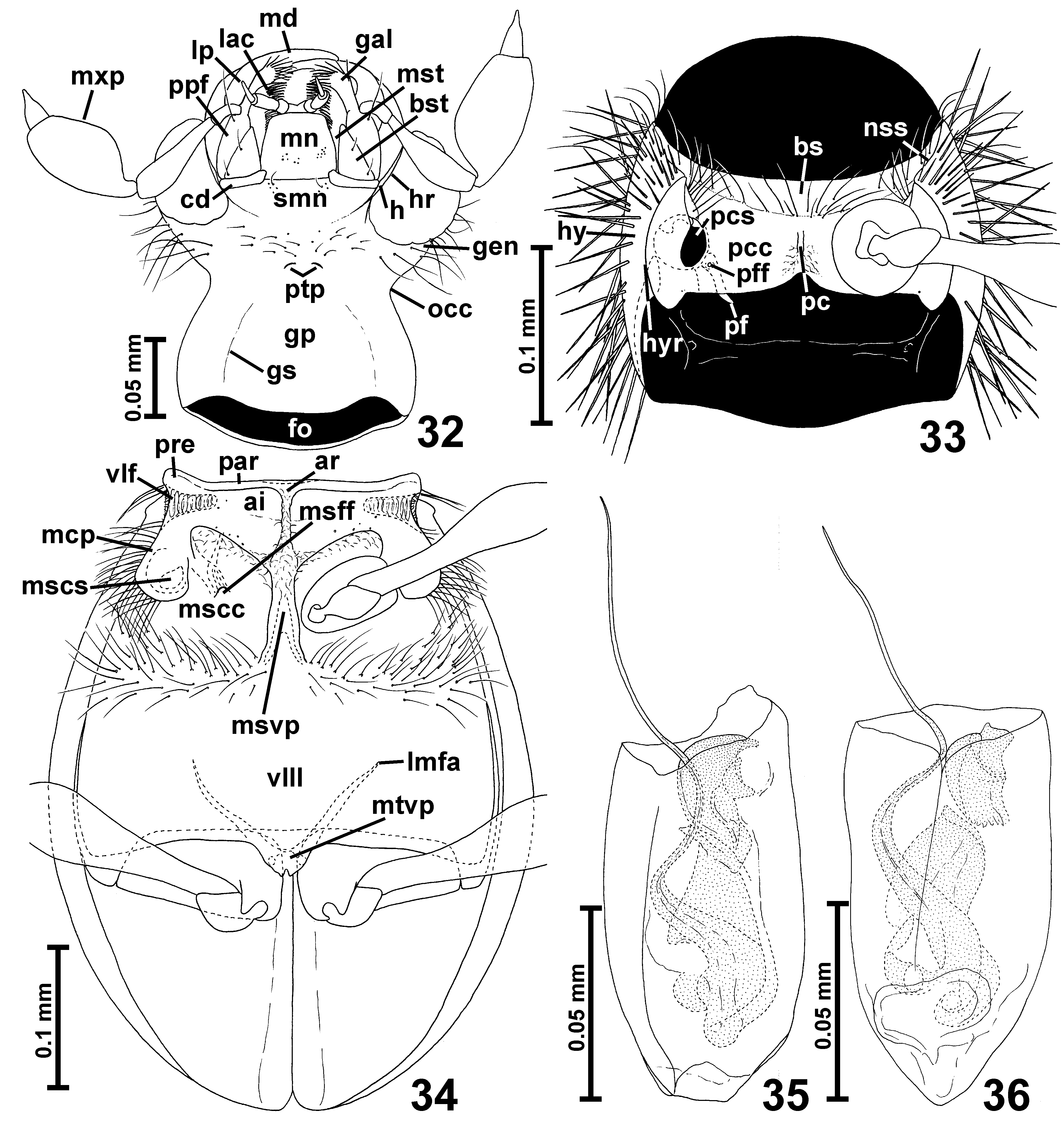
Labrum ( Fig. 26 View FIGURES 26 – 31 ; lb) transverse with rounded anterior margin. Mandibles ( Figs. 26–27 View FIGURES 26 – 31 , 32 View FIGURES 32 – 36 ; md) symmetrical, each with broad basal part, without noticeable prostheca, and with slender and curved distal part, without mesal tooth. Each maxilla ( Figs. 27 View FIGURES 26 – 31 , 32 View FIGURES 32 – 36 ) with subtriangular basistipes ( Figs. 27 View FIGURES 26 – 31 , 32 View FIGURES 32 – 36 ; bst), elongate galea ( Figs. 27 View FIGURES 26 – 31 , 32 View FIGURES 32 – 36 ; gal) and lacinia ( Figs. 27 View FIGURES 26 – 31 , 32 View FIGURES 32 – 36 ; lac) and long maxillary palp ( Figs. 27 View FIGURES 26 – 31 , 32 View FIGURES 32 – 36 ; mxp) composed of relatively long palpomere I, strongly elongate, pedunculate palpomere II, broad and stout palpomere III broadest between middle and basal third, and small, subconical and pointed palpomere IV.
Labium ( Figs. 27 View FIGURES 26 – 31 , 32 View FIGURES 32 – 36 ) with large and transverse submentum ( Figs. 27 View FIGURES 26 – 31 , 32 View FIGURES 32 – 36 ; smn) not demarcated from gular plate ( Figs. 27 View FIGURES 26 – 31 , 32 View FIGURES 32 – 36 ; gp) and laterally fused with postcardinal parts of hypostomae ( Figs. 27 View FIGURES 26 – 31 , 32 View FIGURES 32 – 36 ; h), subtrapezoidal mentum ( Figs. 27 View FIGURES 26 – 31 , 32 View FIGURES 32 – 36 ; mn); and short prementum with small 3-segmented labial palps narrowly separated at bases ( Figs. 27 View FIGURES 26 – 31 , 32 View FIGURES 32 – 36 ; lp). Hypostomal ridges ( Figs. 27 View FIGURES 26 – 31 , 32 View FIGURES 32 – 36 ; hr) posteriorly reaching to half length of submentum.
Gular plate ( Figs. 27 View FIGURES 26 – 31 , 32 View FIGURES 32 – 36 ; gp) large and strongly narrowing anterad; gular sutures ( Figs. 27 View FIGURES 26 – 31 , 32 View FIGURES 32 – 36 ; gs) superficial; posterior tentorial pits ( Figs. 27 View FIGURES 26 – 31 , 32 View FIGURES 32 – 36 ; ptp) distinct, located at base of submentum.
Antennae ( Figs. 19–21 View FIGURES 19 – 21 , 26 View FIGURES 26 – 31 ) with distinct club composed of antennomeres IX–XI.
Pronotum ( Figs. 19–21 View FIGURES 19 – 21 ) in dorsal view approximately oval with strongly rounded lateral margins, moderately distinct anterior and distinct posterior corners; without marginal carinae or edges but with short sub-lateral carinae visible near posterior pronotal corners; base of pronotum with one pair of shallow and small external lateral pits; sides of pronotum with dense, thick and long bristles.
Prosternum ( Figs. 28 View FIGURES 26 – 31 , 33 View FIGURES 32 – 36 ) with short basisternal part ( Figs. 28 View FIGURES 26 – 31 , 33 View FIGURES 32 – 36 ; bs) distinctly demarcated from procoxal cavities ( Figs. 28 View FIGURES 26 – 31 , 33 View FIGURES 32 – 36 ; pcc); median part of sternum with fine intercoxal carina; procoxal sockets ( Figs. 28 View FIGURES 26 – 31 , 33 View FIGURES 32 – 36 ; pcs) closed by lateral lobes of sternum which are fused with internal parts of prothoracic hypomera; hypomera ( Figs. 28 View FIGURES 26 – 31 , 33 View FIGURES 32 – 36 ; hy) elongate; hypomeral ridge ( Figs. 28 View FIGURES 26 – 31 , 33 View FIGURES 32 – 36 ; hyr) complete or nearly complete; pronotosternal sutures ( Figs. 28 View FIGURES 26 – 31 , 33 View FIGURES 32 – 36 ; nss) entire.
Mesoscutellum very small, subtriangular, barely visible between bases of elytra; mesoscuto-scutellar suture indiscernible in slide preparation.
Mesoventrite ( Figs. 29 View FIGURES 26 – 31 , 34 View FIGURES 32 – 36 ) with narrow anterior ridge ( Figs. 29 View FIGURES 26 – 31 , 34 View FIGURES 32 – 36 ; ar) and median projection of anterior ridge ( Figs. 29 View FIGURES 26 – 31 , 34 View FIGURES 32 – 36 ; par); mesoventral intercoxal process ( Figs. 29 View FIGURES 26 – 31 , 34 View FIGURES 32 – 36 ; msvp) narrow and keel-shaped; mesanepisternum with long prepectus ( Figs. 29 View FIGURES 26 – 31 , 34 View FIGURES 32 – 36 ; pre) and posterior part in ventral view visible only near to ventro-lateral fovea; mesepimeron not visible in ventral view; sides of mesothorax with deep ventro-lateral foveae ( Figs. 29 View FIGURES 26 – 31 , 34 View FIGURES 32 – 36 ; vlf); mesoventrite with variously distinctly delimited lateral asetose impressions ( Figs. 29 View FIGURES 26 – 31 , 34 View FIGURES 32 – 36 ; ai), without setose impressions; mesocoxal projections ( Figs. 29 View FIGURES 26 – 31 , 34 View FIGURES 32 – 36 ; mcp) with mesocoxal sockets ( Figs. 29 View FIGURES 26 – 31 , 34 View FIGURES 32 – 36 ; mscs) located on their mesal or meso-ventral surface and with or without posterior lobes ( Fig. 29 View FIGURES 26 – 31 ; pl).
Metaventrite ( Figs. 29 View FIGURES 26 – 31 , 34 View FIGURES 32 – 36 ; vIII) strongly transverse, anteriorly fused with mesoventrite, posteriorly moderately deeply bisinuate and with narrow median subtrapezoidal metaventral intercoxal process ( Figs. 29 View FIGURES 26 – 31 , 34 View FIGURES 32 – 36 ; mtvp) bearing median notch. Metanepisterna and metepimera narrow, only posterior parts of episterna partly visible in ventral view.
Metafurca ( Fig. 34 View FIGURES 32 – 36 ) with very short and broad stem and divergent lateral furcal arms ( Fig. 34 View FIGURES 32 – 36 ; lmfa).
Elytra ( Figs. 19–21 View FIGURES 19 – 21 ) oval, each with single deep and setose basal fovea located in shallow basal impression; humeral calli well-marked and developed as longitudinal protuberances; elytral apices unmodified, separately rounded.
Legs ( Figs. 19–21 View FIGURES 19 – 21 , 28-29 View FIGURES 26 – 31 , 33–34 View FIGURES 32 – 36 ) moderately long and slender; procoxae subglobose, mesocoxae slightly elongate, metacoxae transverse, stout; all trochanters short; all femora weakly clavate; tibiae short and slightly expanded near middle or nearly parallel-sided; tarsi short and stout.
Abdominal sternites ( Fig. 29 View FIGURES 26 – 31 ) unmodified, suture between VII and VIII barely marked.
Aedeagus ( Figs. 30–31 View FIGURES 26 – 31 , 35–36 View FIGURES 32 – 36 ) elongate, thin-walled, with asymmetrical internal armature composed of moderately darkly sclerotized set of complicated sclerites; parameres absent.
Distribution and composition. One species of Parapseudoconnus is known to occur in the north-western part of Brazil and three others in the northern part of Peru.
Remarks. Parapseudoconnus differs from Euconnus s. str. in distinct antennal club composed of three antennomeres (antennae gradually thickening distally in Euconnus s. str.), lack of fronto-clypeal groove (present in Euconnus s. str.), short hypostomal ridges (long in Euconnus s. str.), presence of prosternal intercoxal carina (absent in Euconnus s. str.), postero-lateral (postcoxal) parts of prosternum fused with internal parts of prothoracic hypomera (separated in Euconnus s. str.), asetose impressions of mesoventrite (setose in Euconnus s. str.), nearly contiguous metacoxae (moderately broadly separated in Euconnus s. str.), and the aedeagus without parameres (with parameres in Euconnus s. str.).
Parapseudoconnus differs from Euconnomorphus in distinct antennal club composed of three antennomeres (antennae gradually thickening distally in Euconnomorphus ), short hypostomal ridges (long in Euconnomorphus ), short head with the vertex not expanded dorso-caudad (long head with the vertex subconical in Euconnomorphus ), presence of prosternal intercoxal carina (absent in Euconnomorphus ), postero-lateral (postcoxal) parts of prosternum fused with internal parts of prothoracic hypomera (separated in Euconnomorphus ), deep and setose basal elytral fovea (rudimentary and asetose in Euconnomorphus ), and the aedeagus without parameres (with parameres in Euconnomorphus ).
Parapseudoconnus differs from Venezolanoconnus in distinct antennal club composed of three antennomeres (antennae gradually thickening distally in Venezolanoconnus ), short hypostomal ridges (long in Venezolanoconnus ), presence of prosternal intercoxal carina (absent in Venezolanoconnus ), postero-lateral (postcoxal) parts of prosternum fused with internal parts of prothoracic hypomera (separated in Venezolanoconnus ), deep and setose basal elytral fovea (rudimentary and asetose in Venezolanoconnus ), and the aedeagus without parameres (with parameres in Venezolanoconnus ).
Differences between Parapseudoconnus , Archiconnus and Mexiconnus were described in remarks under the two latter genera.

No known copyright restrictions apply. See Agosti, D., Egloff, W., 2009. Taxonomic information exchange and copyright: the Plazi approach. BMC Research Notes 2009, 2:53 for further explanation.