
Scalpelloniscus nieli, Hosie, Andrew M., 2008
|
publication ID |
https://doi.org/ 10.5281/zenodo.182627 |
|
DOI |
https://doi.org/10.5281/zenodo.6228500 |
|
persistent identifier |
https://treatment.plazi.org/id/C92F87C6-FFE3-FFE6-FF20-E420FC43F882 |
|
treatment provided by |
Plazi |
|
scientific name |
Scalpelloniscus nieli |
| status |
sp. nov. |
Scalpelloniscus nieli View in CoL sp. nov.
( Figs 1 View FIGURE 1 B, 8–10)
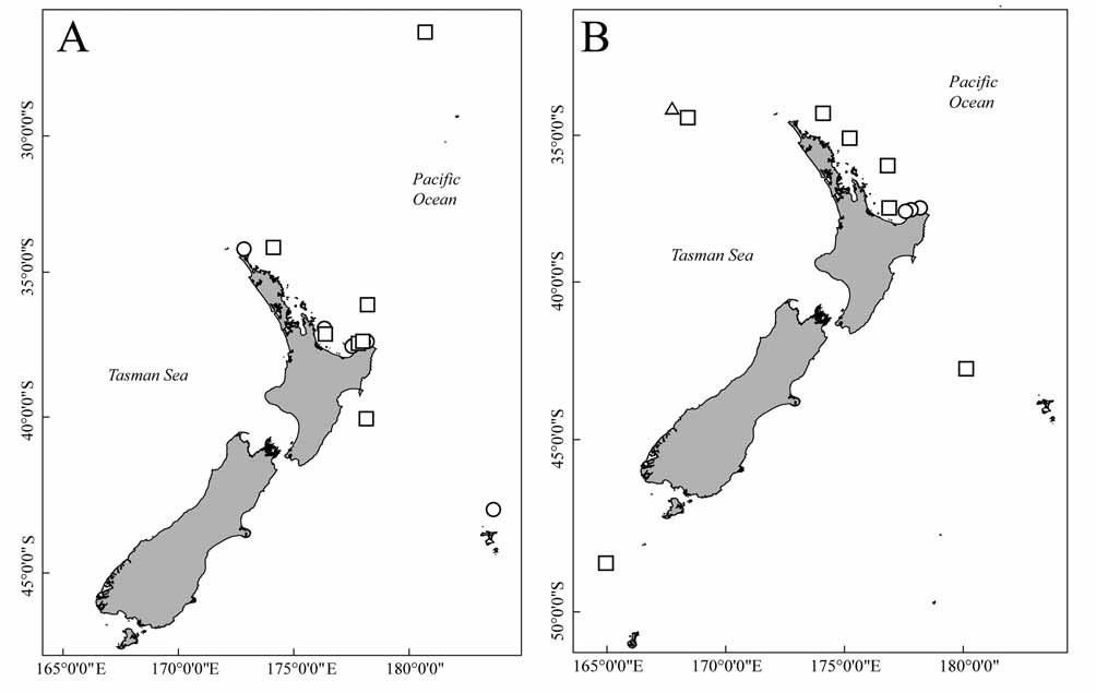
Material examined: All from Amigdoscalpellum costellatum . Holotype: 1ɗ 1.91 mm ( NIWA 35060), Stn F876, 3 Oct 1968, Bay of Plenty, 37°32.50'S, 177°34.00'E, 529 m, coll. R.V. Taranui.
Paratypes: 1ɗ attached to host prosoma on SEM stub ( NIWA 43482) 2ɗ on SEM stub ( NIWA 43483) stn F870, 2 Oct 1968, North of East Cape, 37°25.50'S, 178°10.80'E, 263 m, coll. R.V. Taranui 1Ψ on SEM stub ( NIWA 43484) from type locality. 1Ψ ( WAM C40018), 1ɗ 1.89 mm ( WAM C40019), stn KAH0001/68, 19 Feb 2000, Bay of Plenty, 37°29.38'S, 177°47.80'E, 415 m, coll. R.V. Kaharoa.
Diagnosis: Male body dorsoventrally flattened, widest at pereonite 6. Antennule article 1 with 9 posterior teeth, lateral most tooth much narrower than and abuts onto second tooth. Article 3 rami with 3 terminal setae. Antenna reaches pereonite 6. Pereopods 3–5 propodus with short sharp seta in notch on distal margin, dactylus 0.50–0.60 propodus length. Pereopods 6 and 7 dactylus-propodus length ratio as in pereopods 3–5. Coxal plate tooth formula: 6-5-5-5-5-5-3. Pleotelson posterior lobe occupying 0.80 of posterior margin. Uropod exopod 0.70–0.80 length and 0.40–0.50 width of endopod.
Mature females, with cephalon and pereonites 1–3 retained, remaining segments fused, globose, without limbs.
Description: Male (holotype) body tear-drop shaped, cuticular striations prominent. Total length 1.89 mm widest at pereonite 6 (0.64 mm), cephalon length 0.31 mm, width 0.50 mm.
Antennule article 1 with 9 posterior teeth, lateral most tooth acute extremely narrow, abuts onto second tooth (Under light microscopy, division discernible only as notch or shoulder to second tooth), other teeth blunt, medial tooth truncate. Cuticular striations present near mesial margin. Lateral margin concave forming laterally directed rounded anterior point. Article 2 with heavier cuticular sculpting distally, forming tooth-like scales. Lateral margin with 5 setae, more or less evenly distributed. Article 3 with bundle of aesthetascs placed dorsally to and 1 seta near base of 2 uniarticulate rami; rami both with 3 terminal setae. Posterior ramus longer and wider than anterior ramus and 2 aesthetascs near midpoint.
Antenna originating from beneath posterior teeth of antennule all articles except basis cylindrical, flagellar articles approximately half as wide as terminal peduncular article. Setal formula 0-1-2-5-2-1-2-1-3.
Pereopods 1 and 2 dactylus short, aquiline, approximately 0.4 length of propodus, terminus slots between two multifid spines on propodus. Propodus egg-shaped, two setae near base of dactylus. Carpus triangular, tuft of setae at distal angle. Merus subtriangular, long subterminal seta originating from groove on inner face, single shorter seta present medially. All articles with cuticular striations, propodus with ctenae.
FIGURE 9. Scanning electron micrograph of male Scalpelloniscus nieli sp. nov. paratypes (A, NIWA 43482; B–J, NIWA 43483). A, lateral view of male attached to prosoma of host; B, ventral view of anterior section; C antennule; D, detail of lateral antennule teeth; E, 1st coxal plate; F, pereopod 1; G, pereopods 3–5; H, propodus-dactylus junction of pereopod 5; I, pleopod 1; J, Pleotelson. Scale bars: A, B = 100 μm; C, F, I = 30 μm; D = 10 μm; E = 20 μm; G, J = 50 μm.
FIGURE 10. Male Scalpelloniscus vomicus sp. nov. holotype (NIWA 35063). A, Ventral view; B, antennule; C, antenna (cuticular striations not shown); D–F, 1st, 2nd and 7th coxal plates respectively; G, pereopod 1; H, pereopod 5; I, pereopod 7; J, pleopod 1; K, uropods. Not all setation shown in J and K. Cuticular striations shown are representative only. Scale bars: A = 200 μm; B–K = 50 μm.
Pereopods 3–5 ambulatory, dactylus elongate, 0.50–0.60 length of propodus, distally curved into terminal hook. Propodus distally quadrate, flattened; stout, sharp seta in notch on distal margin, ventral margin slightly concave with 1 stout medial seta. Carpus and merus as in pereopods 1 and 2. Ischium with deep groove. Cuticular striations as in pereopods 1 and 2 except dactylus without striations.
Pereopods 6 and 7 propodus semicylindrical, tapered distally, with notch, terminal seta reduced. Otherwise as in pereopods 3–5.
Coxal plates with rounded posteriorly directed teeth, width of teeth as little as 0.1 length. Striations prominent, absent on all but first tooth, giving an articulated appearance to the remainder. Formula: 6-5-5-5-5-5-3.
Pleopods becoming progressively smaller posteriorly. Pleopods 1–5 basis with 2 long, flattened trifurcate setae, posterior margin extends into bi-lobed lamella covering bases of rami, mesial lobe much smaller than lateral lobe. Exopod with 5 sparsely plumose setae; lateral seta approximately one third length of others, exopod quadrate distally, tapering in basal half. Endopods with 5 (3 in pleopod 5) sparsely plumose setae, quadrate distal margin, ovate basally and internal cuticular ring occupying basal two-thirds. All articles with prominent cuticular striations and ctenae, often overrunning margins.
Ventral abdominal lobe between first pair of pleopods truncate with fringe of fine setules and medial notch.
Pleotelson as long as wide, posterior margin entire, forming broad bell-curve shaped lobe, occupying approximately 0.80 of posterior margin, cuticular striations overextending margin forming small serrations. Uropod basis quadrate with 2 setae on posterolateral angle. Exopod cylindrical approximately 0.7–0.8 length and 0.40–0.50 width of endopod with 4 terminal setae. Endopod triangular, dorsoventrally flattened, tapering distally with six small setae basally on dorsal surface, mesial margin with profuse comb of fine setae and terminally with dense tuft of long setae.
Females 2.7 mm long, 1.1 mm wide. Anterior four segments retained intact, medial and posterior segments fused, forming globose egg sac. Shape of egg sac variable, typically with ventral depression where it is in contact with the host prosoma. Ventral seam absent.
Host: Amigdoscalpellum costellatum ( Withers, 1935) . Host specimens were found attached to the same urchins as the host specimens of Crinoniscus cephalatus sp. nov.
Remarks: The coxal plate formula sets male Scalpelloniscus nieli apart from the other species in the genus. The teeth of the coxal plates are also relatively shorter in ‘immature female’ S. penicillatus and in the ‘male’ spatulate; and are absent in S. binoculis . Relative lengths of the dactylus in pereopod 6 and 7 are also much shorter in S. nieli . The posterolateral angles of the cephalon do not project as they do in S. Penicillatus and S. binoculis . Characters distinguishing S. nieli and S. vomicus are discussed in the Remarks for the latter. Mature females are not known for the previously described species.
Males and females were never found cohabiting within a host, suggesting that males are not restricted to a single host. The females, however, lack effective ambulatory appendages and would not be able to leave the host. Unlike Scalpelloniscus vomicus sp. nov., described below, this species does not burrow or embed itself within the host’s tissue. Instead, the males will attach to the dorsal surface of the prosoma of the barnacle, oriented towards the adductor muscle, much like the males of Crinoniscus cephalatus . During gender transition the male expands dorsoventrally, presumably as ovaries develop. The male would then moult at least the cuticle of the posterior segments, which would then form the egg sac.
Etymology: Named for Dr. Niel L. Bruce of the Museum of Tropical Queensland, Townsville for confirming that there were indeed isopods living within my barnacles.
No known copyright restrictions apply. See Agosti, D., Egloff, W., 2009. Taxonomic information exchange and copyright: the Plazi approach. BMC Research Notes 2009, 2:53 for further explanation.