
Syndesmis patagonica, Brogger, Martín I. & Ivanov, Verónica A., 2010
|
publication ID |
https://doi.org/ 10.5281/zenodo.194977 |
|
DOI |
https://doi.org/10.5281/zenodo.6212240 |
|
persistent identifier |
https://treatment.plazi.org/id/C9555167-FFC6-2705-FF32-FE8F24E5F818 |
|
treatment provided by |
Plazi |
|
scientific name |
Syndesmis patagonica |
| status |
sp. nov. |
Syndesmis patagonica View in CoL n. sp.
( Figs. 1 View FIGURE 1 , 2 View FIGURE 2 )
Type locality: Puerto Madryn (42º46'S; 65º02'W), Argentina.
Type material: Holotype: 1 entire specimen prepared as a whole mount. Paratypes: 3 entire specimens prepared as whole mounts, 1 slide with sagittal section of 1 specimen, and 1 slide with cross sections of 1 specimen (MACN-Pa N 495/1–6).
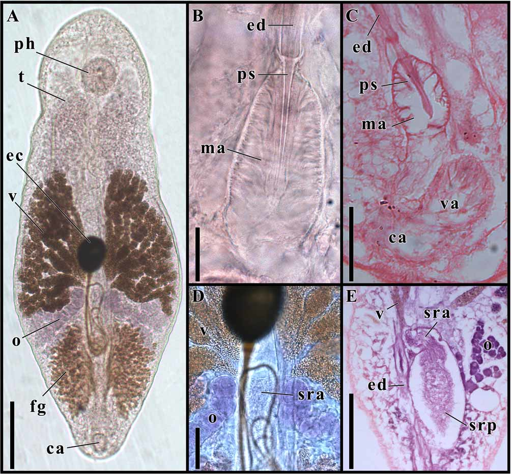
Scale bars: A and E = 200 µm, B = 20 µm, C and D = 50 µm. Abbreviations: ca, common antrum; ec, egg capsule; ed, ejaculatory duct; fg, filament glands; ma, male antrum; o, ovary; sra, anterior portion of seminal receptacle; srp, posterior portion of seminal receptacle; ph, pharynx; ps, penis stylet; t, testis; v, vitellarium; va, vagina.
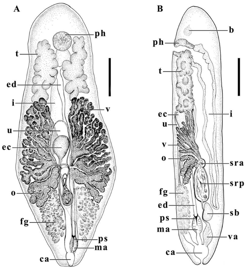
Scale bars: A and B = 200 µm. Abbreviations: b, brain; ca, common antrum; ec, egg capsule; ed, ejaculatory duct; fg, filament glands; i, intestine; ma, male antrum; o, ovary; sb, seminal bursa; sra, anterior portion of seminal receptacle; srp, posterior portion of seminal receptacle; ph, pharynx; ps, penis stylet; t, testis; u, uterus; v, vitellarium; va, vagina.
Other material: Ten whole-mounted specimens. Two sectioned specimens. Type host: Arbacia dufresnii (Echinodermata: Echinoidea).
Site of infection: Digestive tract.
Etymology: The species is named after the geographic region of the type locality (Patagonia, Argentina). Prevalence of infection: 9.2% (11 infected hosts out of 120 specimens examined). Intensity of infection: 1–15 worms per host.
Diagnosis: Pinkish living worms up to 2.3 mm long. Mouth anteroventral, doliiform pharynx, dorsal intestine, extending posteriorly. Anterior paired testes, slightly lobulated. Short hard penis stylet, 25–50 µm long, beginning at collar like structure and projecting into male antrum. Male antrum opening into common genital atrium. Genital pore at posterior tip of body. Vitellaria posterior to testes, with 3–6 primary branches, each divided once or twice. Paired ovaries posterior to vitellaria. Filament glands in posterior quarter of body. Mature egg capsules ovoid, amber, with long whip-like filament on one pole. This species can be clearly distinguished from all other species in the genus by having the shortest penis stylet ever registered in species of Syndesmis .
Description: Based on 14 specimens (whole mounts) and histological sections of 4 specimens. Worms 1.32–2.33 mm (1.68±0.30, n = 14) long, 0.40–0.69 mm (0.59±0.08, n = 14) maximum width at level of middle part of body. Living worms pinkish, slightly reddish at level of intestine. Mouth anteroventral, at end of first 1/7 of body length, opening into a doliiform pharynx. Pharynx 77–126 µm (104±14, n = 14) in diameter. Intestine dorsally, extending posteriorly from pharynx to beginning of last 1/6 of body length.
Paired testes on both sides of anterior body midline, 280–690 µm (390±118, n = 14) long, 100–230 µm (154±38, n = 14) wide, slightly lobulated, extending from posterior half of pharynx to posterior first 1/3 of body length, slightly overlapping vitellaria posteriorly. Ejaculatory duct close to midline, without coils or notorious loops, seminal vesicles not observed. Short hard penis stylet, beginning at collar like structure and projecting into male antrum, 25–50 µm (36±10, n = 13) long, stylet length to body length ratio 1:24–73 (1:50±1:15, n = 13). Male antrum opening into common genital atrium. Genital pore at posterior tip of body.
Vitellaria posterior to testes, 320–640 µm (503±85, n = 14) long, 180–300 µm (250±39, n = 14) wide, having 3–6 primary branches, each divided once or twice. Paired ovaries posterior to vitellaria, longest lobe 180–270 µm (219±35, n = 14) long, with 3–5 terminal lobes. Vitellaria and ovaries enter seminal receptacle at its anteriormost portion. Seminal receptacle formed by a spherical anterior portion, becoming elongated posteriorly, lumen with sperm. Seminal bursa posterior to seminal receptacle. Proximal part of vagina joining seminal bursa through bursal valve. Vagina gradually wider distally, reaching common genital atrium. Uterus ventrally, about 2/3 of the body length. Entry of uterus into common genital atrium ventral to entrances of male antrum and vagina. Filament glands occupying almost whole posterior quarter of body, 220–630 µm (368±119, n = 14) long, 100–170 µm (134±23, n = 14) wide. Mature egg capsules ovoid, 111–148 µm (128±14, n = 7) long, 87–98 µm (93±9, n = 7) wide, amber in color, with long whip-like filament on one pole.
No known copyright restrictions apply. See Agosti, D., Egloff, W., 2009. Taxonomic information exchange and copyright: the Plazi approach. BMC Research Notes 2009, 2:53 for further explanation.