
Tipula ( Lunatipula ) vernalis Meigen, 1804
|
publication ID |
https://doi.org/10.11646/zootaxa.4567.1.5 |
|
publication LSID |
lsid:zoobank.org:pub:621811DE-518F-4CB4-8E74-3ECB95081265 |
|
DOI |
https://doi.org/10.5281/zenodo.5921834 |
|
persistent identifier |
https://treatment.plazi.org/id/C96CC650-1557-B40D-FF1E-F8DBFF0CFA44 |
|
treatment provided by |
Plazi |
|
scientific name |
Tipula ( Lunatipula ) vernalis Meigen |
| status |
|
Tipula ( Lunatipula) vernalis Meigen
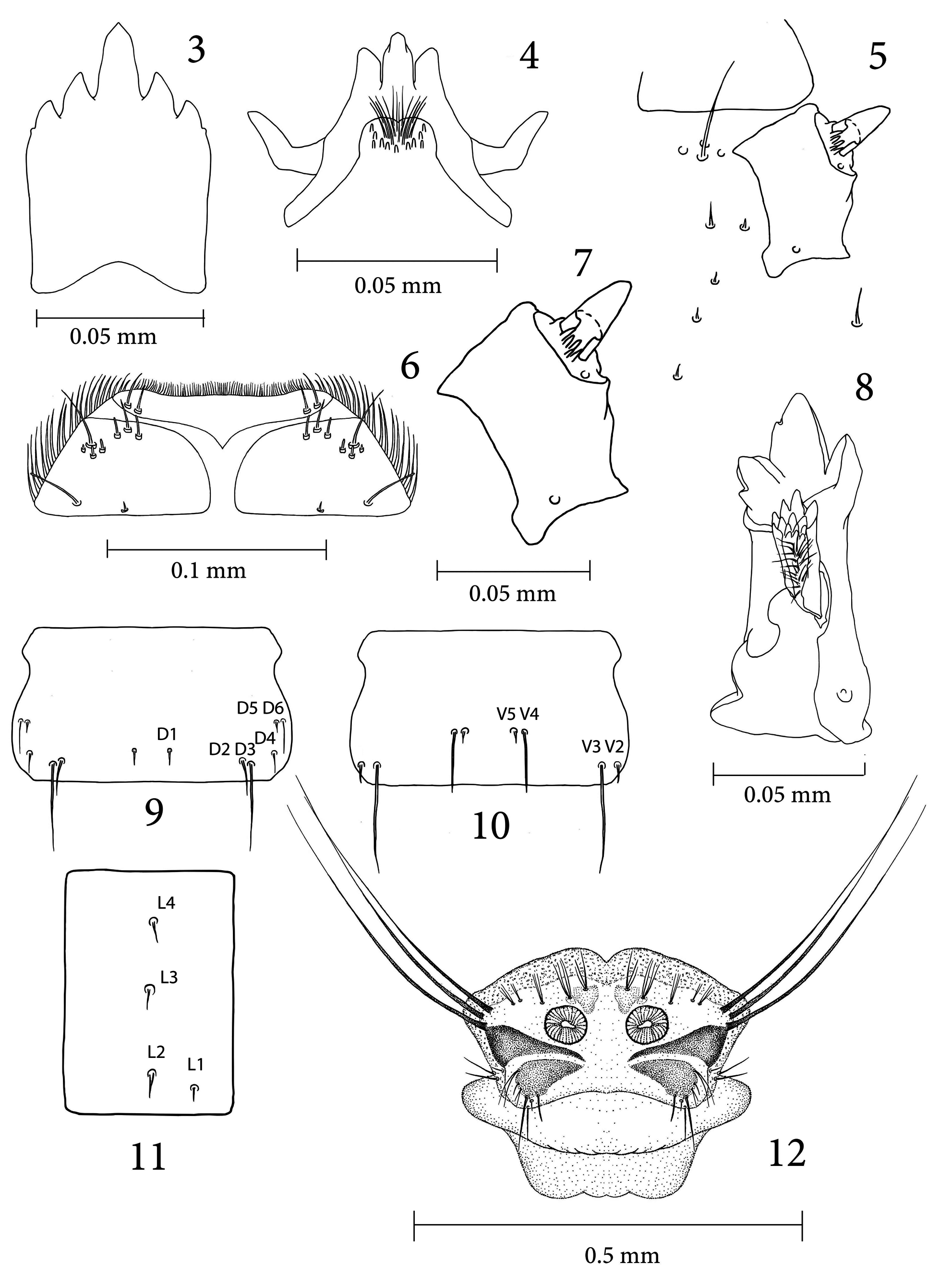
( Figs. 1–12 View FIGURES 1–2 View FIGURES 3–12 )
Examined material: 48 egg-larvae from female captured in Lazdijai district, N54.24063, E23.51554, 29 V 2011,
eggs were laid on 0 1 V1 2011, egg-larvae hatched on 16 VIII 2011; 2 egg-larvae from female captured in Lazdijai district , N54.24063, E23.51554, 29 VI 9 2011 GoogleMaps , eggs were laid on 0 2 V1 2011, egg-larvae hatched on 20 VIII 2011; 167 egg-larvae from female captured in Vilnius district, N54.68662, E25.30054, 26 GoogleMaps V 2011 , eggs were laid on 27 V 2011 , egg-larvae hatched on 4 VIII 2011 .
First instar larva. Length 1.90–2.10 mm, width 0.48–0.49 mm. Body covered with pale microscopic hairs. Cuticle transparent.
Head capsule. Length 0.39–0.41 mm, width 0.23–0.24 mm. Head capsule prognathous, hemicephalic, oval in shape, slightly depressed dorsoventrally and slightly sclerotized ( Figs. 1, 2 View FIGURES 1–2 ). Internolateralia and externolateralia equal in length and separated by incisions, which reaches almost half-length of head capsule. Externolateralia widely separated on the ventral side of capsule ( Fig. 2 View FIGURES 1–2 ). Premaxillary suture separates side plate from the rest of head capsule. Side plate wedge-shaped with anteromedially situated long seta. Hypostomium bears five large sharp teeth, the middle tooth most prominent ( Fig. 3 View FIGURES 3–12 ), basally fused with ventral margins of genae and side plates. Prementum visible from below. It has three large sharp teeth on anterior margin. Labial area bears several peg-like sensillae and cushion of firm bristles ( Fig. 4 View FIGURES 3–12 ). Frontoclypeus fused with internolateralia. Clypeal part of frontoclypeus membranous, separated from labrum by deep fold. One long seta and three sensory pits located on the anterior part of clypeus, one medium long and one short setae located below them, three very short setae located near the grove, one medium long seta below the base of antenna (close to outer margin) ( Fig. 5 View FIGURES 3–12 ). Ecdysial and coronal sutures not visible. Labrum trapezoidal in shape and composed of two wedge-shaped sclerotized plates separated by membranous area ( Fig. 6 View FIGURES 3–12 ). Apical part of labrum and epipharynx covered with numerous short hairs. Two medium long setae present on membranous part on both sides of labrum. Three medium long setae located on anterior part of labrum, one long and three short setae present on anteriolateral part of labral lobe, one long and one short seta located on posterior part of labral lobe. Antenna short, slightly tapering apically. It consists just of one cylindrical segment, which is almost as long as wide ( Fig. 7 View FIGURES 3–12 ). Apically it bears one large cone-shaped (length of sensilla just slightly shorter than length of segment) and five small peg-like sensillae, dorsally it has a sensory pit near the base. Mandible one segmented and more sclerotized than the rest of head capsule. Mandible with five sharp teeth ( Fig. 8 View FIGURES 3–12 ). Second dorsal and second ventral teeth much smaller than apical, first ventral and dorsal teeth. Prostheca or lacinia mobilis present on the dorsal side of mesal mandibular base ( Fig. 8 View FIGURES 3–12 ). Lacinia mobilis archshaped, solid, with more than 10 sharp small teeth on apical part (exact number is difficult to establish) and with cushion of short hairs in the middle. Lateral margin of mandible bears two long setae near the base. A sensory pit present at the base of dorsal side of mandible. Mandibles operate in horizontal plane. Inconspicuous larval eye spots located below the base of mandible. Maxilla with wedge-shaped cardo, bearing two almost equal setae near the distal end ( Fig. 2 View FIGURES 1–2 ). Two sclerites present on stipes, the inner sclerite extends around inner margin of maxilla onto its dorsal surface, the outer sclerite extends around outer margin of maxilla to its dorsal surface. There is a diamond-shaped sclerite near the base of galea. Galea and lacinia are fused and covered with numerous short hairs. Three long sensory structures located on the ventral side of galea.
Thorax. All thoracic segments short and wide, covered with microscopic hairs. The first thoracic segment has no welts.
Abdomen. Abdominal segments longer than wider. They are covered with two types of hairs. Short hairs cover lateral parts of segments and large portions of tergal and sternal parts. Long hairs form transversal lines on tergal and sternal parts. Setae on abdomen light brown. Setae D1 and D5 very short, more than six times as short as the longest seta D3. D2, D4 and D6 more than twice as long as D1 and D5. D2–D3 and D5–D6 very close to each other ( Fig. 9 View FIGURES 3–12 ). V1 absent. V3 very long, twice as long as V4 and almost five times as long as V2 or V5. V2–V3 and V4– V5 very close to each other ( Fig. 10 View FIGURES 3–12 ). All ventral setae similar in length and equidistant to each other. Setae L1, L2 and L3 arranged in longitudinal line ( Fig. 11 View FIGURES 3–12 ).
Hairs on sixth and seventh abdominal segments longer than those on other segments. Third thoracic and first abdominal segments are wider than longer. First and second thoracic and abdominal segments 2–8 are longer than wider. Last or anal abdominal segment constricted.
Spiracular disc. Spiracular field surrounded by four flat round-tipped lobes ( Fig. 12 View FIGURES 3–12 ), which almost completely covered with large, wedge-shaped sclerites. Lateral lobe slightly wider than longer at the base. It has three long stout apical bristles. Each bristle almost seven times as long as the lobe. Length of ventral lobe almost as width at the base. The lobe bears two medium long and three short setae on outer margin, one medium long seta located on inner margin. Medium long setae are as long as the lobe. Lobe also has one of medium-long setae and a short seta on outer margin. Very long seta and very short seta located on the apex of lobe. Triangular-shaped pale sclerite and five tufts of longer and shorter setae located above the each spiracle. The innermost tuft consists of three long setae. Laterally from it is tuft of four long setae. Further laterally, is tuft of three long setae and tuft of two short setae, the outermost tuft consists of three short setae. Spiracles large, circular and close to each other. Distance between them equals to diameter of a spiracle.
Anal field. Anus surrounded by four short, white and fleshy anal papillae. A few long setae present on anal segment.

| VI |
Mykotektet, National Veterinary Institute |
No known copyright restrictions apply. See Agosti, D., Egloff, W., 2009. Taxonomic information exchange and copyright: the Plazi approach. BMC Research Notes 2009, 2:53 for further explanation.
|
Kingdom |
|
|
Phylum |
|
|
Class |
|
|
Order |
|
|
Family |
|
|
SubFamily |
Tipulinae |
|
Genus |