
Draconarius manus, Wang & Zhang, 2018
|
publication ID |
https://doi.org/10.11646/zootaxa.4418.4.7 |
|
publication LSID |
lsid:zoobank.org:pub:27779767-4D52-49F5-975C-E882103A1074 |
|
DOI |
https://doi.org/10.5281/zenodo.5950018 |
|
persistent identifier |
https://treatment.plazi.org/id/CA4F87FA-FFDF-6602-FF0F-C6CC1D5EF802 |
|
treatment provided by |
Plazi |
|
scientific name |
Draconarius manus |
| status |
sp. nov. |
Draconarius manus View in CoL sp. nov. ( 象鼻龙隙蛛)
Figures 1A–D View FIGURES 1 , 2A–D View FIGURES 2 , 3A–B View FIGURES 3 , 4 View FIGURE 4
Type material. Holotype male: CHINA, Sichuan, Chongzhou, Anzihe Natural Reserve , Shaoyaogou Station , Yanjing Observatory , collected by hand, 30°47.8′ N, 103°12.9′ E, elev. 1634m, 0 2 October 2016, L.Y. Wang, K.Y. Xu & Y.C. Wang leg.; GoogleMaps Paratypes: 2 males and 9 females, same data as holotype; 1 male and 1 female, Anzihe Natural Reserve, Maliugou, 30°47.3′ N, 103°13.8′ E, elev. 1494m, 0 3 October 2016, L.Y. Wang, K.Y. Xu & Y.C. Wang leg.
Etymology: The specific name comes from the Latin word ‘ manus ’, meaning elephant trunk, and refers to the shape of the blind ducts of copulatory ducts; noun.
Diagnosis: This new species belongs to the D. pseudocoreaus -group, similar to D. pseudocoreanus Xu & Li, 2008 ( Zhu, Wang & Zhang 2017: 353, figs 220A–E) and D. retrotubularis Zhang, Zhu & Wang, 2017 ( Zhu, Wang & Zhang 2017: 360, figs 227A–B) in having a long patellar apophysis, filiform embolus, extremely elongated cymbial furrow and developed prolateral conductor lamellar of male pedipalps; a pair of small, anterio-laterally located epigynal teeth, strongly convoluted copulatory ducts with blind ducts posteriorly of epigynes, but can be distinguished by the anteriorly elongated patellar apophysis approaching the median part of tibia, distinct RTA, bifurcate end of conductor of male pedipalps ( Figs 1A–B View FIGURES 1 , 2A–B View FIGURES 2 ) and the anteriorly located spermathecal heads of the epigyne ( Figs 1C–D View FIGURES 1 , 2C–D View FIGURES 2 ).
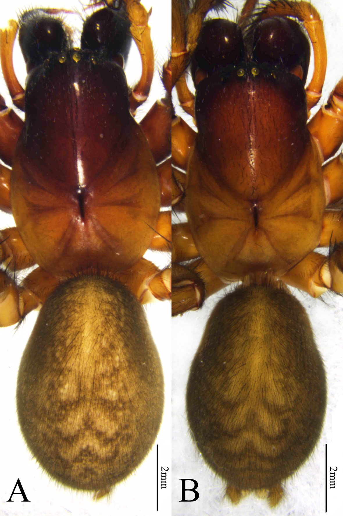
Description: Males total length 9.85–10.73. Male holotype ( Fig. 3A View FIGURES 3 ) total length 10.50: Prosoma 5.41 long, 3.78 wide; opisthosoma 5.09 long, 3.65 wide. Eye sizes and interdistances: AME 0.17, ALE 0.21, PME 0.21, PLE 0.21; AME–AME 0.09, AME–ALE 0.10, PME–PME 0.14, PME–PLE 0.21, ALE–PLE 0.09. MOA 0.58 long, front width 0.42, back width 0.57. Clypeus height 0.20. Chelicerae with 3 promarginal and 2 retromarginal teeth. Leg measurements: I 15.48 (4.10, 5.44, 3.74, 2.20), II 13.52 (3.73, 4.52, 3.22, 2.05), III 11.46 (3.20, 3.67, 2.84, 1.75), IV 14.60 (4.12, 4.67, 4.08, 1.73). Leg formula: 1423.
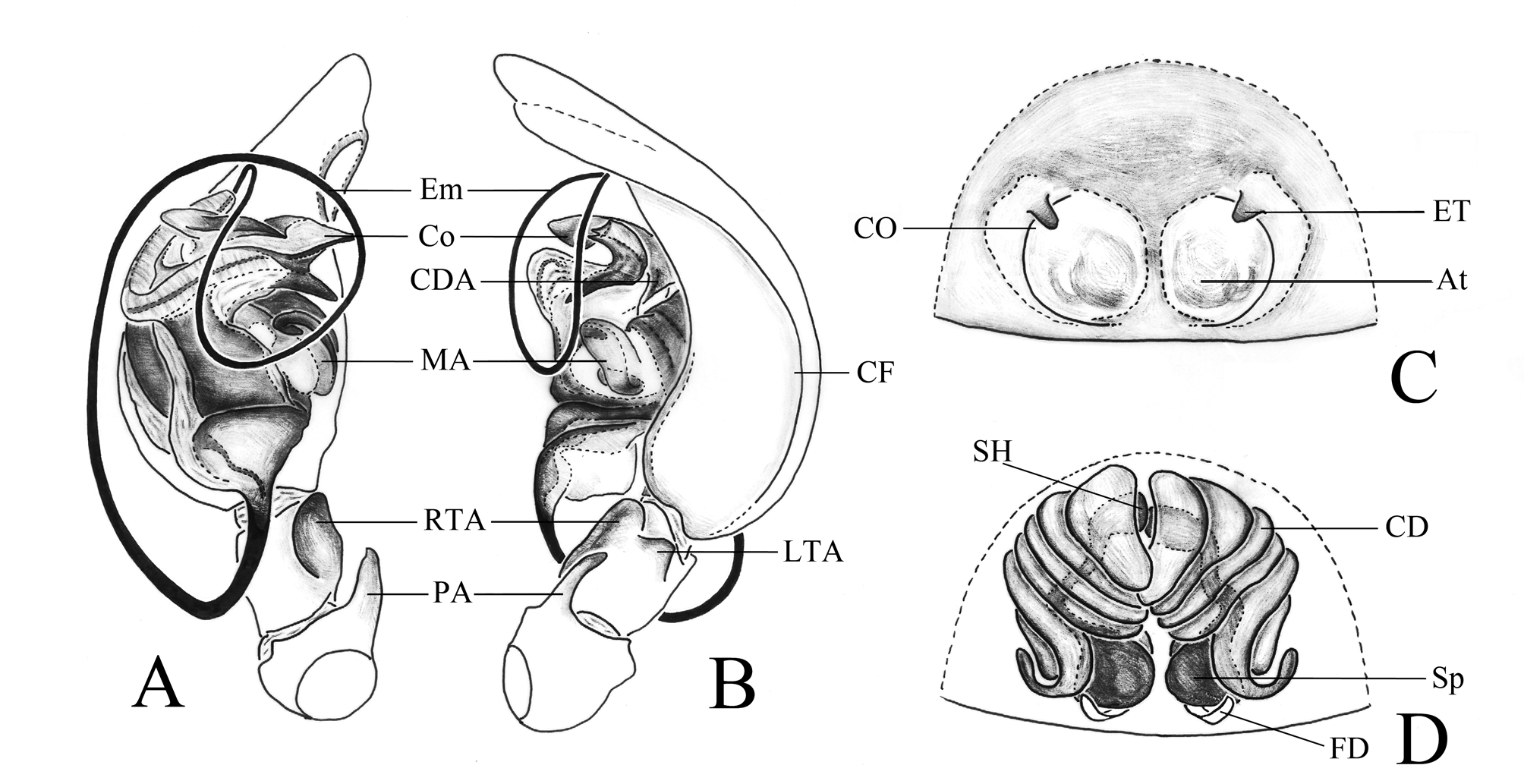
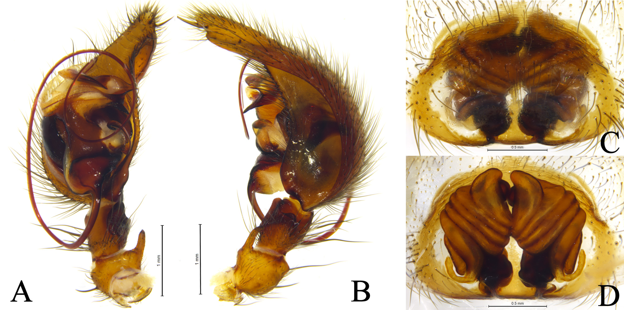
Pedipalp ( Figs 1A–B View FIGURES 1 , 2A–B View FIGURES 2 ). Patellar apophysis long, extending anteriorly and approaching the mid-part of tibia. RTA large and sheet-like, with blunt tip and its length more than half of length of tibia. Lateral tibial apophysis relatively small, located on the retrolateral surface of tibia sub-apically. Cymbial furrow relatively deep, its length much more than half of cymbium. Embolus slender, filiform, originated proximally. Conductor with developed membranous lamellar prolaterally, a bifurcate end, a ventral process and a strong dorsal apophysis. Median apophysis relatively long and spoon-like.
Females total length 8.15–10.99. One of paratypes ( Fig. 3B View FIGURES 3 ) total length 10.88: Prosoma 5.54 long, 3.54 wide; opisthosoma 5.34 long, 3.21 wide. Eye sizes and interdistances: AME 0.16, ALE 0.24, PME 0.22, PLE 0.27; AME– AME 0.10, AME–ALE 0.11, PME–PME 0.13, PME–PLE 0.26, ALE–PLE 0.09. MOA 0.58 long, front width 0.44, back width 0.56. Clypeus height 0.24. Chelicerae with 3 promarginal and 2 retromarginal teeth. Leg measurements: I 13.04 (3.71, 4.58, 2.93, 1.82), II 11.59 (3.28, 3.89, 2.75, 1.67), III 10.26 (3.04, 3.47, 2.48, 1.27), IV 13.54 (3.44, 4.55, 3.74, 1.81). Leg formula: 4123.
Epigyne ( Figs 1C–D View FIGURES 1 , 2C–D View FIGURES 2 ) with a large and shallow atrium. A pair of epigynal teeth originating from anterior margin of atrium laterally. Copulatory openings located beneath the teeth. Copulatory ducts long, spiraled more than four circles, close to each other anteriorly and two blind ducts present posteriorly, like the nose of an elephant. Spermathecal bases posteriorly located, heads anteriorly located and connected to each other, and stalks thin and long. Fertilization ducts originating from spermathecal bases posteriorly.
Distribution. China ( Sichuan) ( Fig. 4 View FIGURE 4 ).
Habitat. The type materials were collected from leaf litters and under stones, in typical microhabitat of most coelotines.
Remarks. Zhu et al. (2017) had erroneously regarded as the blind end of the copulatory ducts as spermathecal heads and on that basis they characterized the D. pseudocoreanus -group as having spermathecal heads that originated posteriorly. However, from this species, we can clearly find that the spermathecal heads are extended anteriorly and those structures in the posterior are in fact the blind endings of the copulatory ducts. Considering the remaining species in this group, the spermathecal heads are anteriorly located and pointed forward in D. retrotubularis but are not seen in D. pseudocoreanus .

No known copyright restrictions apply. See Agosti, D., Egloff, W., 2009. Taxonomic information exchange and copyright: the Plazi approach. BMC Research Notes 2009, 2:53 for further explanation.
|
Kingdom |
|
|
Phylum |
|
|
Class |
|
|
Order |
|
|
Family |
|
|
Genus |