
Diaphanosoma senegal isanensis, Korovchinsky, Nikolai M. & Sanoamuang, La-Orsri, 2008
|
publication ID |
https://doi.org/10.5281/zenodo.180698 |
|
DOI |
https://doi.org/10.5281/zenodo.5661135 |
|
persistent identifier |
https://treatment.plazi.org/id/CB00B647-CA1A-FF85-B0D1-FC7DFB3E248F |
|
treatment provided by |
Plazi |
|
scientific name |
Diaphanosoma senegal isanensis |
| status |
subsp. nov. |
Diaphanosoma senegal isanensis View in CoL ssp. nov.
( Figs 1–24 View FIGURES 1 – 9 View FIGURES 10 – 14 View FIGURES 15 – 20 View FIGURES 21 – 24 )
Parthenogenetic female. Body measurements are shown in Table 1 View TABLE 1 .
Head large (length 39.8–45.5% and height 24.5–31.1% of body length respectively), with strongly developed and protruding dorsal part, its shape may be slightly variable ( Figs 1, 2 View FIGURES 1 – 9 ). Eye large (9.7–11.7% of body length) situated close to ventral side of head. Antennules shifted far backwards and mostly covered by basipodites of swimming antennae; they are relatively long, with short basal part and long and stout sensory seta covering distally by thin densely situated setules ( Fig. 3 View FIGURES 1 – 9 ).
HL: BL, % HH: BL, % DE: BL, % LSA: BL, % LUAB: BaL, % LLAB: BaL, % LPVM: BL, % BL—body length, HL—head length, HH—head height, DE—diameter of eye, LSA—length of swimming antennae, BaL—length of antennal basipodite, LUAB—length of upper antennal branch, LLAB—length of lower antennal branch, LPVM—length of posterior valve margin.
1) general lateral view; 2) head of different shape; 3) antennule; 4) dorsal proximal seta of antennal basipodite; 5) distal part of antennal basipodite, outer side; 6) distal part of proximal segment of upper antennal branch (exopodite); 7) distal part of distal segment of the same branch; 8) ventral valve inflexion; 9) inner setae near posterior valve margin. Scale bars in mm.
10) posterior valve margins of different specimens; 11) deformed posterior valve margin; 12) postero-ventral corner of valve without emargination; 13) deformed postero-ventral corner of valve; 14) postabdomens of different specimens. Scale bars in mm.
Swimming antennae long (66.7–81.1% of body length), reaching or slightly extending posterior valve margins, they are robust with both powerful basipodite and long branches (upper 2-segmented branch (exopodite) 67.5–79.1% of body length, lower 3-segmented branch (endopodite) 50.0–61.5% of body length) ( Fig. 1 View FIGURES 1 – 9 ). Proximal dorsal side of basipodite with long thin seta ( Fig. 4 View FIGURES 1 – 9 ), distal outer end with relatively small thin spine, and opposite to base of upper branch are two prominences, one of them slightly bifid ( Figs 5 View FIGURES 1 – 9 , 15 View FIGURES 15 – 20 ). Apical spine of proximal segment of upper 2-segmented branch long and thin ( Figs 6 View FIGURES 1 – 9 , 16 View FIGURES 15 – 20 ), while that on the end of distal segment of the same branch is rather stout with two prominences: small outer one and larger inner one near its base ( Figs 7 View FIGURES 1 – 9 ). Distal segment of lower antennal branch with small apical spine having outgrowth near its base ( Fig. 18 View FIGURES 15 – 20 ). Formula of antennal setae 4–8 / 0–1–4, their setules seem somewhat flattened ( Fig. 19 View FIGURES 15 – 20 ).
The general shape of mandibular molar plates triangular with a broadened posterior part. Posterior edge of right mandible ( Figs 17, 20 View FIGURES 15 – 20 ) bears two massive sharpened pegs and 4–5 thick pales with somewhat brushed ends. All other pales along all dorsal margin are very densely placed forming almost fused margin. Two long "fishbones" with broadened and coarsely brushed tips closely situated to pegs ( Fig. 17 View FIGURES 15 – 20 ). Central part of molar plate slightly concave with 8–10 parallel rows of diagonals diminishing in size anteriorly ( Fig. 20 View FIGURES 15 – 20 ).
15) distal part of antennal basipodite, outer side; 16) distal part of proximal segment of upper antennal branch; 17) right mandible,view from ventral side (Pe-pegs, Pa-pales, F-"fishbones"); 18) apical part of lower antennal branch (endopodite); 19) antennal setae and setules; 20) right mandible, view from posterior side (see 17, D-diagonals). Scale bars in μm.
Valves elongate and rectangular, their posterior margins almost evenly straight (23.9–28.3% of body length) and armed with 10–29 denticles (on average, 18.1 and 19.5 in Thai and Vietnamese specimens respectively), 8–22 upper of which large and often more sparsely situated ( Fig. 10 View FIGURES 10 – 14 ). These denticles diminish in size ventrally, sometimes evenly but usually abruptly when 3–10 lowermost of them appear to be conspicuously smaller than upper ones ( Fig. 23 View FIGURES 21 – 24 ). More ventrally, along postero-ventral valve margin, these denticles are followed by a row of thin, short setules ( Figs 23, 24 View FIGURES 21 – 24 ) and then, along edge emargination, by 6–10 short feathered setae ( Fig. 10 View FIGURES 10 – 14 ). Sometimes this emargination may be absent on one or both valves of one specimen thus making postero-ventral margin more or less rounded ( Fig. 12 View FIGURES 10 – 14 ), or this part of valves may be deformed ( Figs 11, 13 View FIGURES 10 – 14 ). Ventral valve margins form rather wide inflexion, widening proximally, its edge with long row of about 35 relatively short and finely feathered setae, the proximal of which appear to be more densely situated ( Fig. 8 View FIGURES 1 – 9 ). Two inner small spine-like finely feathered setae near posterior margin of each valve ( Figs 9 View FIGURES 1 – 9 , 22 View FIGURES 21 – 24 ).
21) postabdominal claws; 22) inner setae near posterior valve margin; 23) postero-ventral corner of valve; 24) setules of postero-ventral corner of valve. Scale bars in μm.
Postabdomen comparatively small, with 3–7 small anal denticles, some of which may be duplicated, and groups and rows of minute spinules on each lateral side ( Fig. 14 View FIGURES 10 – 14 ). Terminal claws massive with three robust basal spines. Distally to the latter and more ventrally, a series of spinules along claws' lateral and concave outer margins ( Fig. 21 View FIGURES 21 – 24 ). Setae natatoriae long and sit on strongly developed base.
Body length 0.99–1.76 mm. Most females had 2– 12 eggs or embryos in inflated brood pouches.
Etymology. The subspecies is named for the native name of people (‘isan’) leaved in North-East Thailand.
Differential diagnosis. Specimens from Thailand and Vietnam are very similar morphologically and differ from all others, especially from those known in West Africa and India (see Korovchinsky 1991, 1993), by presence of conspicuously larger, less numerous, and more sparsely situated denticles along posterior valve margins, upper and lowermost of which may differ considerably in their size. Specimens from Bangladesh seem closer to Thai and Vietnamese ones in this respect but, nevertheless, they have more numerous and smaller denticles, thus being intermediate in this feature between the extreme variants of this feature in West African and Indian specimens which represent the nominal subspecies ( D. senegal senegal Gauthier, 1951 ) and representatives of new subspecies from South-East Asia. Specimens from Bangladesh may be tentatively regarded as a nominal subspecies also, until their more detailed investigation in the future.
Type specimens. Holotype—adult female with body length 1.40 mm from the locality NE 245 (temporary pond at Ban Eiat, Muang dt., Maha Sarakham Province, 08.05.1999) contained in a small jar with formalin deposited in Zoological Museum of Moscow State University; Cat. No. Ml 63.
Paratypes— 21 adult females from the same locality in a small jar with formalin deposited in Zoological Museum of Moscow State University, Cat. No. Ml 64.
All other specimens ( paratypes) have been deposited in a personal collection of NMK.
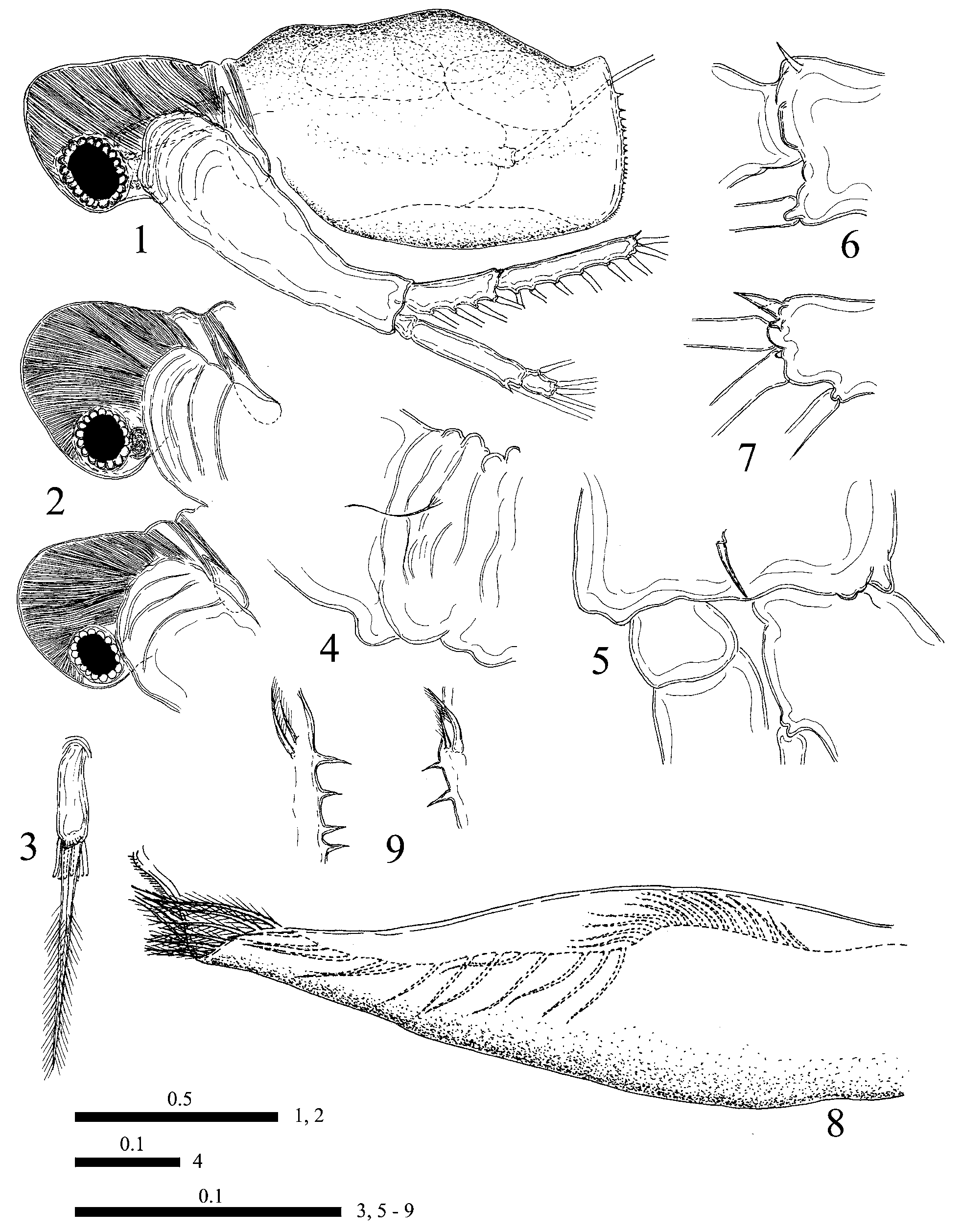

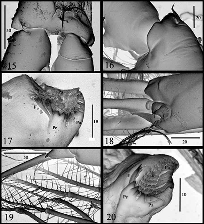
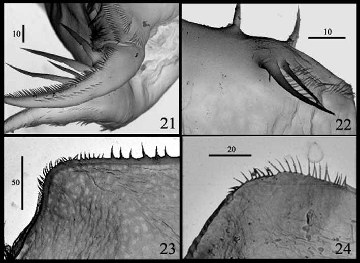
TABLE 1. Measurements of body parts' proportions of adult females of Diaphanosoma senegal isanensis ssp. nov. (n = 17) (in each column from top to down: Range, Mean, SD, CV)
| 39.8–45.5 | 24.5–31.1 | 9.7–11.7 | 66.7–81.1 | 67.5–79.1 | 50.0–61.5 | 23.9–28.3 |
|---|---|---|---|---|---|---|
| 42.8 | 28.1 | 10.8 | 73.4 | 73.2 | 55.3 | 25.6 |
| 1.76 | 1.68 | 0.52 | 3.5 | 3.2 | 3.5 | 1.17 |
| 4.1 | 6.0 | 4.8 | 4.8 | 4.4 | 6.3 | 4.6 |
No known copyright restrictions apply. See Agosti, D., Egloff, W., 2009. Taxonomic information exchange and copyright: the Plazi approach. BMC Research Notes 2009, 2:53 for further explanation.
|
Kingdom |
|
|
Phylum |
|
|
Class |
|
|
Order |
|
|
Genus |