
Ocnophila integra Brunner, 1907
|
publication ID |
https://doi.org/ 10.11646/zootaxa.5296.2.3 |
|
publication LSID |
lsid:zoobank.org:pub:30BAA198-ACE8-4559-871F-4847416EA355 |
|
DOI |
https://doi.org/10.5281/zenodo.7982369 |
|
persistent identifier |
https://treatment.plazi.org/id/CB2E8781-F911-FFE2-D893-77C410C75EC6 |
|
treatment provided by |
Plazi |
|
scientific name |
Ocnophila integra Brunner, 1907 |
| status |
|
Ocnophila integra Brunner, 1907 View in CoL
( Figs 1–7 View FIGURE 1 View FIGURE 2 View FIGURE 3 View FIGURE 4 View FIGURE 5 View FIGURE 6 View FIGURE 7 , but also see Fig. 8 View FIGURE 8 )
Ocnophila integra Brunner, 1907: 313 View in CoL ; Hebard 1919: 163; Brock 1998: 36; Zompro 2001: 234; Otte & Brock 2005: 229; Brock & Büscher 2022: 515.
>> Lectotype, J ( NMW): det. Br.v.W. Ocnophila integra Br ; Coll. Br.v.W., Porto Cabello Thorey ; 1399.; Ocnophila integra Brunner v. W., 1907 J, LECTOTYPUS, det. O.Zompro XI.1997 (NHMW) ( Figs 1–3 View FIGURE 1 View FIGURE 2 View FIGURE 3 ), designated by Zompro, 2001: 234 .
= Parapygirhynchus catenatus Brunner, 1907: 316 View in CoL ; Weidner 1966: 231.
>> Holotype, ♀ ( NMW): Porto Cabello , Venezuela, W. Sievers leg. Geogs. “Ysidid.”, 6.X.1892.; Parapygirhynchus catenatus Br. ♀. C Brunner v. W., determ. 1899, Typus, public. 1906-08., Bestimm.-Verz. Nr. 39.; Parapygirhynchus catenatus Brunner v. W., HOLOTYPUS det. O.Zompro III.2001.
= Ocnophila signatior Brunner, 1907: 315 View in CoL ; Brock 1998: 58; Otte & Brock 2005: 230; Brock & Büscher 2022: 515. syn. nov.
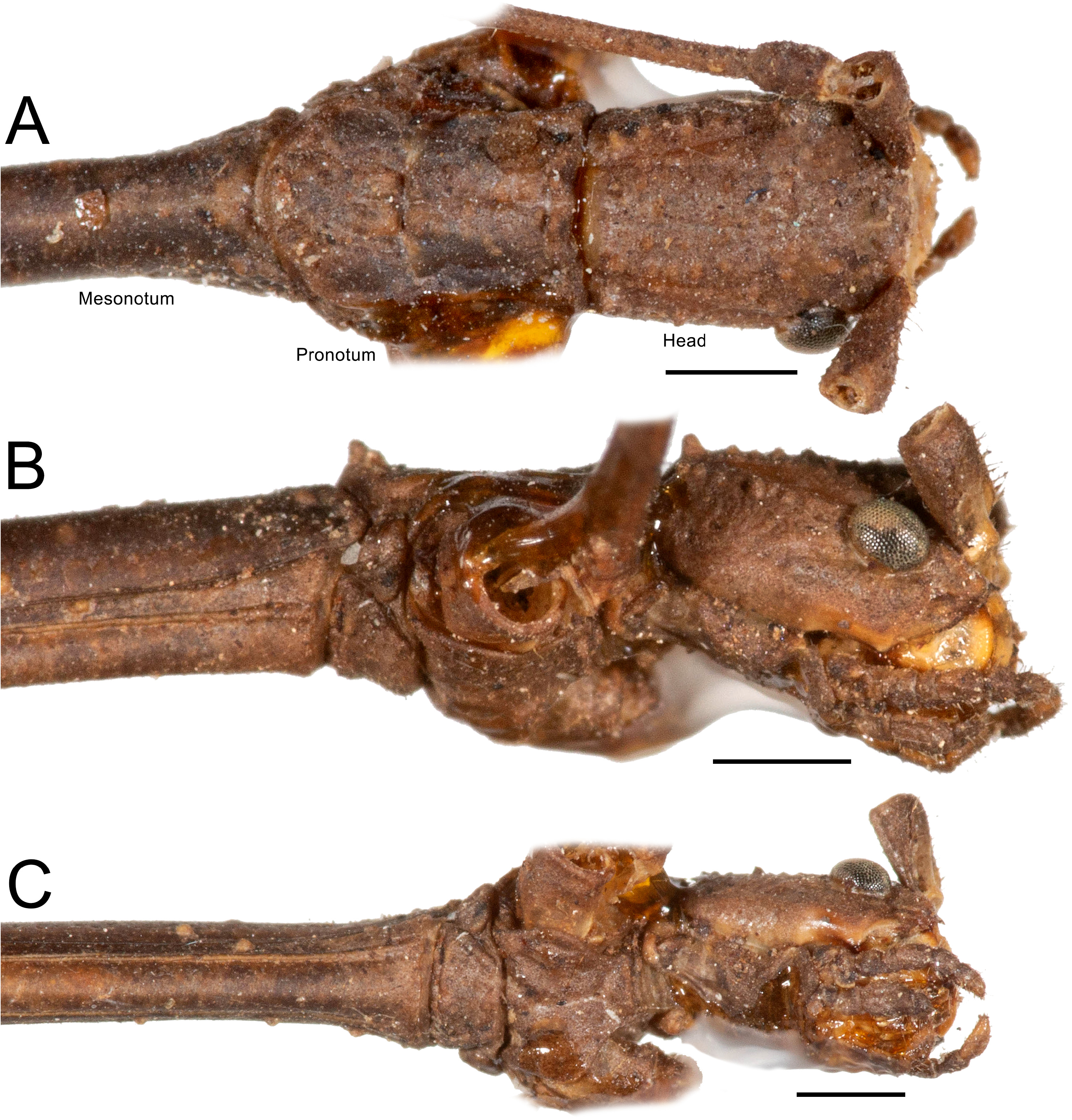
>> Holotype, J (NMW): det. Br.v.W. Ocnophila signatior ; Coll. Br.v.W., Venezuela, Thorey ded. ; 20.862; 629. ( Figs 4 View FIGURE 4 , 7A–C View FIGURE 7 )
Further material:
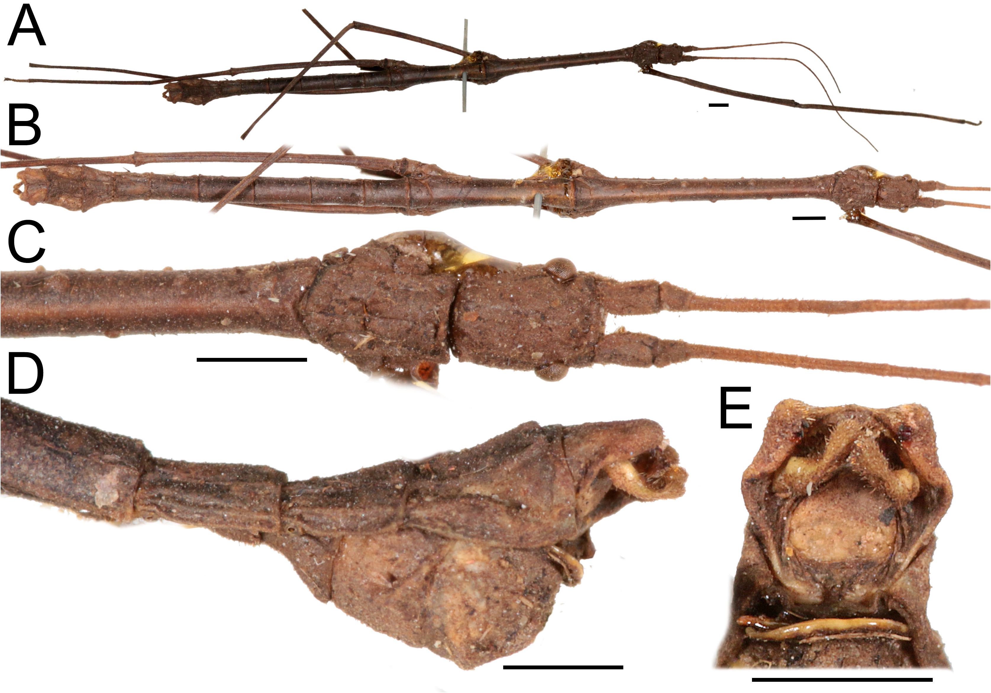
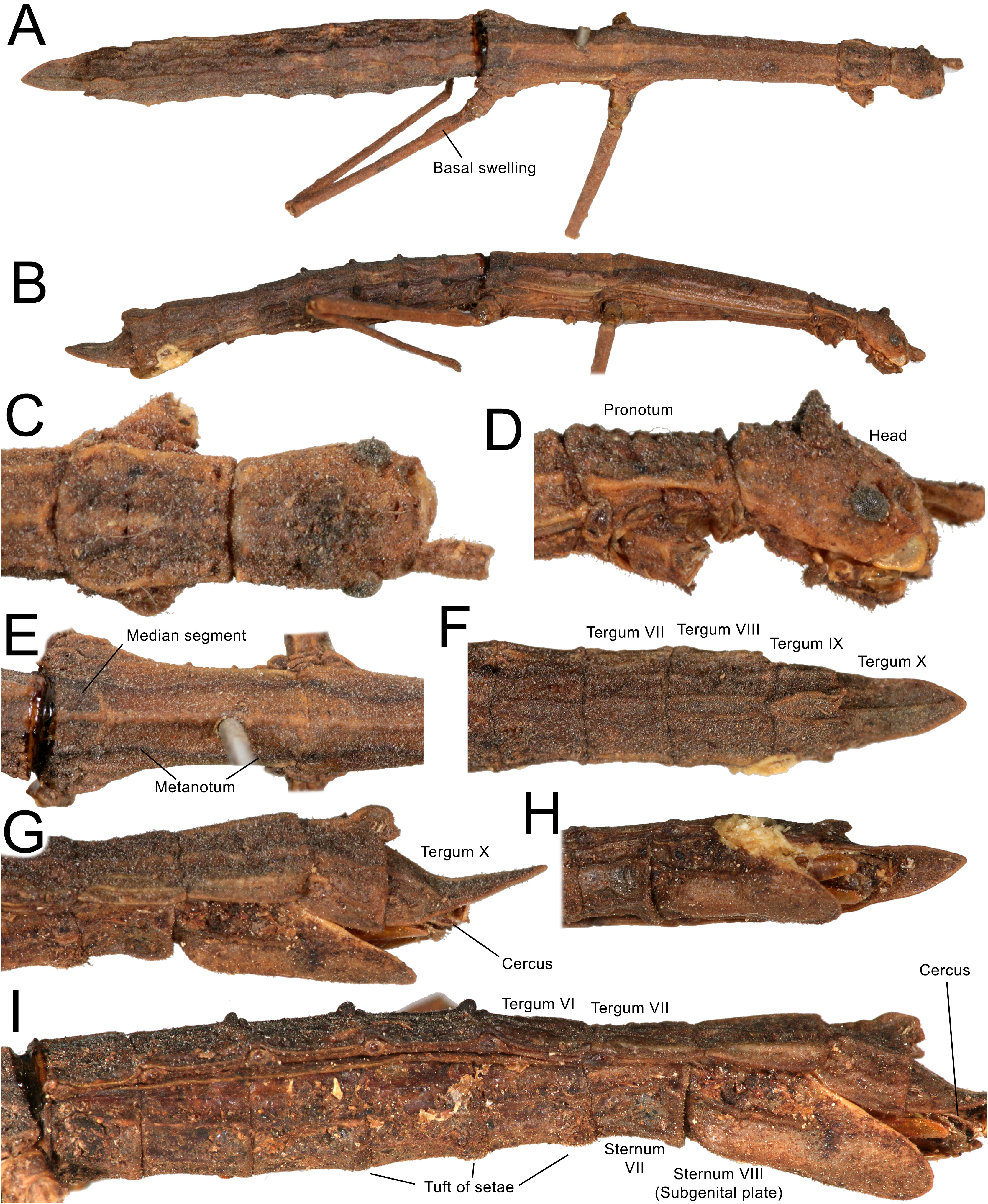
♀ (NMW): det. Br.v.W. Parapygirhynchus iphiclus Br ; Coll. Br.v.W., Porto Cabello Thorey ; 1401; 632. ( Fig. 6 View FIGURE 6 ); J( NMW): det. Hebard, 1926. Parapygirhynchus iphiclus (Westw.) ; Ssn Esteban X–XI 1910 Ven MA Carriker Jr.; Porto Cabello; 632. ( Figs 5 View FIGURE 5 , 7D–F View FIGURE 7 ).
Diagnosis. Ocnophila integra can be differentiated from the other two species of the genus by its slender, more elongate body (mesothorax around 5x in females and 13x in males longer than wide; in other species 3x in females and 6x in males longer than wide or less). Furthermore, females of O. integra can be differentiated from females of O. serrata sp. nov. by the much less serrated legs, less scalloped abdomen and terga VIII–IX slightly shorter in height. Males of O. integra can be further differentiated from males of O. iphicla by the longer legs, smoother surface of the body, smooth femora and shorter tergum X.
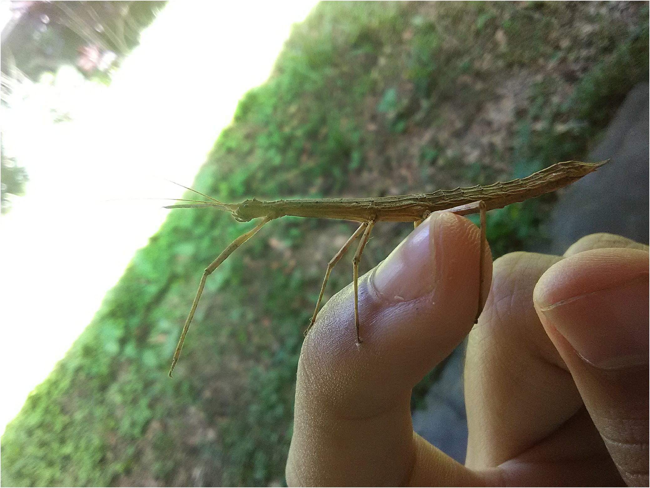
Remarks. A record made by Javier Uzcátegui and uploaded to iNaturalist ( Uzcátegui 2022) of a female stick insect from Caracas ( Fig. 8 View FIGURE 8 ) is tentatively assumed to represent a female of O. integra , or at least to represent an undescribed species of Ocnophila which would be more closely related to O. integra than to other species of the genus, as it shares the most striking diagnostic feature of this species, i.e., the slenderer, more elongate body especially seen at the thorax. To the best of our knowledge this is the only photo of a living Ocnophila with that feature.
Redescription.
Males ( Figs 1–5 View FIGURE 1 View FIGURE 2 View FIGURE 3 View FIGURE 4 View FIGURE 5 , 7 View FIGURE 7 ).
Colour ( Figs 1 View FIGURE 1 , 4 View FIGURE 4 , 5 View FIGURE 5 ). Entirely brown, with lighter stains and tubercles over body creamish.
Head ( Figs 2 View FIGURE 2 , 4C View FIGURE 4 , 5C–D View FIGURE 5 ). Subrectangular, ca. 1.5x longer than wide, vertex gently convex; with several irregular granules dorsally mainly in two paramedial rows, larger posteriorly ( Fig. 2B View FIGURE 2 ). Frontal convexity developed, frontal suture deep and curved. Eyes large, hemispherical. Labial and maxillary palp segments wide. Antennae filiform, reaching tergum VI, barely exceeding front legs. Scapus dorsoventrally compressed, wide. Pedicel more than half the length of scapus, longer than wide. Antennae with around 50 articles.
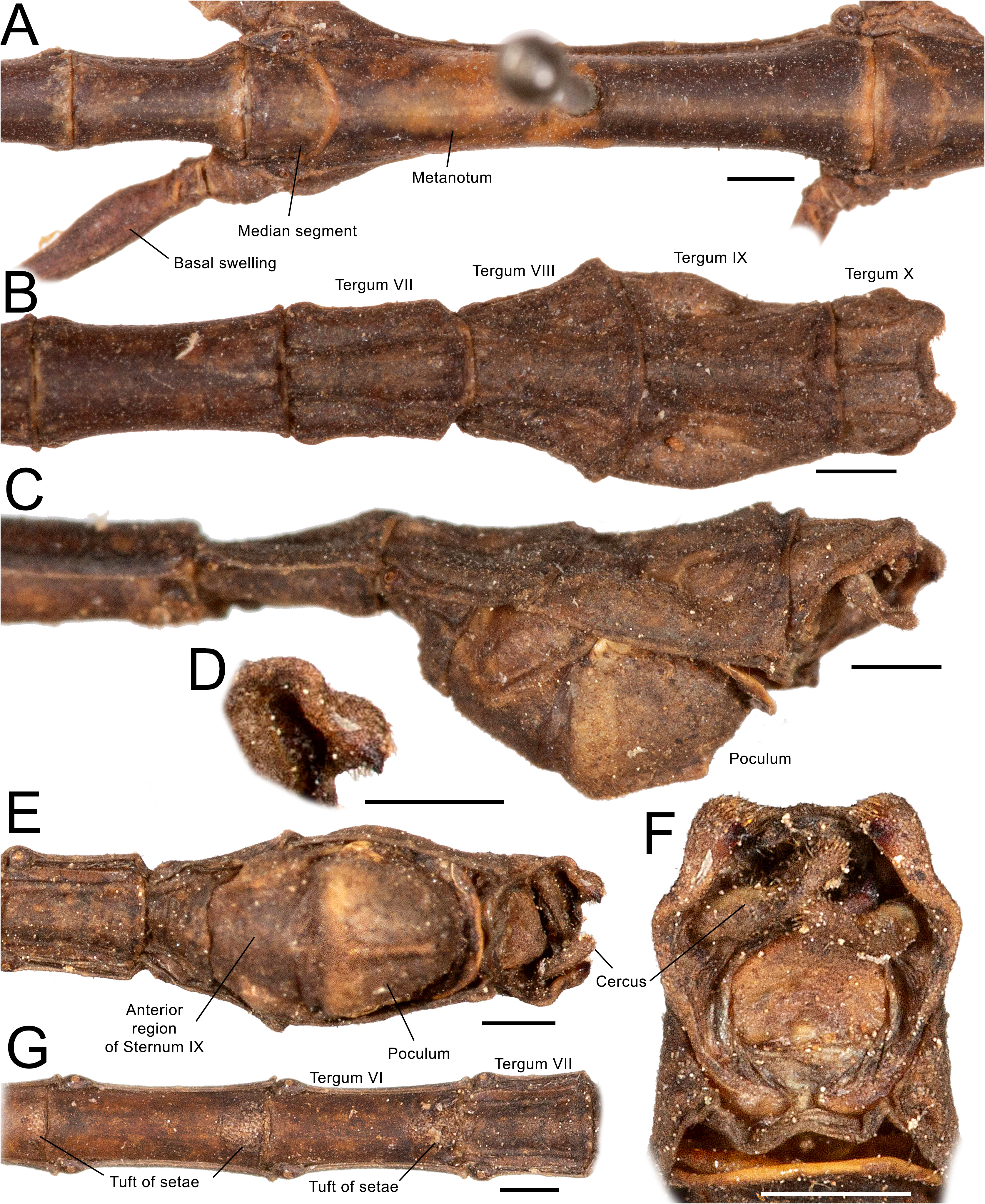
Thorax ( Figs 1–5 View FIGURE 1 View FIGURE 2 View FIGURE 3 View FIGURE 4 View FIGURE 5 ). Smooth surface with sparse minute setae and small to large irregular granules. Pronotum longer than wide, slightly constricted premedially, slightly narrower anteriorly; rugose, with several irregular granules, a paramedial pair near the posterior margin the largest ( Figs 2A–B View FIGURE 2 , 4C View FIGURE 4 , 5C–D View FIGURE 5 ). Probasisternum with longitudinal sulcus, anterior margin narrower, posterolateral margins round. Profurcasternum trapezoid, covered with short stiff setae ( Fig. 2C View FIGURE 2 ). Mesothorax 5–6x longer than prothorax; mesonotum with 8–15 irregular granules varying in size along its length, mesepisternum with 5–6 irregular small granules along its length ( Fig. 1D View FIGURE 1 ). Metathorax with very few irregular granules in dorsal surface mostly in mid length and 8–10 small irregular granules laterally scattered along each metepisternum. Mesothorax ca. 1.8–1.9x longer than metathorax. Meso and metanotum with faint, thick, smooth longitudinal medial carina. Mesofurca in “Y” shape and metafurcae as “Ψ” both also with a posteriormost longitudinal sulcus. Metanotum 5.8–6x the length of median segment.
Legs ( Figs 1B,C View FIGURE 1 , 3A View FIGURE 3 , 4A View FIGURE 4 , 5A View FIGURE 5 . Hindlegs slightly longer than forelegs; midlegs shortest. Coxae smooth. Profemur slightly longer than mesothorax.Mesofemur ca. 0.8x the length of profemur.Profemur with basal curvature occupying around one fourth of the segment ( Fig. 1B View FIGURE 1 ). Metafemur with a strong basal swelling occupying around one sixth to one seventh of the length of the segment ( Figs 1B View FIGURE 1 , 3A View FIGURE 3 , 5A View FIGURE 5 ). Carinae of femora and tibiae with sparse setae between them and each bearing a row of porrect thick setae larger in the tibiae. Carinae of tibiae with ca. 3–5 small, short spines at the apex mainly in the two ventrolateral and the ventral carinae, with more spines in mid and hind tibiae. Basitarsi short, around the same size as the respective following three tarsomeres combined; ventrally with tuft of stiff, porrect setae mainly in mid and hind basitarsi. Euplantulae well developed and present in all tarsomeres.
Abdomen ( Figs 1A–C View FIGURE 1 , 3 View FIGURE 3 , 4 View FIGURE 4 , 5 View FIGURE 5 ). Smooth surface, without granules but with carinae faint in anterior segments and well-marked from segment VII. Median segment well marked by a sulcus, ca. 0.15x the length of the metanotum. Terga II–VII longer than wide, VIII–IX as long as wide, X wider than long. Terga II–VII constricted except at anterior and posterior margins, forming a series of scallops laterally at dorsal view; VII only slightly constricted. Terga II–X and sterna II–VIII with a pair of paramedial longitudinal carinae, only faint until VI and well-marked in terga VII–X and sterna VII–VIII; terga VIII–IX with a furthermore central paramedial pair of carinae; tergum X further with a medial carina, strong, raised. Sterna IV–VI with dense tuft of setae near posterior margin, that of IV shorter and elliptical, in V–VI round ( Fig. 3G View FIGURE 3 ). Posterior margin of tergum X elevated and with a round and wide emargination forming two round prominent lobes bearing thorn pads at the ventral surface ( Figs 3B View FIGURE 3 , 5E View FIGURE 5 ). Tergum X shorter than IX; IX shorter than VIII. Tergum VIII widening towards posterior and IX tapering towards posterior; X wider centrally with convex lateral prominences. Poculum well developed, round, with a medial carina running from near the margin with anterior part of sternum IX to slightly more than mid the length until posterior margin ( Figs 3C, D View FIGURE 3 , 4D View FIGURE 4 , 5F View FIGURE 5 ). Poculum with posterior margin round and labellate, i.e., continuing shortly towards posterior at an angle of around 90º ( Fig. 3C, D, F View FIGURE 3 ). Anterior part of sternum IX long, almost as long as poculum. Cerci short, shorter than tergum X, slightly surpassing its posterior margin; strongly incurved, dorsoventrally compressed, apex round ( Fig. 4E View FIGURE 4 ). Thorn pads small, slightly displaced towards the centre, bearing only 1–3 strong, large slightly incurved spines ( Figs 3E, F View FIGURE 3 , 4E View FIGURE 4 ). Vomer well developed.
Internal genitalia – phallic organ ( Fig. 7 View FIGURE 7 ). Composed of dorsal sclerite, dorsal lobe, ventral or longitudinal lobe, basal lobe and a basal sclerite. Dorsal sclerite large, in “b” shape, i.e., a subquadrate anteriormost area with a thin, somewhat straight prolongation at right side towards posterior. Basal sclerite on the right side, curled, claw-shaped, with flat base, located between the basal and longitudinal lobes. Basal lobe short, round. Longitudinal lobe large, with several foldings and with fine granulation, denser in the large posteriormost round part. Dorsal lobe large, originating ventrally to the base of the prolongation of the dorsal sclerite, round, extending to the longitudinal lobe; anteriorly bearing short stiff setae ( Fig. 7F View FIGURE 7 ).
Female ( Fig. 6 View FIGURE 6 , but also see Fig. 8 View FIGURE 8 ).
Colour ( Fig. 6 View FIGURE 6 ). Entirely brown, with lighter stains to darker stains, posterior region of mesonotum, metanotum and median segment dorsally light brown bordered by paramedial dark sinuous lines.
Head ( Fig. 6C–D View FIGURE 6 ). Subrectangular, ca. 1.4x longer than wide, vertex gently convex; dorsally with several irregular granules and a central pair of conical rugose prominences. Frontal convexity developed, frontal suture poorly marked. Eyes relatively small, less than hemispherical. Labial and maxillary palp segments wide. Antennae filiform, broken, reaching at least metanotum. Scapus dorsoventrally compressed, wide. Pedicel more than half the length of scapus, longer than wide. Antennae with at least 26 articles.
Thorax ( Fig. 6A–E View FIGURE 6 ). Fairly to strongly rugose surface, with sparse minute setae, thick carinae and irregular granules of varying size. Pronotum slightly longer than wide, slightly constricted premedially, slightly narrower anteriorly; rugose, with several irregular granules, may present a paramedial pair of large conical granules near the posterior margin ( Fig. 6C–D View FIGURE 6 ). Probasisternum with longitudinal sulcus, anteriorly slightly narrow, posterolateral margins round. Profurcasternum rectangular, short. Mesothorax slightly widening towards posterior. Mesothorax ca. 3.6–4x longer than prothorax; mesonotum with few to several irregular round granules varying in size along its length; mesepisternum with ca. 6 irregular small granules along its length ( Fig. 6A View FIGURE 6 ). Metathorax with few to several irregular granules in dorsal surface and few small irregular granules laterally scattered along each metepisternum ( Fig. 6A, E View FIGURE 6 ). Mesothorax around 2x longer than metathorax. Meso and metanotum with thick, rough carinae, a longitudinal medial one and a paramedial pair which is sinuous, curving towards the centre, near posterior margin ( Fig. 6E View FIGURE 6 ). Mesofurca in “Y” shape and metafurcae in “Ψ” shape, both also with a posteriormost longitudinal sulcus. Metanotum 3.3–3.8x the length of median segment.
Legs ( Fig. 6A–B View FIGURE 6 ). Hindlegs slightly longer than forelegs; midlegs shortest. Hindlegs exceeding end of the abdomen. Coxae somewhat rugose. Profemur slightly longer than mesothorax. Profemur with basal curvature occupying slightly less than one third of the segment. Metafemur with a basal swelling occupying around one fifth of the length of the segment ( Fig. 6A View FIGURE 6 ). Carinae of femora and tibiae with sparse setae between them, bearing small granules or serrations along its length and row of short porrect spiniform setae larger and conical in the tibiae. Carinae of tibiae presenting around 3–5 small, short spines at the apex mainly in the two ventrolateral and the ventral carinae, with more spines on mid and hind tibiae. Basitarsi short, about the same size than the respective following three tarsomeres combined; ventrally with tuft of stiff, porrect setae mainly in mid and hind basitarsi. Euplantulae well developed and present in all tarsomeres.
Abdomen ( Fig. 6A–B, E–I View FIGURE 6 ). Surface rugose to strongly rugose, with several paramedial carinae, granules absent or present in some individuals. Median segment well marked by a sulcus, around 0.3x the length of the metanotum, continuing the three carinae of metanotum. Tergum II–VII and IX wider than long, VIII as long as wide to longer than wide, X much longer than wide. Terga II–VII slightly to very slightly and discreetly constricted except at anterior and posterior margins, forming a series of small scallops laterally at dorsal view ( Fig. 6A View FIGURE 6 ). Terga II–IX with three to four conspicuous pairs of paramedial longitudinal carinae, X with two pairs and a single medial longitudinal carina. Tergum X anteriorly tectiform and sloping down, posteriorly dorsoventrally compressed and lanceolate, with roundly acute posterior margin, resembling a duck’s bill, 1.6x longer than wide; paramedial carinae short, not extending more than half the length of tergum, the lateralmost shorter ( Fig. 6F–H View FIGURE 6 ). Sterna bearing thick paramedial carinae; sterna IV–VI with dense tuft of setae near posterior margin, that of IV shorter and elliptical, in V–VI denser, round ( Fig. 6I View FIGURE 6 ). Praeopercular organ swollen and rugose. Subgenital plate gently convex, with two strong paramedial carinae in posterior half, with round apex, reaching only less than one third the length of tergum X ( Fig. 6G–H View FIGURE 6 ). Cerci short, compressed, with slightly lanceolate apex ( Fig. 6H–I View FIGURE 6 ).
Distribution. Known from Parque Nacional San Esteban and Puerto Cabello, in Carabobo state, and Maracay, Aragua state, in northern Venezuela. Based on iNaturalist records the species also possibly occurs in Caracas, Venezuela ( Carnesrojas 2019; Maleno 2019; Uzcátegui 2021; Sharpe 2022).


| NMW |
Austria, Wien, Naturhistorisches Museum Wien |
No known copyright restrictions apply. See Agosti, D., Egloff, W., 2009. Taxonomic information exchange and copyright: the Plazi approach. BMC Research Notes 2009, 2:53 for further explanation.
|
Kingdom |
|
|
Phylum |
|
|
Class |
|
|
Order |
|
|
Family |
|
|
Genus |
Ocnophila integra Brunner, 1907
| Ghirotto, Victor Morais, Engelking, Phillip Watzke & Crispino, Edgar Blois 2023 |
Ocnophila integra
| Brock, P. D. & Buscher, T. H. 2022: 515 |
| Otte, D. & Brock, P. D. 2005: 229 |
| Zompro, O. 2001: 234 |
| Brock, P. D. 1998: 36 |
| Hebard, M. 1919: 163 |
| Brunner von Wattenwyl, K. 1907: 313 |
Parapygirhynchus catenatus
| Weidner, H. 1966: 231 |
| Brunner von Wattenwyl, K. 1907: 316 |
Ocnophila signatior
| Brock, P. D. & Buscher, T. H. 2022: 515 |
| Otte, D. & Brock, P. D. 2005: 230 |
| Brock, P. D. 1998: 58 |
| Brunner von Wattenwyl, K. 1907: 315 |