
Styela clavata ( Pallas, 1774 )
|
publication ID |
https://doi.org/10.11646/zootaxa.4232.3.1 |
|
publication LSID |
lsid:zoobank.org:pub:F8F512BA-DD07-467E-B3C1-840155C70692 |
|
DOI |
https://doi.org/10.5281/zenodo.6049274 |
|
persistent identifier |
https://treatment.plazi.org/id/CC0787BC-FFD5-0705-6EDE-FDA1FDB0E946 |
|
treatment provided by |
Plazi |
|
scientific name |
Styela clavata ( Pallas, 1774 ) |
| status |
|
Styela clavata ( Pallas, 1774)
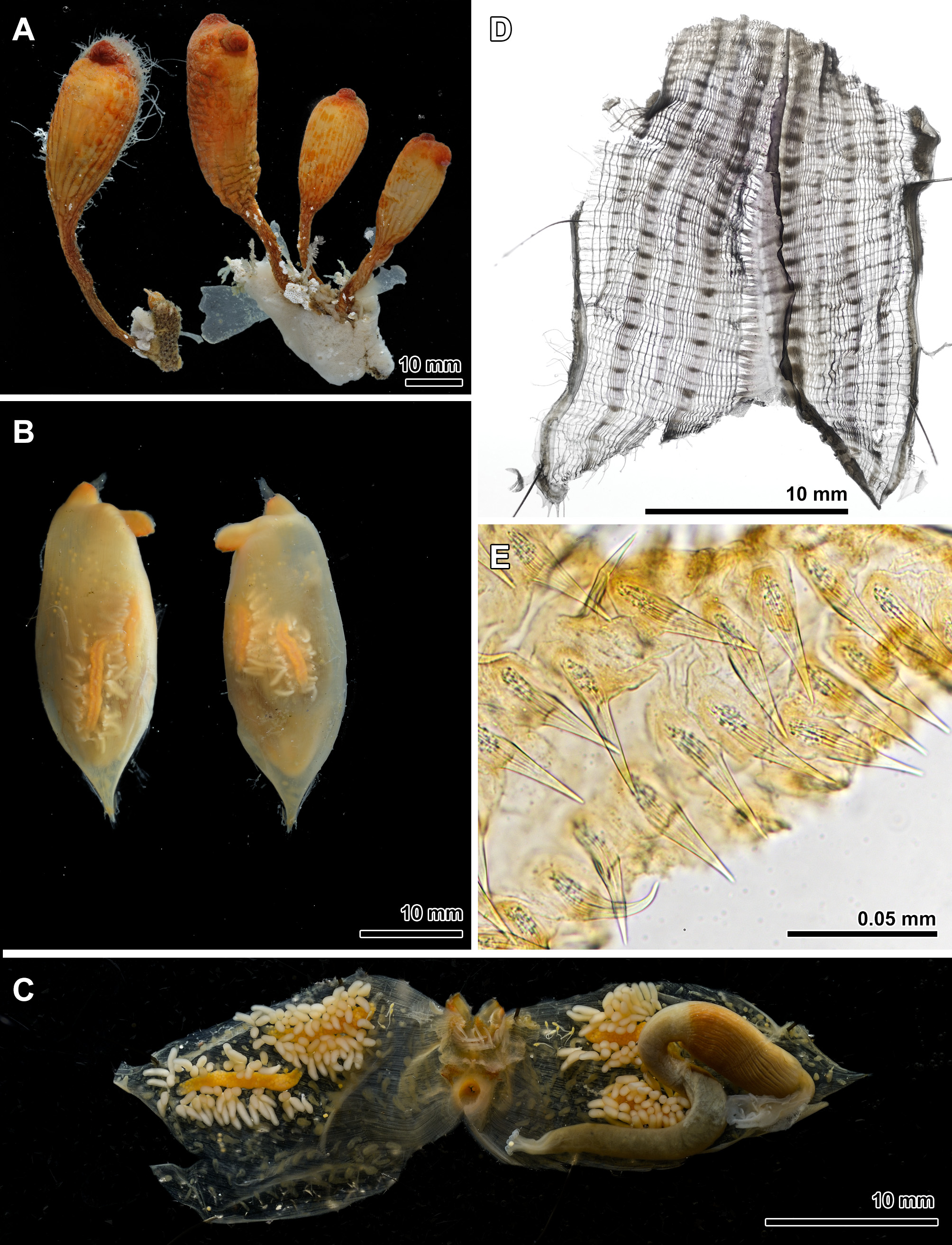
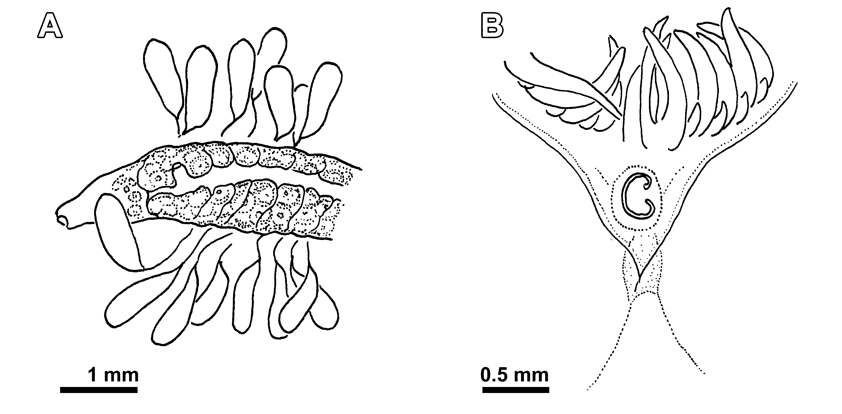
( Figures 4 View FIGURE 4 , 5 View FIGURE 5 )
Ascidia clavata Pallas, 1774: 25 .
Katatropa clavata: Redikorzev, 1916: 204 View in CoL ; 1941: 85. Styela clavata: Van Name, 1945: 316 (and synonymy); Sanamyan, 2000: 68. Botryorchis clava: Redikorzev, 1941: 187 (part). Styela greeleyi Ritter, 1899: 516 .
Not Styela yakutatensis Ritter, 1901: 239 .
Material examined. Matua Island, Point Kluv and Point Crocodile, several tens of specimens in eight lots, collected from intertidal zone to 16 m.
Description. Collected specimens range from several millimeters to about 8 см long ( Figure 4 View FIGURE 4 A). The elongated cylindrical body tapers either gradually, or rather abruptly, to a thin firm stalk attached to substratum. In general the stalk attains about a half length of the body, but may be much shorter or much longer in some specimens. The test is firm, leathery, in the preserved specimens typically longitudinally wrinkled and furrowed. Epibionts (mostly bryozoans and algae) may present on the test, especially on older individuals and especially on the stalk, but usually not in great quantity. Living specimens are uniformly red, the colour is retained in formaline for years.
Branchial and atrial apertures are on the short siphons located on the top of the body, close together. The branchial siphon is directed upward, the atrial curved down and opens downward. The test on the inner surface of the siphons is covered by crowded spines. The spines are narrow and elongated, with sharply pointed tips and with flat oval base, about 50 µm long and 10 µm wide at the base ( Figure 4 View FIGURE 4 E). Thin parallel longitudinal lines are visible inside each spine.
Body wall is muscular, almost opaque ( Figure 4 View FIGURE 4 B). About 15 large branchial tentacles regularly alternating with the same number of small tentacles are present. The dorsal tubercle slit is C shaped directed to the left and with the horns rolled inside ( Figure 5 View FIGURE 5 B). The prepharyngeal band consists of a single lamina running in a close proximity to a ring of tentacles and making shallow V around the dorsal tubercle. The neural ganglion is close to the dorsal tubercle. High dorsal lamina has plain edge. The branchial sac has four rather low folds, the internal longitudinal vessels in dissected specimen are distributed as follow: EN2(7)4(12)4(8)3(17)1DL(14)4(8)4(7)5(7)3EN. Stigmata straight and long, each row is crossed by a parastigmatic vessel.
Gut forms vertical S-shaped loop ( Figure 4 View FIGURE 4 C). The short oesophagus, at the posterior end of the body, is curved at almost 180° before to enter the stomach. The stomach is cylindrical and large, somewhat shorter than a half of the body length. It is lying more or less parallel to the longitudinal axis of the body, its internal wall has at least 30 well defined regular parallel longitudinal folds. Intestine bends posteriorly, forming tight closed primary loop. The secondary loop is open anteriorly, the rectum is long and straight. Anal border has well defined round lobes, about 15 were counted in dissected specimen.
Two gonads are on the each side of the body. Ovaries are straight, not long, with ventrally bent distal ends. They are parallel to each other and to the longitudinal axis of the body. On each side of the body one ovary lies along the longitudinal midline and is located in the posterior half of the body, while another ovary, lying closer to the endostyle, is located more anteriorly (closer to siphons). The oviducts are short. Numerous male follicles surround each ovary from the sides and posteriorly. They are long, cylindrical, sausage-shaped, not branched, freely projecting for their whole length into the peribranchial cavity being attached to the body wall, on some distance from the ovary, by their narrow distal ends only. Wide common sperm duct runs along the mesial surface of each ovary and ends in a short male papilla in the anterior part of the ovary ( Figure 5 View FIGURE 5 A).
The endocarps are numerous. In the form they resemble male follicles (sausage-shaped, attached by narrow ends) but are smaller.
The specimens occurs solitarily or in groups of several individuals, but do not form dense settlements.
Remarks. Although original description of this species lacks any information on its internal features there is no doubt in its identity, the exterior of this species is very characteristic and it is the most common solitary ascidian on the east coasts of Kamchatka ( type locality of the species). In NW Pacific Styela clavata has been mistaken several times with another species, Styela clava Herdman, 1881 (e.g. by Redikorzev, 1941). These two species have similar appearance and similar names but nothing in common in their internal features, and, contrary to the statement of Redikorzev (1941), based on erroneous identification (see remarks under S. clava in Sanamyan, 2000 ) their geographical ranges do not overlap. The distribution of S. clavata is probably limited by Bering Sea, east coast of Kamchatka, and extends to the south to at least to central Kuril Islands .
Styela greeleyi from Pribilof Islands (Bering Sea) is a well established synonym. Van Name (1945: 318) expressed an opinion that S. yakutatensis Ritter, 1901 , another externally similar species known from Yakutat Bay, southern Alaska, and Vancouver Island may be conspecific, but kept them separate. According to him S. yakutatensis has lobed male follicles (he, however, doubted that this is a valid feature separating it from S. clavata ) and probably different shape of the siphonal spines ("very short and squarely truncated"). At our opinion both features, if correctly reported, especially the shape of the spines, may be significant, so we refuse from including S. yakutatensis in the synonymy of S. clavata .
No known copyright restrictions apply. See Agosti, D., Egloff, W., 2009. Taxonomic information exchange and copyright: the Plazi approach. BMC Research Notes 2009, 2:53 for further explanation.
|
Kingdom |
|
|
Phylum |
|
|
Class |
|
|
Order |
|
|
Family |
|
|
Genus |
Styela clavata ( Pallas, 1774 )
| Sanamyan, Karen & Sanamyan, Nadya 2017 |
Katatropa clavata :
| Sanamyan 2000: 68 |
| Van 1945: 316 |
| Redikorzev 1941: 187 |
| Redikorzev 1916: 204 |
| Ritter 1899: 516 |
Styela yakutatensis
| Ritter 1901: 239 |
Ascidia clavata
| Pallas 1774: 25 |