
Thagria decussata Fan & Dai
|
publication ID |
https://doi.org/ 10.11646/zootaxa.3918.4.1 |
|
publication LSID |
lsid:zoobank.org:pub:14FD40D0-1D41-461A-B5E6-02FD4F996266 |
|
DOI |
https://doi.org/10.5281/zenodo.5261466 |
|
persistent identifier |
https://treatment.plazi.org/id/CD5187C4-E32B-FFC0-B89D-FB0177E4FBB6 |
|
treatment provided by |
Plazi |
|
scientific name |
Thagria decussata Fan & Dai |
| status |
sp. nov. |
Thagria decussata Fan & Dai View in CoL sp. nov.
( Figs. 60–84 View FIGURES 60 – 69 View FIGURES 70 – 84 )
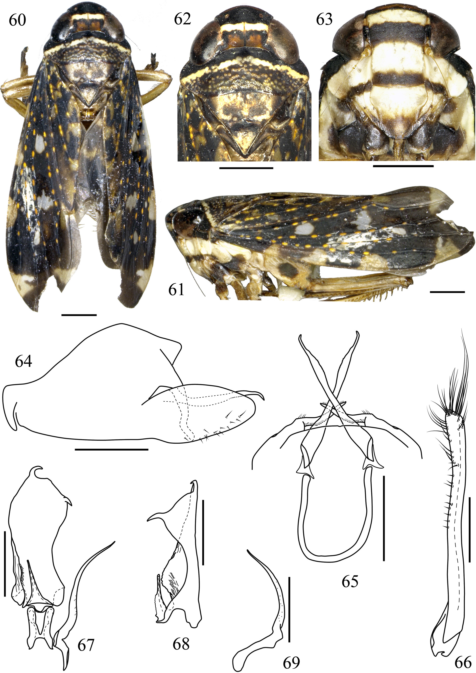
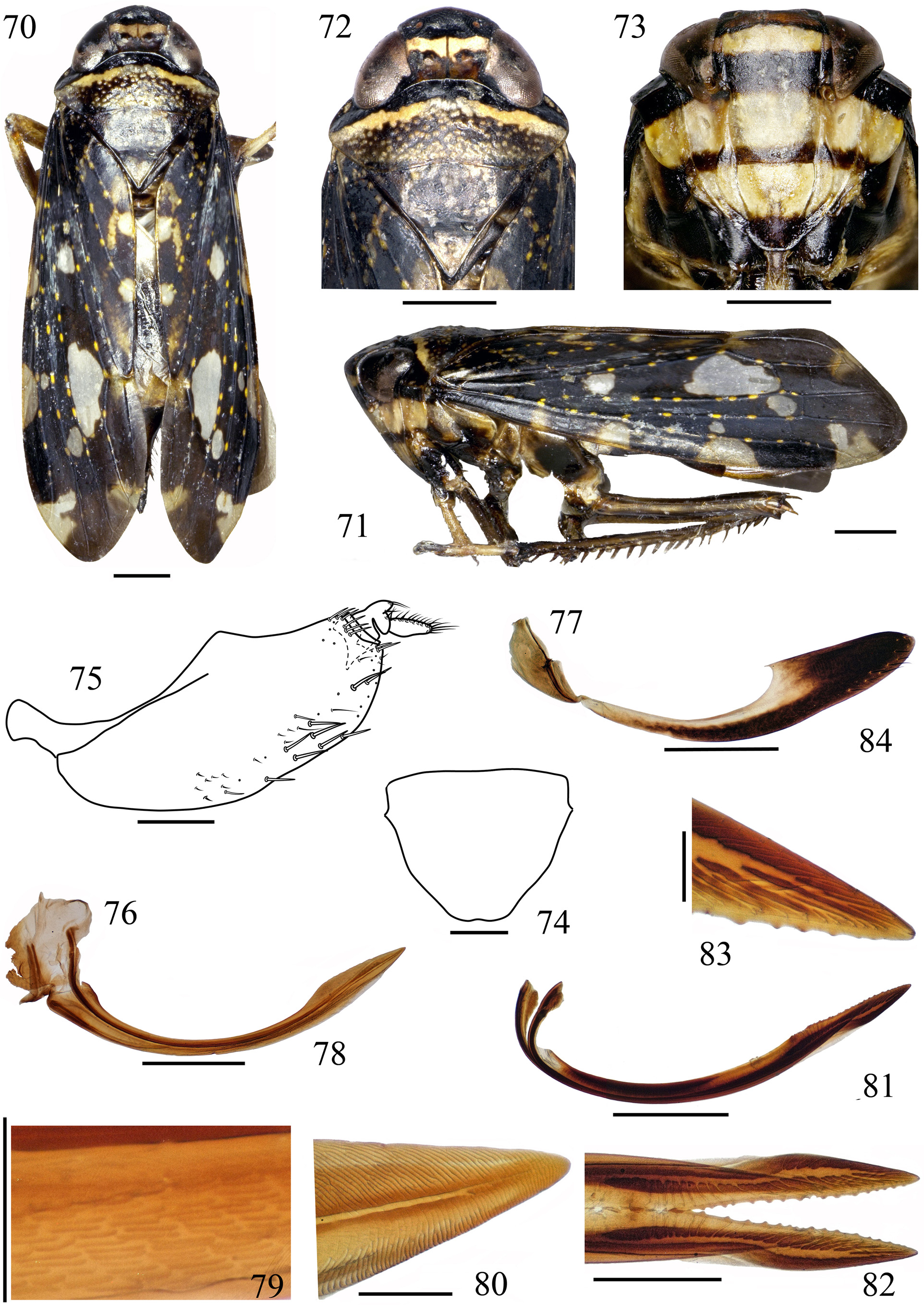
Description. Body length of male 8.2 mm, female 9.5 mm.
Body slightly robust, medium ( Figs. 60 View FIGURES 60 – 69 , 70 View FIGURES 70 – 84 ). Crown with four transverse bands, from base to apex brown, black, ivory and black; ocelli and eyes brown ( Figs. 62 View FIGURES 60 – 69 , 72 View FIGURES 70 – 84 ). Face with clypeus with three black-brown bands, anterior band narrowest, middle widest, distal longest crossing lora and genae; clypellus apical half blackish brown, remainder of face light yellow in male or deep yellow in female ( Figs. 63 View FIGURES 60 – 69 , 73 View FIGURES 70 – 84 ). Pronotum densely covered with yellow-brown nubs and traversed by three bands: black anteriorly, ivory medially, posteriorly brown ( Figs. 62 View FIGURES 60 – 69 , 72 View FIGURES 70 – 84 ). Mesonotum brown, with yellowish white spots ( Figs. 62 View FIGURES 60 – 69 , 72 View FIGURES 70 – 84 ). Forewing fuscous, with numerous transparent ivory and ivory-yellow patches, venation black with orange spots ( Figs. 60, 61 View FIGURES 60 – 69 , 70, 71 View FIGURES 70 – 84 ).
Head in dorsal view anteriorly rounded, crown wider than eye width, produced about 1/4 of crown midline anterad of eyes, coronal suture evident, about half of median length, disk flat ( Figs. 62 View FIGURES 60 – 69 , 72 View FIGURES 70 – 84 ). Face with clypeus long, lateral margins slightly convex; clypellus short, base broad, inflated (male) or slightly inflated (female), wider than clypeus at juncture of clypeal suture, apical half constricted, apex truncate ( Figs. 63 View FIGURES 60 – 69 , 73 View FIGURES 70 – 84 ). Crown, pronotum and mesonotum midline ratio about 1:1:1.5 ( Figs. 62 View FIGURES 60 – 69 , 72 View FIGURES 70 – 84 ). Forewing long, apex rounded, appendix narrowed ( Figs. 60, 61 View FIGURES 60 – 69 , 70, 71 View FIGURES 70 – 84 ).
Male genitalia. Pygofer in lateral view with caudoventral lobe long, broad, narrowed distally, ventral margin folded near middle, caudodorsal margin with paired processes, bent subapically ( Fig. 64 View FIGURES 60 – 69 ). Segment X ventral processes slender, exceeding margin of pygofer, apex hooked ventrad in lateral view ( Fig. 64 View FIGURES 60 – 69 ). Subgenital plate long and narrow, segmented subbasally, marginal setae fine long or short ( Fig. 66 View FIGURES 60 – 69 ). Dorsal connective broad Ushaped, stem absent, rami long, attaching to base of segment X ventral processes by membrane ( Fig. 65 View FIGURES 60 – 69 ). Aedeagus in dorsal view symmetrical, short, about 1/3 as long as paraphysis, broad basally, gradually narrowed apically ( Figs. 67, 68 View FIGURES 60 – 69 ); paraphysis in dorsal view asymmetrical, broad throughout, narrowest near midlength, right margin with serrate fold from base to middle, opposite side expanded distally with short, retrorse spine subapically at broadest point, apex with hooked spine, in lateral view X-shaped, both spines twisted dorsally, right margin expanded subbasally and narrowed apically, outer margin narrowed basally and expanded distally ( Figs. 67, 68 View FIGURES 60 – 69 ). Style long, exceeding the midlength of paraphysis, base broad, tapered to apex, subbase twisted ( Figs. 67, 69 View FIGURES 60 – 69 ).
Female genitalia. Abdominal sternite VII in ventral view with base broad, narrowed distally, anterior margin nearly straight, posterior margin with shallow emargination medially, lateral margins convex ( Fig. 74 View FIGURES 70 – 84 ). Internal sternite VIII completely membranous. Pygofer in lateral view, moderately produced posteriorly, dorsoposterior margin obliquely truncate, with macrosetae extending to ventral margin in posterior half ( Fig. 75 View FIGURES 70 – 84 ). Valvifer I in lateral view, broad, posterodorsal margin with a small lobe ( Fig. 76 View FIGURES 70 – 84 ). Valvifer II length-width ratio 3:1, in lateral view, nearly fusiform, with small group of clustered setae above articulation point, articulation point located on submedian ( Fig. 77 View FIGURES 70 – 84 ). Valvulae I long, in lateral view curved, medial 1/2 narrowed, distal 1/4 broadened and gradually narrowed towards acute apex ( Fig. 78 View FIGURES 70 – 84 ), with horizontally nephroid sculptured sclerotized area on the basal half of shaft ventrally ( Fig. 79 View FIGURES 70 – 84 ), and with obliquely striated sclerotized area on dorsal margin 1/4 and on ventral margin near apex ( Fig. 80 View FIGURES 70 – 84 ). Valvulae II long, basal 3/4 with membrane joining two sides, distal 1/4 detached and narrowed gradually to tip, shaft bearing 18 or 20 teeth over posterior 1/4 of each blade, and one shared tooth at apex of joint area, teeth rounded, widely spaced, apicoventral margin without teeth, dorsal margin of blade without secondary denticles, ducts well delimited, extending to teeth and towards apical blade portion in lateral and dorsal views ( Figs. 81–83 View FIGURES 70 – 84 ). Valvulae III in lateral view, expanded at apical half, apex obtuse, surface with fine setae mostly distributed on apical portion and extending anteriorly along ventral margin of apical half ( Fig. 84 View FIGURES 70 – 84 ).
Type material. Holotype: ♂, CHINA: Guangxi, Damingshan, Alt. 800–1300 m, 5–10 Aug. 2011, coll. Yang Zaihua ( GUGC). Paratype: ♀, same data as holotype ( GUGC).
Etymology. The new species name is derived from the Latin word “ decussata ” meaning “the figure X” for the X-shaped paraphysis in lateral view.
Remarks. This new species is superficially similar to T. multipars (Walker, 1858) , but easily distinguished by the combination of the two spines of the paraphysis ( Figs. 67, 68 View FIGURES 60 – 69 ); pygofer caudoventral lobe broad and segment X ventral processes hooked apically ( Fig. 64 View FIGURES 60 – 69 ). It also resembles T. fossa Nielson, 1977 , but can be distinguished from the latter by the following: clypeus with longest band crossing lora and genae ( Fig. 63 View FIGURES 60 – 69 ) and two spines of paraphysis at apex and subapex, respectively ( Fig. 67 View FIGURES 60 – 69 ).
No known copyright restrictions apply. See Agosti, D., Egloff, W., 2009. Taxonomic information exchange and copyright: the Plazi approach. BMC Research Notes 2009, 2:53 for further explanation.