
Exechonella spinosa Osburn, 1940
|
publication ID |
https://doi.org/10.11646/zootaxa.4305.1.1 |
|
publication LSID |
lsid:zoobank.org:pub:1192C3A0-5CCB-4A86-903C-A2B82906A5F9 |
|
DOI |
https://doi.org/10.5281/zenodo.6017352 |
|
persistent identifier |
https://treatment.plazi.org/id/CF0AB852-FFC7-E912-FF03-F98B9471E3E8 |
|
treatment provided by |
Plazi |
|
scientific name |
Exechonella spinosa Osburn, 1940 |
| status |
|
Exechonella spinosa Osburn, 1940 View in CoL
( Fig. 24 View FIGURE 24 , Table 23)
Exechonella spinosa new. var.: Osburn 1940, p. 367‒368 (part), pl. 4, fig. 35.
Material examined: Lectotype: USNM 11849 About USNM , three small fragments. Atlantic Ocean , Bermuda, May 1936.
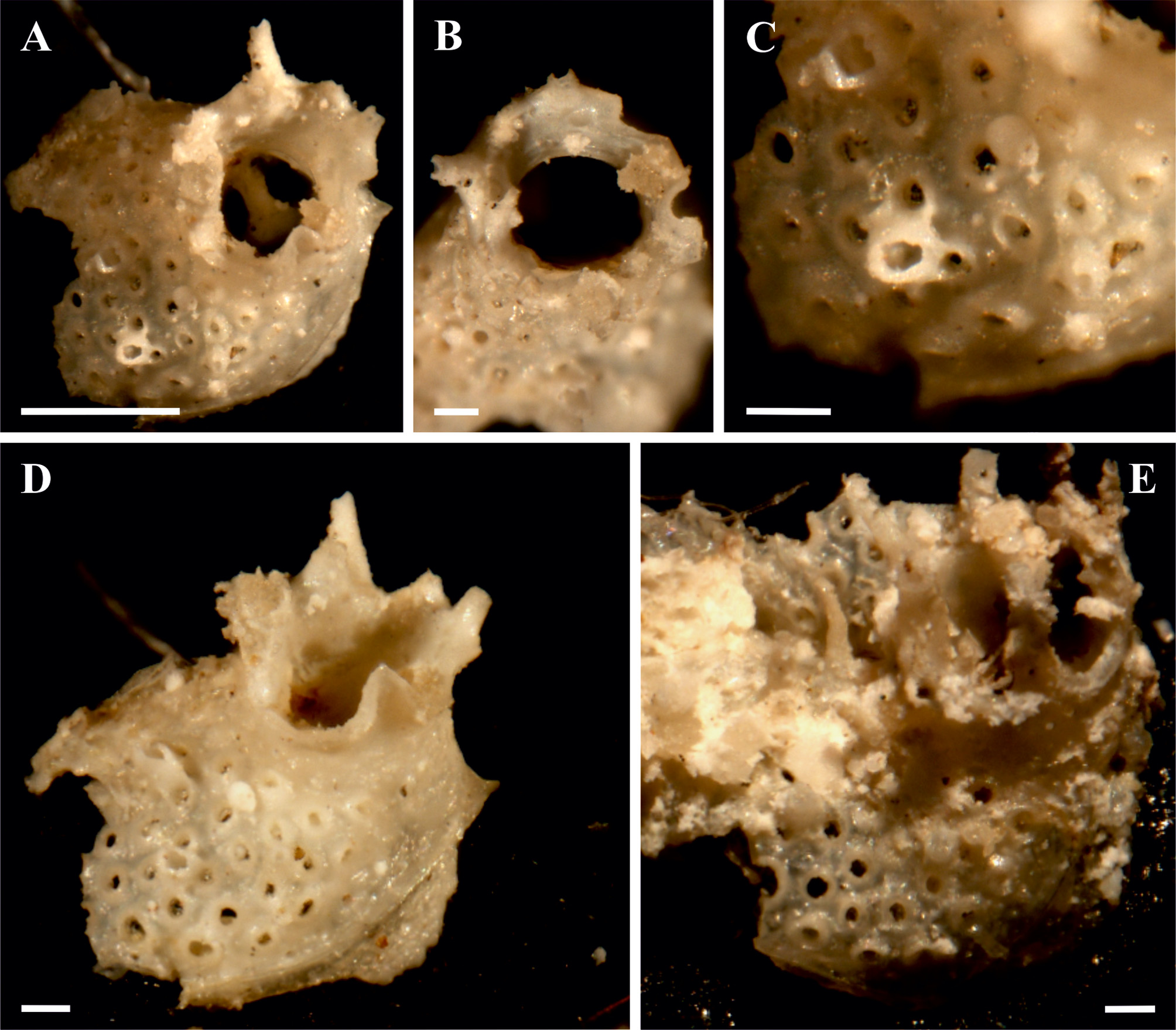
Description. Colony encrusting, unilaminar, multiserial. Autozooids convex, oval in shape. Primary orifice oval, wider than long. Peristome short and thick with 5–7 long and tubular projections around, which may stay simple or bifurcated at the end. Frontal shield slightly pustulose being perforated by about 40 spaced, circular to subcircular foramina, each with gymnocystal rim. Fusions between foraminal rims were not seen. Autozooids have long, spire-like hollow processes on the frontal shield. Their formation involves the fusion of the gymnocystal rims of several foramina, whose openings are distinguished near the process base. Vertical zooidal walls narrow and obviously represented by multiporous mural septula. Lateral avicularia and adventitious kenozooids were not seen in the studied fragments.
Bermuda, Atlantic Ocean
m±sd r n AzL 750 700‒800 2 AzW 663 625‒700 2 OrL 246 ‒ 1 OrW 300 ‒ 1 FoN 38 ‒ 1 FoD 57±2.1 53–60 10 OD 24±3.0 19–28 10 Remarks. Despite the above redescription requires improvement based on the better preserved material from the type locality, E. spinosa is a ‘good species’ characterized by its frontal shield with numerous rounded foramina, spire-like, thinner hollow processes associated with foramina of the frontal shield and thicker, often branching processes on the peristome.
Osburn in 1940, together with the description of E. antillea , described and figured, E. antillea new var. s pinosa (p. 367–368, pl. 4, fig. 35) mentioning the only difference between them the presence of “tall unjointed spines” on the frontal shield and peristome. In fact, Osburn noticed that had two specimens, one from Bermuda and another from Jamaica, that he initially considered as different species because of the absence of the frontal spines in the second specimen. Finally this author decided that they belong to one species, because of the similarity in size.
Comparison of the Osburn’s material from Bermuda ( USNM 11849 About USNM , selected here as lectotype consisting of three tiny fragments of presumably one colony) and the material from Port Antonio , Jamaica ( USNM 9911 About USNM , labeled as E. pumicosa , also represented by two small specimens loaned from Dr. Bassler) showed that the published illustration ( Osburn 1940, p. 4, fig. 35) corresponds more to the Bermuda's fragments because of the presence of the frontal spines. Such spines were broken, however, in the specimens from Jamaica one of which also shows broken peristomial spines. The foraminal luminae are not round, but slit-like and their number (about 50) is higher in comparison to the specimen depicted by Osburn. We suggest that these specimens belong to a separate (while very similar) species.
Examination of four SEM-images of the colony fragment from Bocas del Toro, Panama (currently nonnumbered, kept in the collection of the Virginia Museum of Natural History , USA), also showed its close similarity to the fragments from Bermuda. We suggest it could belong to E. spinosa although better preserved material from the Bermuda, Jamaica and Bocas del Toro is required for better comparison.
Describing material (as Lagenipora tuberculata MacGillivray ) from Indian waters Thornely (1905, p. 113) included a comment as “there are also among [the studied colonies] some that have the hollow tubercles very much lengthened, ending in points” and “simple or branched spines round the margin of the much raised peristome”. While no illustrations were given, this description clearly shows a similarity of the Thornely’s material (kept at the Natural History Museum, London) with E. spinosa , and should be compared in the future.
No branching spines were seen on the peristomes in the studied specimens in E. verrucosa and E. kleemanni n. sp. (see below), although better preserved material should be obtained to make more precise comparison between the species.
Distribution. Until more specimens will be obtained, distribution of Exechonella spinosa should be restricted to Bermuda.
| USNM |
Smithsonian Institution, National Museum of Natural History |
No known copyright restrictions apply. See Agosti, D., Egloff, W., 2009. Taxonomic information exchange and copyright: the Plazi approach. BMC Research Notes 2009, 2:53 for further explanation.
|
Kingdom |
|
|
Phylum |
|
|
Class |
|
|
Order |
|
|
Family |
|
|
Genus |