
Exechonella vavrai, Cáceres-Chamizo & Sanner & Tilbrook & Ostrovsky, 2017
|
publication ID |
https://doi.org/10.11646/zootaxa.4305.1.1 |
|
publication LSID |
lsid:zoobank.org:pub:1192C3A0-5CCB-4A86-903C-A2B82906A5F9 |
|
DOI |
https://doi.org/10.5281/zenodo.6017348 |
|
persistent identifier |
https://treatment.plazi.org/id/CF0AB852-FFCB-E91F-FF03-FBBA91D0E629 |
|
treatment provided by |
Plazi |
|
scientific name |
Exechonella vavrai |
| status |
sp. nov. |
Exechonella vavrai n. sp.
( Fig. 22 View FIGURE 22 , Table 21)
Material examined. Holotype: DPUV 2012-0005-0001 , on mollusc shell (mounted on the SEM stub and coated with gold). Red Sea, the Northern Bay of Safaga, south of Ras Abu Soma , depth 1–20 m, September 1992 . Paratype: DPUV 2012-0005-0002 , ancestrula on coral rubble. Red Sea, the Northern Bay of Safaga , 31 July, 1987.
Etymology. Named after palaeontologist Dr. Norbert Vávra for his life-long contribution to the study of bryozoans.
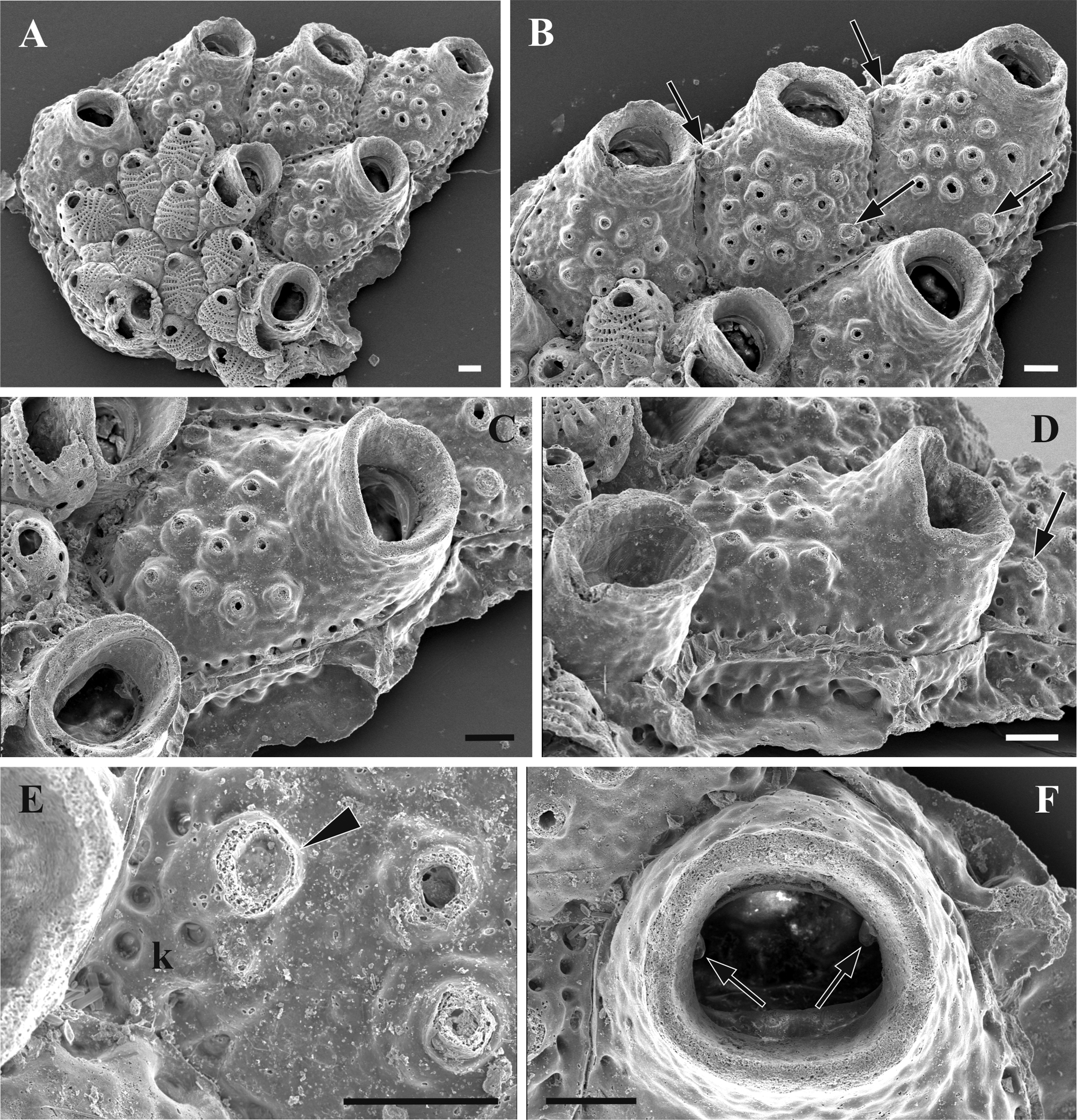
Northern Bay of Safaga, Red Sea Description. Colony encrusting, unilaminar, multiserial. Autozooids pentagonal, hexagonal or oval in shape, separated by deep grooves. Primary orifice oval, wider than long, with proximal shelf (a distalmost part of the zooidal frontal shield proximally surrounded by a wall of the peristome), smooth centrally and with wrinkled lateral areas. Anter wall (one-third of primary orifice) underlain by an inner lamina, which ends form thick and rounded condyles pointed downwards. A tubular thick-walled peristome, externally pustulose, with characteristic proximal lip separated from the distal part of the peristomial edge by a shallow ‘incision’. Frontal shield softly pustulose, with 10–14 conical foramina with wide round basal part and narrow distal tip (mostly destroyed in our material) terminated by a circular opening. Foramina distributed predominantly in the central part of the frontal shield. In contrast with the others, two lateralmost foramina are ‘isolated’ and have cylindrical shape. Their opening is occluded presumably by the avicularian mandible, but preservation of the material was not sufficient to give more details. These foramina are associated with adventitious kenozooids, recognized by 3–5 small pores. Round marginal pores small and distinct. Vertical zooidal walls narrow, represented by multiporous mural septula with communication pores arranged in one, sometimes two rows. Ancestrula autozooidal, smaller than the rest of autozooids.
Remarks. E. vavrai n. sp. is characterized by its thick peristome with a prominent proximal lip and incisions that are not known in any other species. The conical frontal foramina that are present in E. vavrai n. sp. are only comparable with those found in E. albilitus Tilbrook, 2006 , although calcification in the former species is stronger.
Distribution. E. vavrai n. sp. is only known in the Red Sea, Northern Bay of Safaga.
No known copyright restrictions apply. See Agosti, D., Egloff, W., 2009. Taxonomic information exchange and copyright: the Plazi approach. BMC Research Notes 2009, 2:53 for further explanation.
|
Kingdom |
|
|
Phylum |
|
|
Class |
|
|
Order |
|
|
Family |
|
|
Genus |