
Leeonychiurus analis, Park, 2017
|
publication ID |
https://doi.org/ 10.11646/zootaxa.4329.3.7 |
|
publication LSID |
lsid:zoobank.org:pub:848Bafaa-67C8-4105-B12E-F5C8B18Ff689 |
|
persistent identifier |
https://treatment.plazi.org/id/CF1687E1-FFF4-FB4B-269A-2C33FBD8D6CD |
|
treatment provided by |
Plazi |
|
scientific name |
Leeonychiurus analis |
| status |
sp. nov. |
Leeonychiurus analis sp. nov.
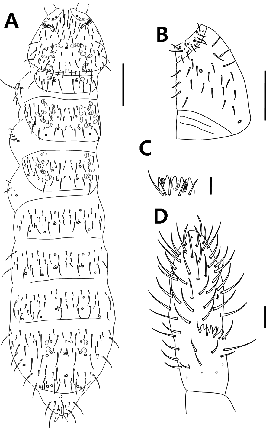
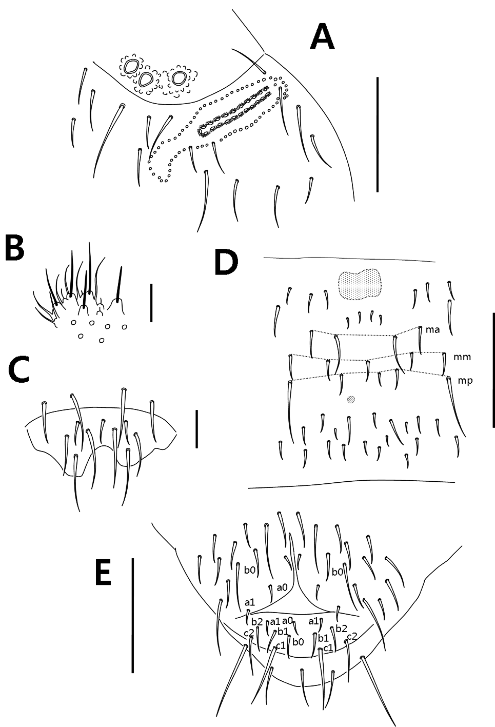
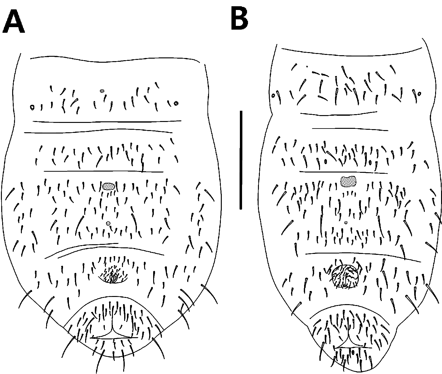
( Figures 3–4 View FIGURE 3 View FIGURE 4 , 5B View FIGURE 5 )
Diagnosis. Dorsal pseudocellar formula 32/12(3)2/33343, ventral 11/000/0100; subcoxa 1 of legs I–III with one pseudocellus each. Postantennal organ with 18–22 compound vesicles. Abd. I–III terga with 3+3 chaetae along axial line. Th. I–III sterna with 0+0, 1+1, 1+1 chaetae respectively. Chaeta c0 on upper anal valve absent, but chaetae a1 present.
Type material. Holotype: female on slide, Korea, Sinwol-ri, Dongsang-myeon, Wanju-gun , Jeollabuk-do, (N127°17´23´´ E35°56´40´´), 27–III– 2015 in roots of grass, collected by K.H. Park. Paratypes: 4 females and 1 male on slides, same data as holotype.
Description. Body length (excluding antennae) of adults: male 1.65mm, females 1.80–2.12mm. Color white in alcohol. Cuticle granulation more or less uniform. Dorsal pseudocellar formula 32/12(3)2/33343 ( Fig. 3A View FIGURE 3 ), ventral 11/000/0100; subcoxa 1 of legs I–III with one pseudocellus each. Parapseudocellar formula ventrally 0/000/ 101001m ( Fig. 5B View FIGURE 5 ), dorsal psx absent. Subcoxa 1 of legs I–III with 1 psx each. Psp formulas: 00/011/11110 dorsally and 00/111/0001m0 0 ventrally ( Fig. 5B View FIGURE 5 ).
Head: Antennal bases well marked ( Fig. 3A View FIGURE 3 ). Antennae slightly shorter than head; ratio antenna/head=0.83– 0.88. Ant. IV subapical organite with globular apex; invaginated apical bulb absent; microsensillum in lateroexternal position approximately one-third length from the base; sensilla not well distinguish from ordinary chaetae ( Fig. 3D View FIGURE 3 ). Ant. III sensory organ composed of five papillae, five guard chaetae, 2 rods and 2 granulated sensory clubs ( Fig. 3C View FIGURE 3 ); lateral ms just behind sensory organ. Ant. II with 16 chaetae. Ant. I with 9 chaetae. Postantennal organ with 18–22 compound vesicles arranged in two rows along axis of organ ( Fig. 4A View FIGURE 4 ). Head with unpaired dorsal chaeta d0. 4+4 p-chaetae between two inner posterior pso on head, p1 anterior to others. Mandible with strong molar plate and four apical teeth, maxilla bearing 3 teeth and 6 lamellae. Maxillary palp simple with 1 basal chaeta and 2 sublobal hairs. Labral formula as 4/342 ( Fig. 4C View FIGURE 4 ). Labial palp of type AC ( Fig. 4B View FIGURE 4 : After Fjellberg, 1999), labial papillae A, B, C, D and E with 1, 4, 0, 3, 3 guard chaetae respectively ( Fig. 4B View FIGURE 4 ). Labium with 6 proximal, 4 basomedian and 6 basolateral chaetae. 4+4 postlabial chaetae present along ventral groove ( Fig. 3B View FIGURE 3 ).
Body chaetotaxy: s-chaeta formula as 2/012/222120 dorsally. Tiny and blunt ms, present on Th. II and III. Th. I tergum with 8+8 chaetae on each side. Th. II–Abd. III with 3, 3, 3, 3, 3 chaetae respectively on both sides of axial line and without unpaired axial chaetae, sometimes with asymmetrical chaetae. Abd. IV tergum with unpaired axial chaeta p0, sometimes with m0 chaetae; Abd. V tergum with axial chaeta m0; Abd. VI tergum with 2 unpaired axial chaetae: a0, p0 ( Fig. 3A View FIGURE 3 ). Th. I, II and III sterna with 0+0, 1+1 and 1+1 chaetae between legs, respectively. Subcoxae 1 of legs I–III with 4, 5, 5 chaetae respectively, subcoxa 2 with 1, 4, 4 chaetae.
Appendages: Tibiotarsi I, II and III with 22 (11, 8, 3), 21 (11, 8, 2) and 21 (11, 8, 2) chaetae, respectively. Claw without teeth. Empodial appendage with basal lamella, appendage length shorter than inner edge of unguis. Ventral tube with 8–9+8–9 distal chaetae, without anterior and basal chaetae. Furca reduced to a finely granulated area, with four small dental chaetae in one row posterior to furcal rudiment; three manubrial rows of chaetae present ( Fig. 4D View FIGURE 4 ).
Anal valves chaetotaxy: each lateral valve with chaetae a0, 2a1, b0 and 2b1; upper valve with chaetae a0, 2a1, b0, 2b1, 2b2, 2c1 and 2c2 ( Fig. 4E View FIGURE 4 ). Anal spines set on distinct papillae, as long as inner edge of hind unguis.
Ecology. Found in roots of grass.
Etymology. The name of the species refers to the anal lobe that has specific chaetotaxy. Korean name of the species: Dongsang-lee-eori-tok-to-gi.
TABLE]. Major characters of all known species in the genus Leeonychiurus .
North Distribution Korea Korea China China China Korea (cave) America References: 1) Sun & Zhang 2O12; 2) Sun & Arbea 2O14; 3) Lee & Park 1986; 4) Bernarđ 2O15
Discussion. This new species is most similar to L. koreanus sp. nov. having identical number of ventral pseudocelli (11/000/0100) and the same set of unpaired axial chaetae on Abd. IV–VI. Nevertheless it can easily be distinguished due to the presence of dorsal pso on Th. I (absent in L. koreanus sp. nov.) and three dorsal pso on Abd. III (only two pso in L. koreanus sp. nov.), chaetotaxy of upper anal valve appears to be also rather specific (Table.1). The new species is also close to L. gulinensis ( Sun & Zhang, 2012) and L. antennalis ( Sun & Zhang, 2012) as sharing same dorsal pseudocellar formula, but it can be distinguished from two latter species by the ventral pseudocellar formula on abdomen (0100 in L. analis sp. nov. vs. 0 101 in L. gulinensis & L. antennalis ) and the absence of chaetae c0 on upper anal valve (present in L. gulinensis & L. antennalis ).
No known copyright restrictions apply. See Agosti, D., Egloff, W., 2009. Taxonomic information exchange and copyright: the Plazi approach. BMC Research Notes 2009, 2:53 for further explanation.