
Cretojapyx huangi, Wang & Huang & Cai, 2023
|
publication ID |
https://doi.org/ 10.11646/zootaxa.5396.1.12 |
|
publication LSID |
lsid:zoobank.org:pub:EBC29EC0-BB77-46F1-A715-B8724796741F |
|
DOI |
https://doi.org/10.5281/zenodo.10441205 |
|
persistent identifier |
https://treatment.plazi.org/id/D007EA01-7328-2B72-FF3A-FD60FA06FE99 |
|
treatment provided by |
Plazi |
|
scientific name |
Cretojapyx huangi |
| status |
sp. nov. |
Cretojapyx huangi sp. nov.
Figs 1–5 View FIGURE 1 View FIGURE 2 View FIGURE 3 View FIGURE 4 View FIGURE 5
Material. Holotype, genitalia not visible, developmental stage and gender unknown, NIGP203387 View Materials . The amber specimen generally yellow and relatively transparent, with many bubbles inside and some around C. huangi .
Etymology. The specific epithet ‘ huangi ’ is dedicated to the famous entomologist, Fusheng Huang, who made remarkable discoveries of Gigasjapyginae in China with Prof. Yao Zhou, and presented the extant japygids for our study.
Diagnosis. As for the genus.
Locality and horizon. Amber mine located near Noije Bum Village , Tanai Township , Myitkyina District , Kachin State, northern Myanmar (26°15′N, 96°36′E); upper Albian to lower Cenomanian, mid-Cretaceous GoogleMaps .
Description. Body elongated, ca. 9.9 mm in length from the forefront of head the end of abdominal segment X ( Fig. 1A, B View FIGURE 1 ). Abdominal segment I–VII with nearly parallel sides and narrowing from the segment VIII. Body surface smooth under optical microscope, with s, sM, M. Cuticle unpigmented, with lacinia apex; dorsal head, abdominal segment VIII–X and cerci sclerotized ( Fig. 1A, B View FIGURE 1 ).
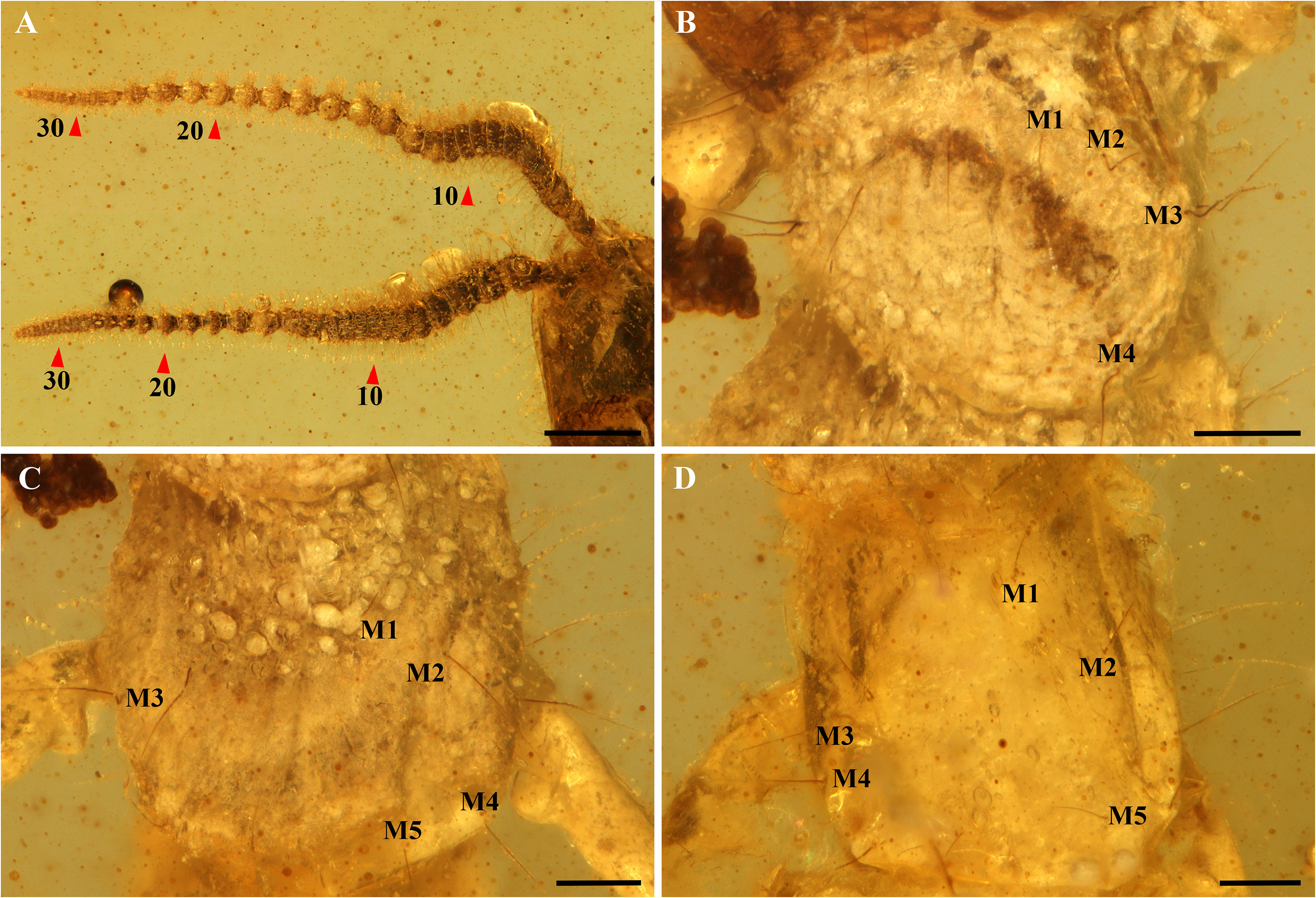
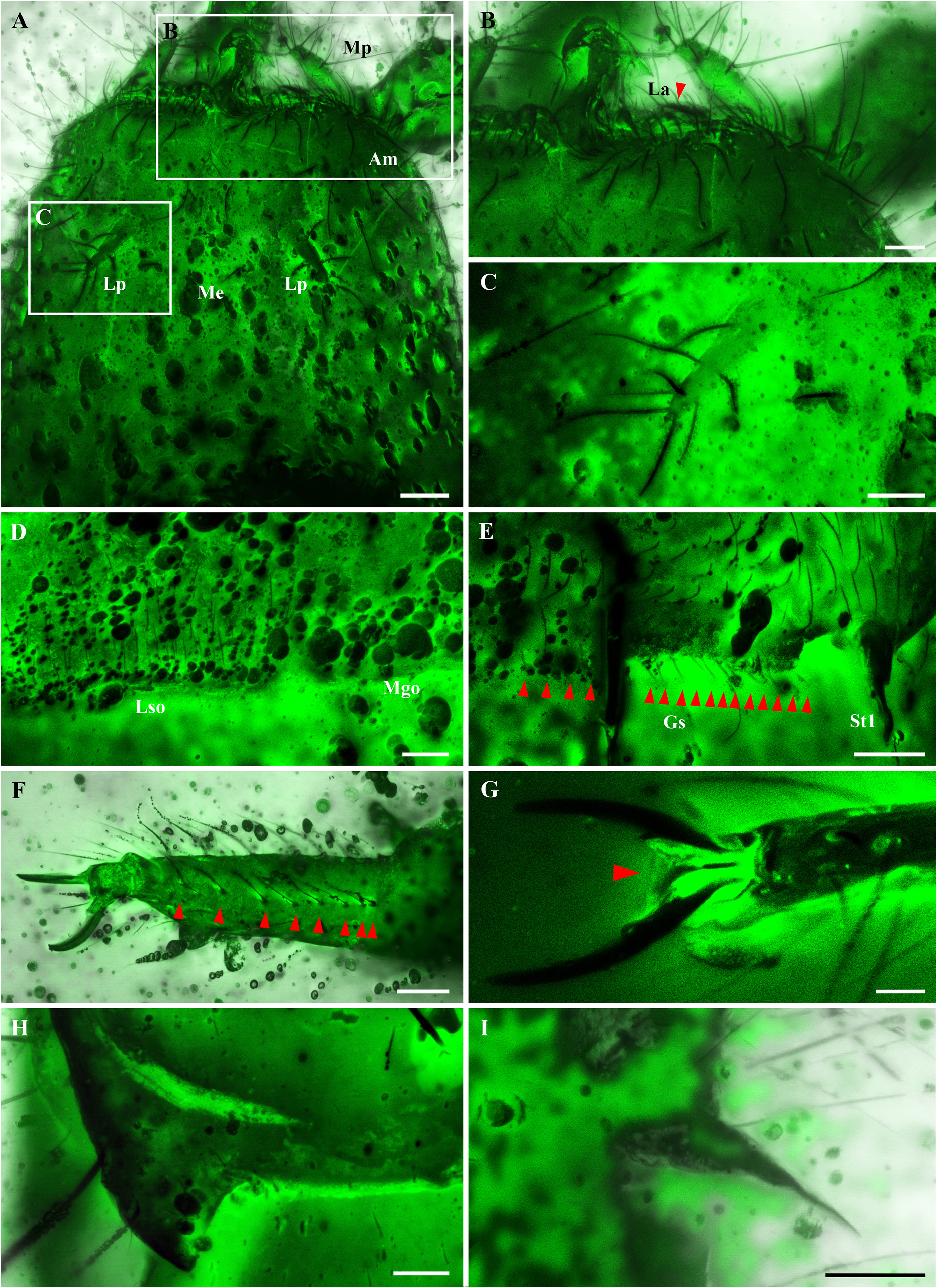
Antennae ca. 3.2 mm in length, 0.3× as long as body, the left antenna with 38 antennomeres, the right with 39 ones ( Fig. 2A View FIGURE 2 ); antennomeres telescopic, 2 nd antennomere longest, 1.6× as long as wide. All antennomeres covered dense s and macrosetae; 3 rd –5 th antennomeres with several long macrosetae ( Fig. 2A View FIGURE 2 ). Distribution pattern of trichobothria and placoid sensilla not observed due to decay of antennal surface. Head covered with s, sM and M dorsally; Ventral side with less setae and macrosetae, admentum with 12+12 macrosetae, mentum with 2+2 macrosetae ( Fig. 5A View FIGURE 5 ). Maxillary palps with some macroseta on the distal half ( Fig. 5B View FIGURE 5 ). Labial palps elongated and tapering towards the distal, with 1 proximal, 5 medial and 2 distal macrosetae. Lacinia apex sharp, curved inwardly and sclerotized strongly ( Fig. 5B View FIGURE 5 ).
Thoracic segment I shortest, gradually elongatated towards segment III. Thorax contracted, prescuta not visible; pronotum with 4+4M1–4 ( Fig. 2B View FIGURE 2 ), mesonotum and metanotum each with 5+5 M1–5 ( Fig. 2C, D View FIGURE 2 ), Ventral side of thorax significantly decay, the structures anterior to the meta-intersternum indistinct, Y-shaped cuticular structure in the metasternite visible. Legs normal, dorsal side with numerous s, sM and M. Tarsus with 2 rows of setae ventrally, 8 setae in each row ( Fig. 5F View FIGURE 5 ); hind leg ca. 2.1 mm, reaching middle of segment IV. Pretarsus with 2 unequal claws and 1 short and sharp medial unguiculus; the posterior claw 1.3× than the length of the anterior ( Fig. 5G View FIGURE 5 ).
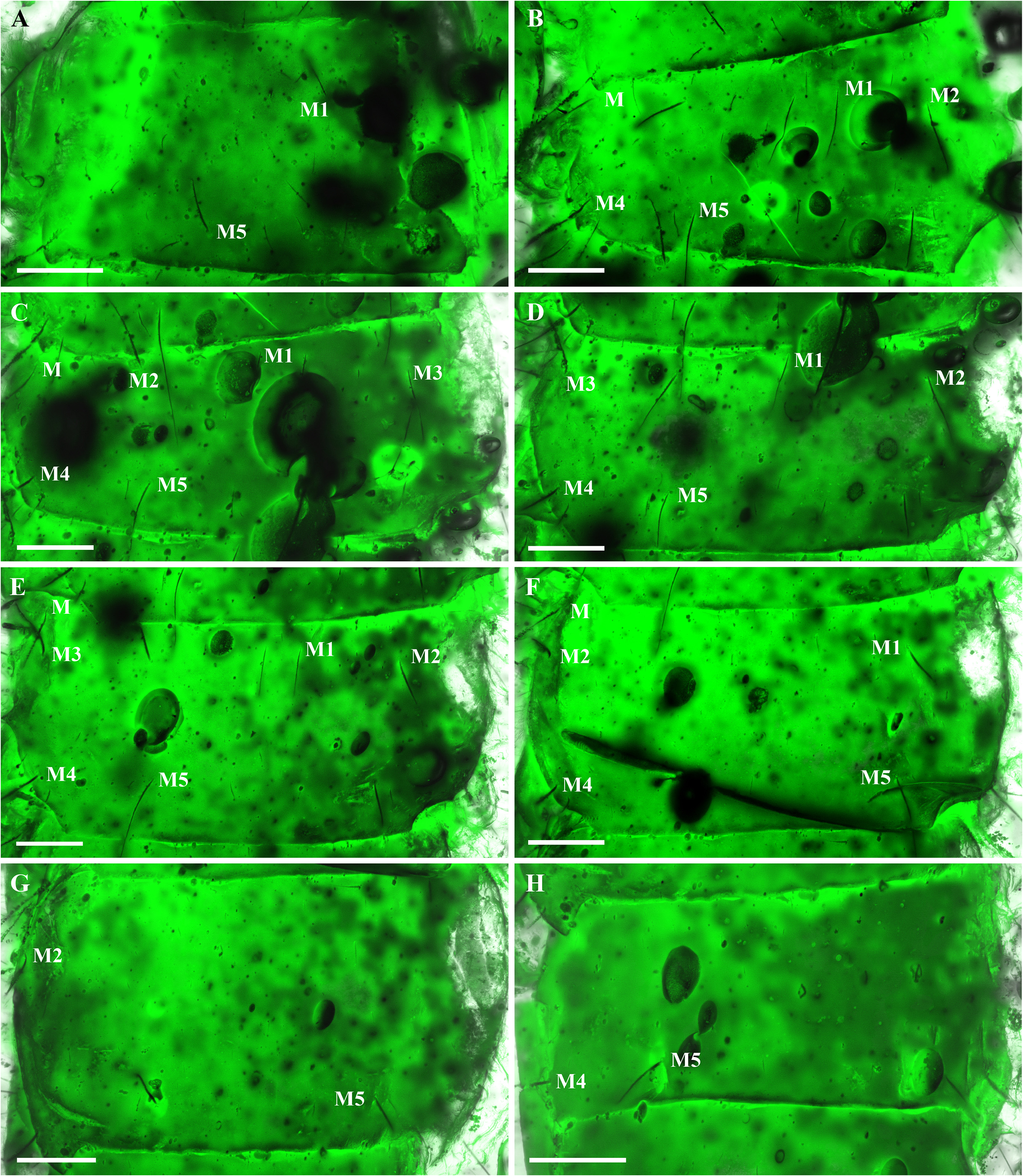
Abdominal tergites with M and a few sM. Abdomen contracted, prescuta not visible. Urotergite I with 2+2 macrosetae (M1, M5) ( Fig. 3A View FIGURE 3 ); urotergite II, VI each with 4+4 macrosetae (M1–2, M4–5) plus 1 anterior lateral M ( Fig. 3B, F View FIGURE 3 ); urotergite IV with 5+5 M1–5 ( Fig. 3D View FIGURE 3 ); urotergite III, V each with 6+6 M ( Fig. 3C, E View FIGURE 3 ); urotergite VII with 2+2 (M2, M5) ( Fig. 3G View FIGURE 3 ); urotergite VIII with 2+2 (M4, M5) ( Fig. 3H View FIGURE 3 ). Segment X 1.4× as long as wide, with distinct carinae; carinae subparallel and converging towards the posterior margin of the segment X until disappearing; dorsal side with 2+2 M between the carinae; acropygium round. Urotergite VII protruding backward, forming pointed angles and each with 1 macroseta ( Fig. 5H View FIGURE 5 ). Urosternites with dense macrosetae. Median glandular organ invaginated ( Fig. 5D View FIGURE 5 ), lateral subcoxal organ with 1 row of glandular seta ( Fig. 5E View FIGURE 5 ); lateral subcoxal organ occupying 0.3× of interstylar area. Styli tapering towards apex ( Fig. 5E, I View FIGURE 5 ). Cerci asymmetric, ca. 0.96 mm in length, slightly shorter than Segment X (ca. 1 mm); basal 2/3 of lateral margin straight and curved inward rapidly in the distal, the distal 1/3 slightly upward, end sharp and hook-like ( Fig. 4A View FIGURE 4 ); carinae arising from the dorsal and ventral acetabular articulations and disappearing at the basal 2/3 ( Fig. 4A View FIGURE 4 ). Right cercus with 1 postmedial tooth; predental margin slightly concaved near the tooth, postdental margin with a row of at least 10 small round denticles gradually decreasing in size until disappearing before the hook ( Fig. 4C View FIGURE 4 ). Left cercus slenderer than the right and strongly concaved in the distal 1/3 of inner margin, relatively smooth without distinct tooth or denticles ( Fig. 4B View FIGURE 4 ).


No known copyright restrictions apply. See Agosti, D., Egloff, W., 2009. Taxonomic information exchange and copyright: the Plazi approach. BMC Research Notes 2009, 2:53 for further explanation.
|
Kingdom |
|
|
Phylum |
|
|
Class |
|
|
Family |
|
|
Genus |