
Kodaianellissus, Zhang, 2017
|
publication ID |
https://doi.org/ 10.11646/zootaxa.4312.2.10 |
|
publication LSID |
lsid:zoobank.org:pub:618B7E19-Af76-4Bcb-Bb95-2B31F1E4Bc9C |
|
DOI |
https://doi.org/10.5281/zenodo.6032764 |
|
persistent identifier |
https://treatment.plazi.org/id/D06CCB3B-FFE2-4F30-D6C6-FDEEE8A64E45 |
|
treatment provided by |
Plazi |
|
scientific name |
Kodaianellissus |
| status |
gen. nov. |
Kodaianellissus View in CoL View at ENA gen. nov.
Type species: Kodaianellissus intorqueus sp. nov., here designated.
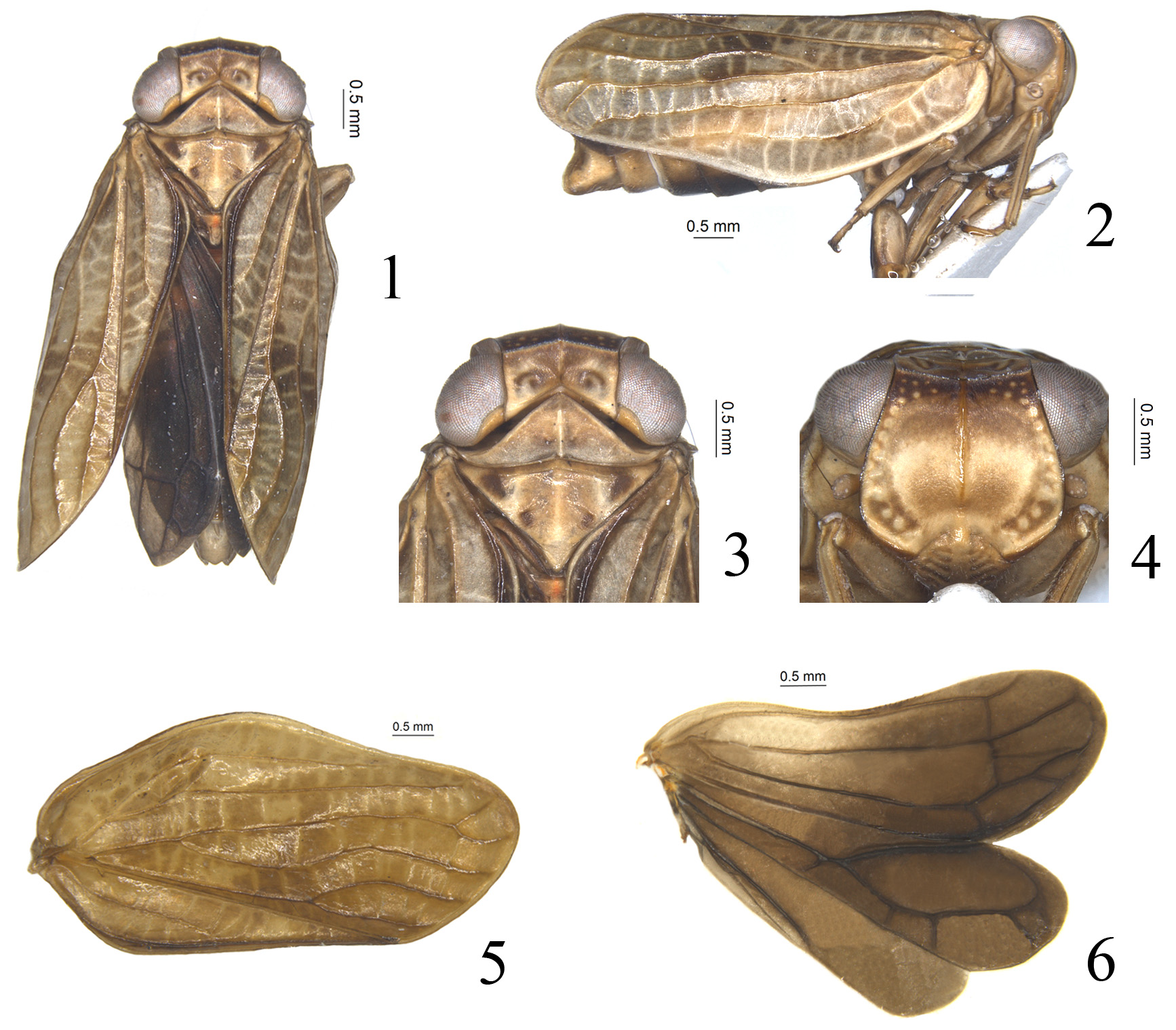
Diagnosis. This genus is very similar to the genus Kodaianella Fennah, 1956 in general appearance, but differs by: 1) ScP-R-MP-Cu lobe of hindwing around 1.5 times wider than Pcu-A1 lobe ( Fig. 6 View FIGURES 1 – 6 ), but the latter much narrower, 4 times wider than Pcu-A1 lobe ( Gnezdilov, 2013, fig. 6); 2) The forewing, in lateral view, broadest at basal 1/3 ( Fig. 2 View FIGURES 1 – 6 ), but broadest from basal 1/3 to apex in Kodaianella ( Gnezdilov, 2013, fig. 3); 3) Frons slightly narrower than in Kodaianella , frons just 1.3 times broader at widest part than long in midline, but 1.5 times in Kodaianella .
Description. Head with compound eyes almost as wide as thorax ( Figs. 1, 3 View FIGURES 1 – 6 ). Vertex rectangular, obviously broader than long, median carina present; anterior margin slightly convex, posterior margin strongly angular concave medially ( Figs. 1, 3 View FIGURES 1 – 6 ). Frons roundly widened below antenna, apical margin almost straight, apical and lateral margins carinate and elevated, median carina obviously elevated from apex extending to near base, but not reaching frontoclypeal sulcus ( Fig. 4 View FIGURES 1 – 6 ); frons with apical and lateral area distributed with numerous tubercles, tubercles change from small to large from apical to lateral part ( Fig. 4 View FIGURES 1 – 6 ). Frontoclypeal suture strongly convex ( Fig. 4 View FIGURES 1 – 6 ). Clypeus with several transverse thin stripes on lateral sides ( Fig. 4 View FIGURES 1 – 6 ). Rostrum short, reaching midcoxae. Gena in lateral view flattened and oblique, with small protuberance near base ( Fig. 2 View FIGURES 1 – 6 ). Pronotum triangular, with two small incisions on disc, median carina present ( Figs. 1, 3 View FIGURES 1 – 6 ). Mesonotum smooth, without carina ( Figs. 1, 3 View FIGURES 1 – 6 ). Forewings elongate, distinctly longer than wide, with very narrow hypocostal plate, longitudinal veins obvious and elevated, forewing slightly broadened near the basal 1/3 ( Figs. 1, 2, 5 View FIGURES 1 – 6 ). Vein ScP+R first separating near base, ScP+RA vein extremely long, surpassing the apical 1/3 of costal margin, the terminal of RP vein almost reaching the outer margin of forewing; MP vein first fork near the basal 1/4, MP1+2 straight, fork again at apical 1/9, MP3+4 sinuate, without fork; CuA vein first separate at apical 1/3; MP first fork before the first separation of CuA ( Fig. 5 View FIGURES 1 – 6 ). Clavus obvious, veins Pcu and A1 fused at basal 1/2 of clavus ( Fig. 5 View FIGURES 1 – 6 ). Forewings with numerous transverse veins ( Figs. 1, 2, 5 View FIGURES 1 – 6 ). Hindwing well developed, 3-lobed; Pcu-A1 lobe distinctly thinner than ScP-R-MP-Cu lobe; A2 lobe narrow with anterior and posterior margins subparallel and distinctly surpassing half length of Pcu-A1 lobe; A2 not branched ( Fig. 6 View FIGURES 1 – 6 ). Hind tibia with 2 lateral spines on apical half.
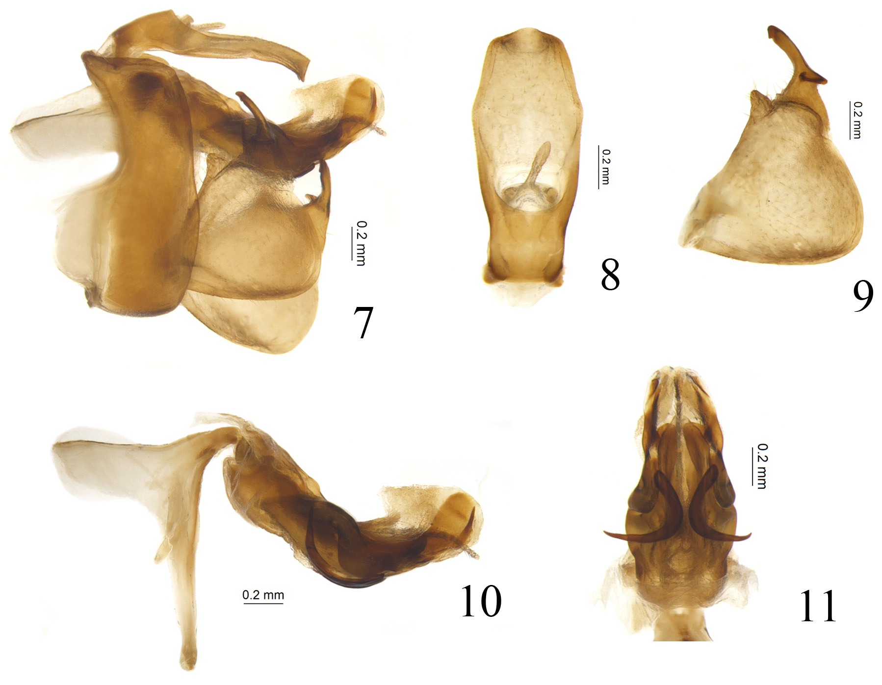
Male terminalia. Anal tube in lateral view long and narrow ( Fig. 7 View FIGURES 7 – 11 ). Gonostyli triangular in profile, becoming broader from base to apex, caudo-ventral angle rounded, dorso-lateral margin with process; capitulum very long and narrow, with auricular process ( Figs. 7, 9 View FIGURES 7 – 11 ). Pygofer rectangular in lateral view, apparently longer than width, posterior margin not obviously caudally produced ( Fig. 7 View FIGURES 7 – 11 ). Periandrium symmetrical, basally tubular, apical part divided into dorso-lateral lobe and ventral lobe ( Fig. 10 View FIGURES 7 – 11 ). Dorso-lateral lobe longer than ventral lobe ( Fig. 11 View FIGURES 7 – 11 ). Aedeagus with pair of ventral processes directed dorso-basad.
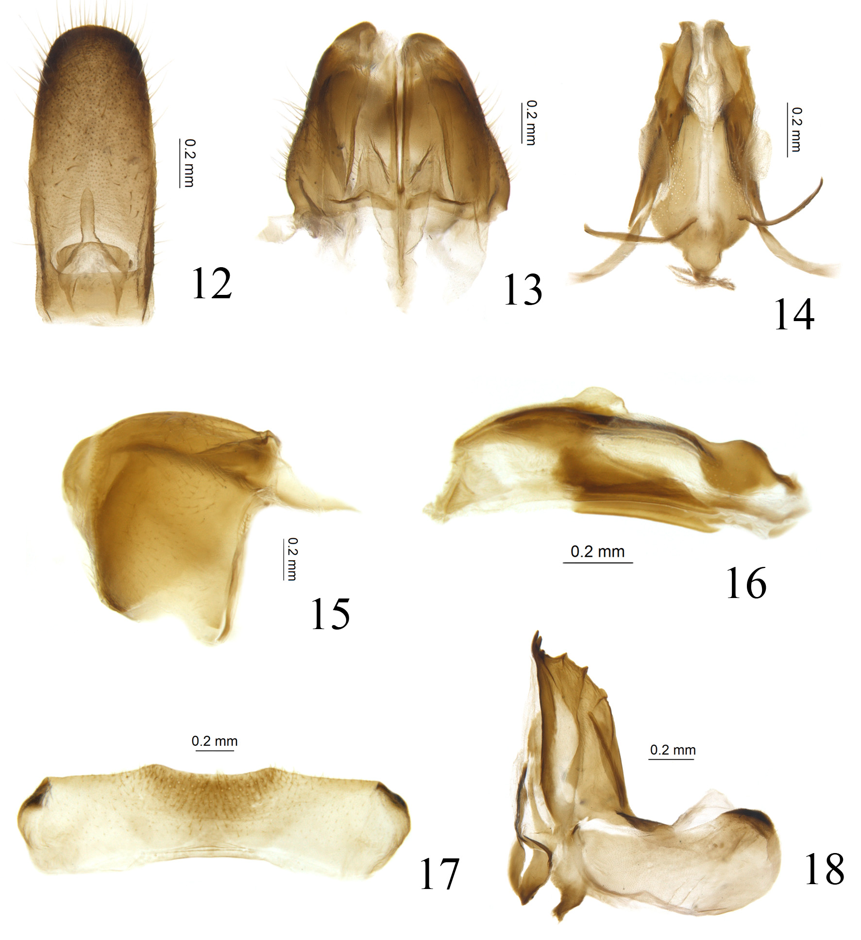
Female terminalia. Anal tube relatively long in lateral view, reaching level of posterior margin of gonoplac. Anterior connective lamina of gonapophysis VIII with teeth at apex and outer lateral margin, inner lateral margin without teeth ( Fig. 18 View FIGURES 12 – 18 ). Gonocoxa VIII subquadrangular, connected to gonapophysis VIII with rectangular angle ( Fig. 18 View FIGURES 12 – 18 ). Gonapophysis IX in lateral view boat-shaped ( Fig. 16 View FIGURES 12 – 18 ). Gonoplac subrectangular in lateral view ( Fig. 15 View FIGURES 12 – 18 ), fused at middle near base, widest at base ( Fig. 13 View FIGURES 12 – 18 ). Hind margin of sternite VII concave medially ( Fig. 17 View FIGURES 12 – 18 ).
Etymology. The name of this new genus is an arbitrary combination between two generic names of issids: Kodaianella and Issus , indicating this genus has a close relationship with the genus Kodaianella . The gender is masculine.
Note. This genus refers to the taxon “ gen. nov. apud Kodaianella ” on the molecular phylogeny trees of Wang et al. (2016). The position of this genus within Kodaianellini is supported by both the molecular phylogenetic results and the morphological characters listed above.
No known copyright restrictions apply. See Agosti, D., Egloff, W., 2009. Taxonomic information exchange and copyright: the Plazi approach. BMC Research Notes 2009, 2:53 for further explanation.