
Longieusarima, Zhang, 2017
|
publication ID |
https://doi.org/ 10.11646/zootaxa.4312.2.10 |
|
publication LSID |
lsid:zoobank.org:pub:618B7E19-Af76-4Bcb-Bb95-2B31F1E4Bc9C |
|
DOI |
https://doi.org/10.5281/zenodo.6032772 |
|
persistent identifier |
https://treatment.plazi.org/id/D06CCB3B-FFE4-4F3A-D6C6-FCDDEB714DC8 |
|
treatment provided by |
Plazi |
|
scientific name |
Longieusarima |
| status |
gen. nov. |
Longieusarima View in CoL gen. nov.
Type species: Longieusarima lunulia sp. nov., here designated.
Diagnosis. Longieusarima gen. nov. looks similar to Eusarima Yang, 1994 , but differs by: 1) Vertex nearly as wide as long ( Fig. 29 View FIGURES 27 – 32 ), while more wider than long in Eusarima ( Chan & Yang, 1994: fig. 45a); 2) The first fork of MP vein occurs well before the fork of CuA vein on forewing ( Fig. 31 View FIGURES 27 – 32 ), while in Eusarima , almost in the same level ( Chan & Yang, 1994: fig. 45c); 3) both median carina and sublateral carinae of frons only visible at basal half ( Fig. 30 View FIGURES 27 – 32 ), while both are very clear from base to apex in Eusarima ( Chan & Yang, 1994: fig. 45b). This genus is also very similar to Parasarima Yang, 1994 , from which it differs by: 1) ScP+RA vein of forewing obvious shorter than Parasarima ; 2) The ratio of length and width of vertex is 1.0 in this genus ( Fig. 29 View FIGURES 27 – 32 ) but less than 0.5 in Parasarima ( Chan & Yang, 1994: fig. 39a); 3) MP vein on forewing forks first before CuA vein ( Fig. 31 View FIGURES 27 – 32 ), while at the same level in Parasarima ( Chan & Yang, 1994: fig. 39c).
This genus is also close to Sinesarima Yang, 1994 and Neosarima Yang, 1994 , but the longer ScP+RA forewing branch and the longer median and sublateral carinae of frons in Longieusarima allow it to be separated from these two genera.
Description. Head with compound eyes very slightly wider than thorax ( Figs. 27, 29 View FIGURES 27 – 32 ). Vertex nearly quadrate, as long as wide, median carina absent; anterior margin obviously and angularly convex, posterior margin roundly concave at middle ( Figs. 27, 29 View FIGURES 27 – 32 ). Frons angularly expanded outwardly below antenna; apical margin slightly angularly convex; sublateral carinae obviously elevated at apical half, reaching middle level of compound eyes; median carina present but weak, extending to near middle of frons ( Fig. 30 View FIGURES 27 – 32 ). Frontoclypeal suture dorsally convex ( Fig. 30 View FIGURES 27 – 32 ). Clypeus flattened, without carina ( Fig. 30 View FIGURES 27 – 32 ). Rostrum short, reaching mesocoxae. Gena in lateral view flattened and oblique, vertex and frons meeting at acute angle ( Fig. 28 View FIGURES 27 – 32 ). Pronotum triangular, anterior and posterior margins elevated, median area with two small incisions, without median carina ( Figs. 27, 29 View FIGURES 27 – 32 ). Mesonotum with weak median carina present in basal half ( Figs. 27, 29 View FIGURES 27 – 32 ). Forewings elongate, apparently longer than width, hypocostal plate present; forewing expanded outwardly at basal third, longitudinal veins obvious and elevated ( Figs. 27, 28, 31 View FIGURES 27 – 32 ); vein ScP+R first separating near base, ScP+RA short, reaching middle of costal margin, the terminal of RP vein reaching the outer margin of forewing; MP vein first fork at basal third, MP1+2 fork again at apical 1/5, MP3+4 simple, not forking again; CuA vein first bifurcate at middle; MP first fork before CuA ( Figs. 27, 28, 31 View FIGURES 27 – 32 ); clavus obvious, veins Pcu and A1 fused near middle, transverse veins of forewing well developed ( Figs. 27, 31 View FIGURES 27 – 32 ). Hindwing well developed, 3-lobed, Pcu-A1 lobe as wide as ScP-R-MP-Cu lobe, anal lobe broad; Pcu single, Pcu and A1 anastomosing for long distance; A2 non branched ( Fig. 32 View FIGURES 27 – 32 ). Hind tibia with 2 lateral spines on apical half.
Male terminalia. Anal tube long ( Figs. 33 View FIGURES 33 – 37 , 38 View FIGURES 38 – 40 ). Gonostylus triangular in profile, broadening from base to apex, widest at apical 1/3, caudo-ventral angle rounded, capitulum finger-shaped ( Figs. 33, 35 View FIGURES 33 – 37 , 38 View FIGURES 38 – 40 ). Pygofer with dorsal margin apparently narrower than ventral margin, posterior margin obviously and caudally produced ( Figs. 33 View FIGURES 33 – 37 , 38 View FIGURES 38 – 40 ). Periandrium shallowly U-shaped, Aedeagus with pair of ventral processes derived from apical 1/9 and directed basad ( Figs. 36 View FIGURES 33 – 37 , 38 View FIGURES 38 – 40 ). Apical part of periandrium divided into dorsal lobe, pair of lateral lobes and ventral lobe.
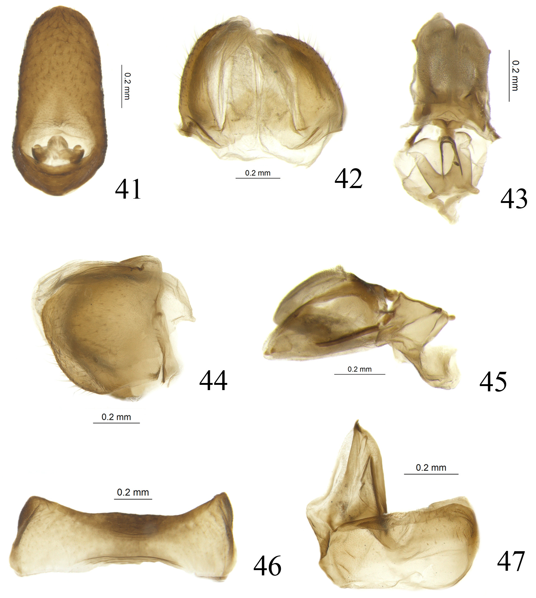
Female terminalia. Anal tube relatively short in lateral view. Anterior connective lamina of gonapophysis VIII with teeth at apex, outer lateral margin and inner lateral margin without teeth ( Fig. 47 View FIGURES 41 – 47 ). Gonocoxa VIII subquadrangular, oblique connected with gonapophysis VIII ( Fig. 47 View FIGURES 41 – 47 ). Posterior connective lamina of gonapophysis IX with large special structure the top of which is rough on surface ( Figs. 43, 45 View FIGURES 41 – 47 ). Gonoplac rounded in lateral view ( Fig. 44 View FIGURES 41 – 47 ), fused at middle, widest at basal half ( Fig. 42 View FIGURES 41 – 47 ). Hind margin of sternite VII almost straight at middle ( Fig. 46 View FIGURES 41 – 47 ).
Etymology. The name is a combination of Latin prefix “longi-” and the genus name Eusarima , indicating this genus with vertex obviously protruded forward in the Eusarima group. The gender is feminine.
Note. This genus refers to the taxon “ gen. nov. apud Eusarima ” on the molecular phylogenic analyses ( Wang et al., 2016).


No known copyright restrictions apply. See Agosti, D., Egloff, W., 2009. Taxonomic information exchange and copyright: the Plazi approach. BMC Research Notes 2009, 2:53 for further explanation.