
Phyllopodopsyllus tenuis Wells and Rao, 1987
|
publication ID |
https://doi.org/10.1080/00222933.2015.1038329 |
|
DOI |
https://doi.org/10.5281/zenodo.4330205 |
|
persistent identifier |
https://treatment.plazi.org/id/D112879B-0638-EE1F-D8DC-FE4E5210F356 |
|
treatment provided by |
Carolina |
|
scientific name |
Phyllopodopsyllus tenuis Wells and Rao, 1987 |
| status |
|
Phyllopodopsyllus tenuis Wells and Rao, 1987 View in CoL
( Figures 1A, B View Figure 1 , 2A–C View Figure 2 , 3A–D View Figure 3 , 4A–F View Figure 4 , 5A–C View Figure 5 , 6A–C View Figure 6 , 7A–D View Figure 7 )
Phyllopodopsyllus T. Scott, 1906 in Gómez and Morales-Serna (2014)
Locality
Off Tabasco State (not Campeche State as in Gómez and Morales-Serna 2014, Appendix 2: 121); 18.6169° N, 93.5° W; 36 m depth GoogleMaps ; 18.6189° N, 94.00° W; 24 m depth GoogleMaps .
Material examined
One adult female, dissected (EMUCOP-11–00); one adult male, dissected (EMUCOP-12–00).
Partial redescription
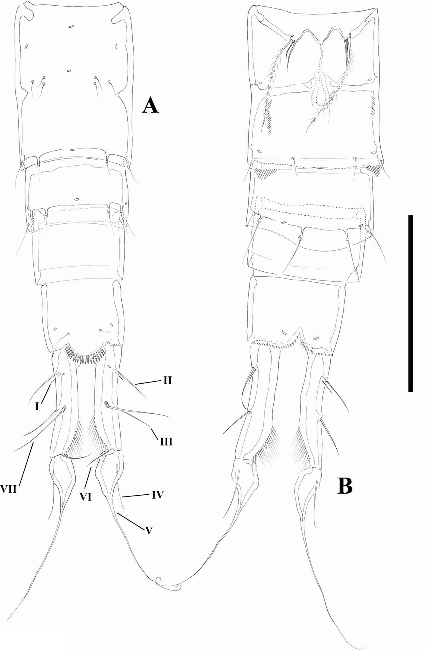
Female. Habitus fusiform; tapering from posterior margin of cephalothorax to anal somite. Second and third urosomites fused forming genital double-somite, with remains of former division ventrally, completely fused dorsally ( Figure 1A, B View Figure 1 ). Genital double-somite and following urosomite with ventral and dorsal pores and sensilla as figured ( Figure 1A, B View Figure 1 ). Fifth urosomite without pores and sensilla. Anal somite ( Figure 1A, B View Figure 1 ) as long as preceding somite; dorsally ( Figure 1A View Figure 1 ) with two pores and two sensilla, ventrally ( Figure 1B View Figure 1 ) with two pores close to medial cleft; with spinules along posterior margin ventrally; with rounded anal operculum ornamented with spinules along posterior margin ( Figure 1A View Figure 1 ). Caudal rami as figured, elongate, about 3.4 times as long as broad, with longitudinal row of inner setules along distal third; with seven setae as follows: seta I and II in proximal fifth, the former small and ventral to seta II, the latter about four times as long as the former; seta III about halfway along outer margin of caudal ramus, as long as seta II; seta IV as long as seta III, fused to seta V, the latter with swollen proximal part; seta VI inserted on inner distal corner of caudal ramus, half as long as seta IV; seta VII longer than seta III, inserted dorsally at same level as seta III, biarticulated.
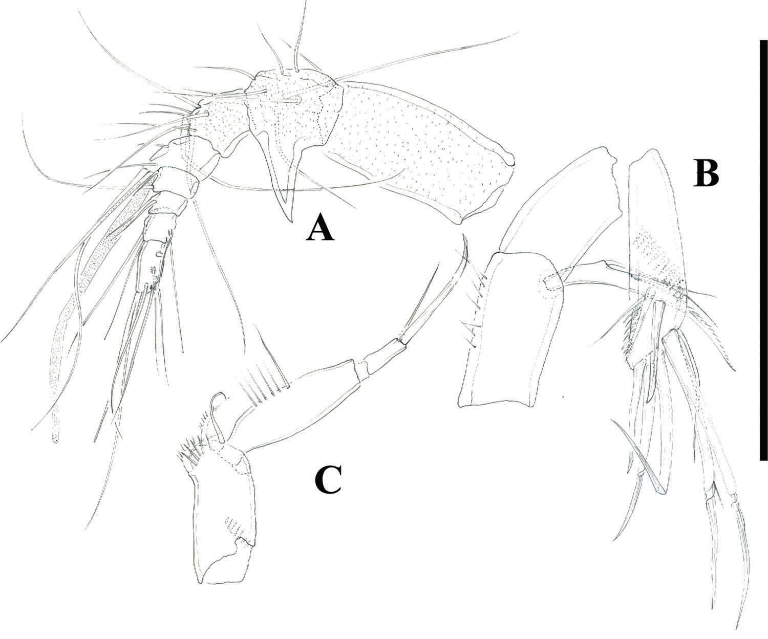
Antennule ( Figure 2A View Figure 2 ). Eight-segmented; first segment elongate, about 2.3 times as long as broad; second segment slightly longer than broad, with strong acute process; all setae bare; armature formula as follows: 1(1); 2(7); 3(8); 4(3+(1+ae)); 5(2); 6(3); 7(3); 8(4+(1+ae)).
Antenna ( Figure 2B View Figure 2 ). With basis about 1.7 times as long as broad, with inner longitudinal row of setules; exopod one-segmented, with three elements; distalmost element fused to exopod. First endopodal segment without armature; second endopodal segment with medial and distal hyaline frills; with one slender seta and two spines laterally, and with six elements distally as shown.
Maxilliped ( Figure 2C View Figure 2 ). Syncoxa with spinules and with three inner elements as figured. Basis with some setules and with one seta along inner margin as shown; endopodal segment small, elongate, about three times as long as broad, with long and slender claw and one seta.
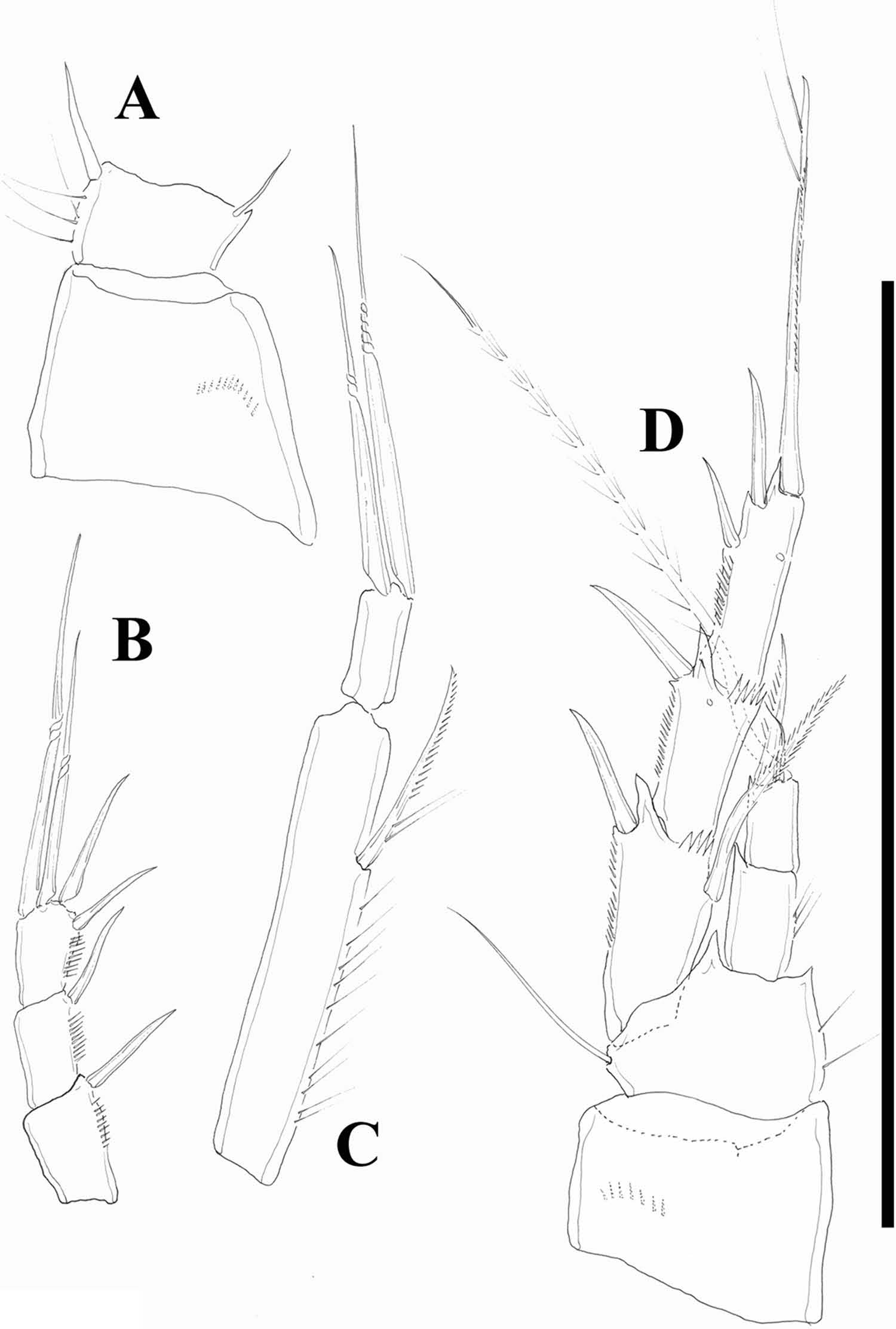
P1 ( Figure 3 View Figure 3 A-C). Only one leg was recovered. Coxa ( Figure 3A View Figure 3 ) large, with some minute spinules medially. Basis ( Figure 3A View Figure 3 ) with outer and inner element as shown. Exopod ( Figure 3B View Figure 3 ) three-segmented; first and second segments with outer minute spinules and outer spine; third segment with four elements. Endopod ( Figure 3C View Figure 3 ) two-segmented; first segment elongate, about 5.7 times as long as broad, with one inner element on distal third; second segment small, about twice as long as broad, with two apical elements.
P2 ( Figure 3D View Figure 3 ). Only the left P2 was recovered completely. Coxa large with spinules as shown. Basis with some inner setules, with outer long seta. Exopod three-segmented; first segment with, second segment without inner seta; third segment with four elements in all (innermost distal seta probably lost during dissection). Endopod twosegmented, reaching about middle of second exopodal segment; first segment unarmed; second segment with three elements (one minute innermost and one medial long seta, and one outermost spiniform element).
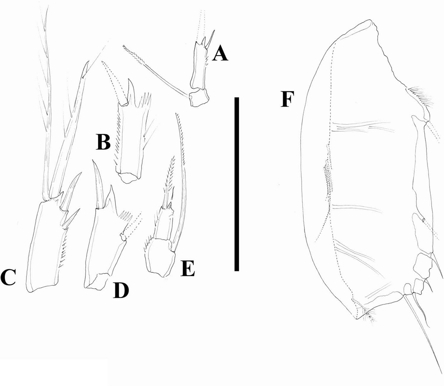
P3 ( Figure 4 View Figure 4 B-E). Both legs badly damaged. Exopod three-segmented; first segment with inner seta ( Figure 4D View Figure 4 ), second segment ( Figure 4B View Figure 4 ) without inner seta; third segment ( Figure 4C View Figure 4 ) with four elements. Endopod two-segmented ( Figure 4E View Figure 4 ); first segment with one inner seta; second segment with three elements (one lost during dissection).
P4. Badly damaged. Endopod ( Figure 4A View Figure 4 ) two-segmented; first segment with one inner seta; second segment with two elements.
P5 ( Figure 4F View Figure 4 ). Large, foliose, forming brood pouch; with 10 elements.
P6 ( Figure 1B View Figure 1 ). Each leg represented by small lobe bearing one long outer seta and two inner elements.
Male. Habitus ( Figure 5A, B View Figure 5 ) fusiform; tapering from posterior margin of cephalothorax to anal somite. Body length measured from anterior tip of rostrum to posterior margin of caudal rami, 270 µm. Rostrum minute ( Figure 5A View Figure 5 ). Surface ornamentation of urosomites as in female. Anal somite and anal operculum
( Figure 5C View Figure 5 ) as in female. Caudal rami sexually dimorphic, elongate, about 11 times as long as wide, with seven setae ( Figure 5C View Figure 5 ).
Antennule ( Figure 6A View Figure 6 ). seven-segmented, subchirocer; first segment elongate, about 1.3 times as long as wide; second segment nearly as long as wide, with strong acute process. Armature formula difficult to define, most probably as follows: 1(1); 2(11); 3(3); 4(9+(1+ae)); 5(0); 6(0); 7(10+(1+ae)?).
Antenna ( Figure 6B View Figure 6 ). As in female.
P1 ( Figure 6C View Figure 6 ). Badly damaged, as in female.
P2. Badly damaged. Exopod ( Figure 7A View Figure 7 ) as in female. Endopod lost during processing.
P3. Badly damaged. Exopod lost during processing. Endopod ( Figure 7C View Figure 7 ) twosegmented, sexually dimorphic, first segment with one element, second segment with one strong bifid and one slender seta.
P4. Badly damaged. Endopod ( Figure 7B View Figure 7 ) two-segmented; first and second segments with one seta each.
P5 ( Figure 7D View Figure 7 ). Baseoendopod of both legs confluent, with outer basal seta and with three elements on inner endopodal lobe. Exopod with five setae as shown.
Remarks
See below.


| V |
Royal British Columbia Museum - Herbarium |
| VI |
Mykotektet, National Veterinary Institute |
No known copyright restrictions apply. See Agosti, D., Egloff, W., 2009. Taxonomic information exchange and copyright: the Plazi approach. BMC Research Notes 2009, 2:53 for further explanation.
|
Kingdom |
|
|
Phylum |
|
|
Class |
|
|
Order |
|
|
Family |
|
|
Genus |