
Mesostoma jilinensis Wang & Huang, 2023
|
publication ID |
https://doi.org/ 10.11865/zs.2023205 |
|
publication LSID |
lsid:zoobank.org:pub:04888FCF-480A-4198-9638-7AD4F8034E9F |
|
DOI |
https://doi.org/10.5281/zenodo.10941776 |
|
persistent identifier |
https://treatment.plazi.org/id/D14887D4-FF9B-2726-FF7C-4445B0E2E34A |
|
treatment provided by |
Felipe |
|
scientific name |
Mesostoma jilinensis Wang & Huang |
| status |
sp. nov. |
3.2 Mesostoma jilinensis Wang & Huang , sp. nov. ( Figs 3–4 View Figure 3 View Figure 4 )
Etymology. The species is named after the province of its typical locality.
Diagnosis. The body of adult is in dark brown color, whilst the body of juvenile is in white color. A prepharyngeal pit is present, with two main excretory ducts running transversely behind the pharynx. The seminal receptacle, oviduct and ovary are developed in a typical manner of the genus Mesostoma . The common vitelloduct connects to the ductus communis. The copulatory organ is characterized by a relatively large penis papilla. A pair of uteri open towards the common genital atrium.
Material examined. Holotype. IZCAS PLA-B001 (whole mount), a lotus pond (125°29'E, 43°46'N; water temperature 23.5℃, pH 8.7), Jingyuetan, Changchun , Jilin, elev. 236 m, July 2019, leg. Xiaozhou Hu and Weixuan Li. Paratypes. IZCAS PLA-P002–005 , same data as holotype GoogleMaps .
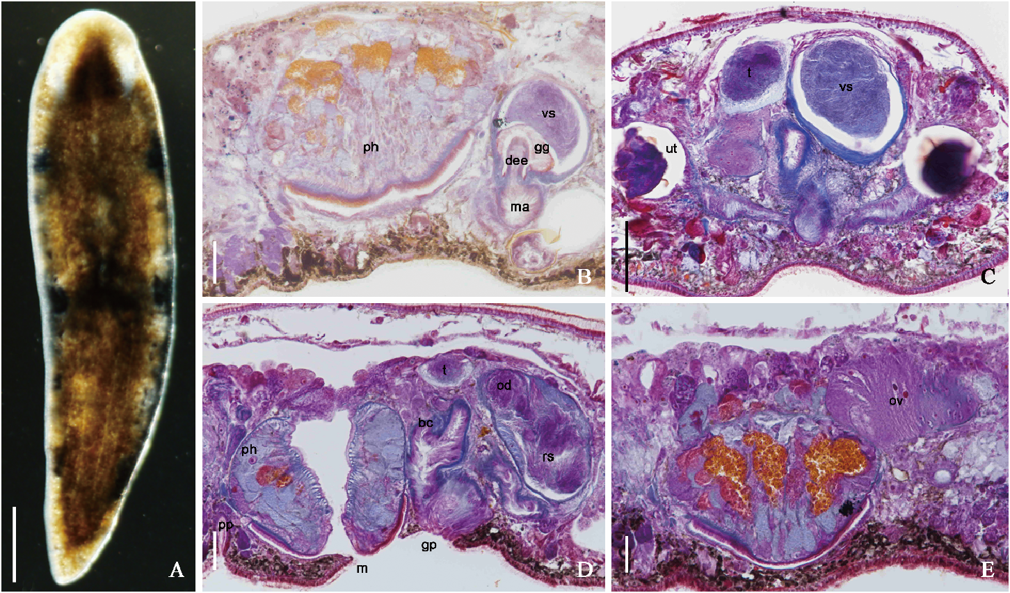
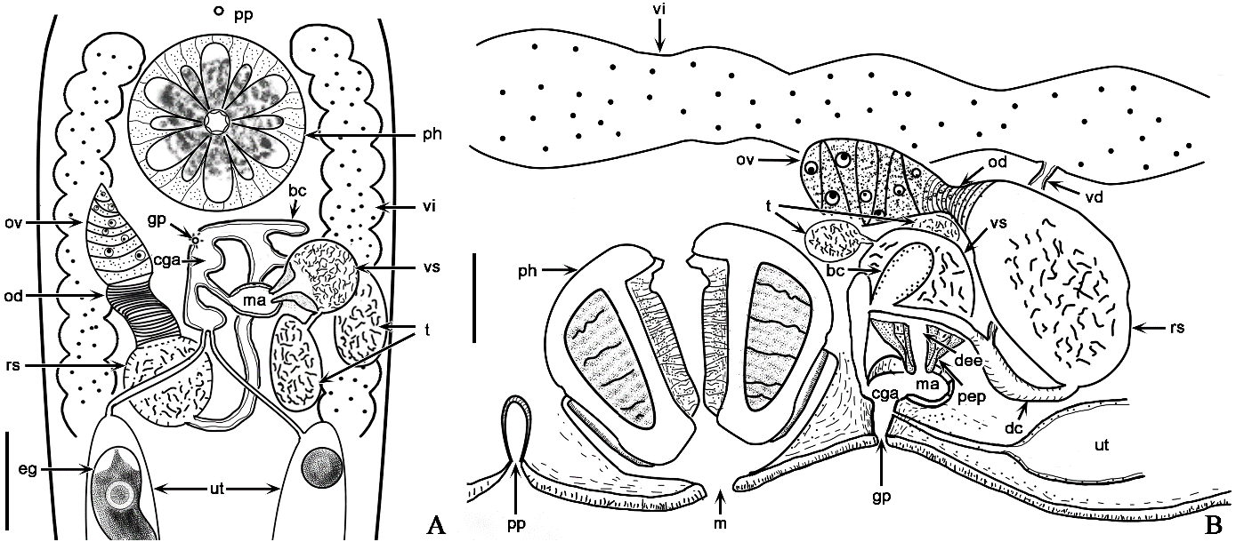
Description. Mature individual 2.14 ± 0.13 mm (n = 5) in length and 0.55 ± 0.05 mm (n = 5) in width, with round anterior end and sharply pointed posterior end. Adult individuals in dark brown color, while juveniles in white ( Fig. 3A View Figure 3 ). Pigmentation absent at two elliptic regions symmetrically located at anterior end ( Fig. 3A View Figure 3 ). Rosulatus-shaped pharynx situated at front 1/3 of body ( Figs 3A View Figure 3 , 4A View Figure 4 ), typical within species of Mesostoma Ehrenberg, 1837 . Prepharyngeal pit, mouth and genital pore located at ventral body, with prepharyngeal pit located anterior to pharynx. Two main excretory ducts run transversely behind pharynx ( Figs 3D View Figure 3 , 4B View Figure 4 ).
Simultaneously hermaphroditic. Female reproductive system consists of an ovary, a seminal receptacle, a pair of vitellaria and a common vitelloduct, a copulatory bursa, a pair of uteri, a superior genital atrium and a gonopore. Long ovoid shape ovary located posterior to pharynx. Transverse muscles present on wall of oviduct. Ball-shaped seminal receptacle usually filled with sperms ( Figs 3D–E View Figure 3 , 4A–B View Figure 4 ). A pair of follicle-shaped vitellaria extend at both sides of body, between pharynx and caudal end. A pair of vitelloducts converge anteriorly as a common vitelloduct in center of body, and then further connects to ductus communis ( Figs 3D View Figure 3 , 4B View Figure 4 ). Single copulatory bursa with two lobes, with a thick epithelium. A pair of uteri, with longitudinal muscle on wall and large number of immature eggs inside, open towards common genital atrium ( Figs 3C View Figure 3 , 4A–B View Figure 4 ).
Paired testes egg-shaped and situated dorsally around seminal vesicle ( Figs 3C–D View Figure 3 , 4A–B View Figure 4 ). A pair of vas deferentia connect respectively to seminal vesicle. Ball-shaped copulatory organ filled with prostate secretion from extracapsular prostate glands, and contains a large intracapsular seminal vesicle in its proximal part. Copulatory organ attaches to male atrium through continuing walls of penis papilla. Penis papilla relatively large, occupying a considerable portion of atrium. Ejaculatory duct surrounded by a plasmatic tissue and prostate secretion ( Figs 3B View Figure 3 , 4A–B View Figure 4 ).
Habitat. The free-living individuals often swim or move upside down at the air-water interface. There were a lot of lotus leaves at the surface water of the sampling site.
Distribution. China (Jilin). So far only known from the type locality.
Remarks. According to the overall body configuration, morphology and location of the pharynx, the paired vitellaria and common vitelloduct, it is evident that these specimens belong to Mesostominae . The seminal receptacle, oviduct and the ovary are developed in a typical manner of the genus Mesostoma (Noreña-Janssen & Faubel, 1992) . With a copulatory bursa and a penis papilla, the new species should be arranged into the M. lingua species group (Noreña-Janssen & Faubel, 1992).
The new species is characteristic by: (1) a ductus communis connects the seminal receptacle and copulatory bursa, (2) a relatively large subapical penis papilla, and (3) the copulatory bursa is smaller than the seminal receptacle. The morphology comparison of M. jilinensis Wang & Huang , sp. nov. and seven species of M. lingua group is shown in Table 4. Among them, four species namely M. sibollae Kolasa, 1976 , M. zariae Kolasa & Mead, 1981 , M. appinum Kolasa & Schwartz, 1988 and M. magnum Kolasa & Schwartz, 1988 , possess a large penis papilla with thick muscular tissue. They can be readily distinguished from each other by body pigmentation and the shape of the copulatory organ. For instance, M. appinum is a yellow-brown colored species with an irregular-shaped copulatory organ; M. magnum is a brown colored species with a pear-shaped copulatory organ; M. sibollae is a green colored species with an oval-shaped copulatory organ, while M. zariae is essentially colorless with a flattened copulatory organ. Moreover, these four species have eye pigmentation at the anterior body. And their two main sperm ducts converge into one before reaching the copulatory organ. Furthermore, the genital pore and mouth of M. magnum open into the same ventral pore in the middle of the body. As for M. jilinensis Wang & Huang , sp. nov., the specimens are in dark brown color without eye pigmentation. In addition, the species has two main excretory ducts, and the two sperm ducts connect to the seminal vesicle directly, therefore, M. jilinensis Wang & Huang , sp. nov. is distinct from these four species. As for M. africanum Kolasa, 1976 , M. viaregginum Kolasa, 1976 and M. lingua Abildgaard, 1789 , they all have a small penis papilla with thin muscular tissue. In terms of the size of the body, M. lingua is similar to M. jilinensis Wang & Huang , sp. nov. However, the seminal receptacle of M. lingua connects to the common genital atrium directly. In contrast, the copulatory bursa of M. jilinensis Wang & Huang , sp. nov. is in the midst between the seminal receptacle and the common genital atrium. Taking these differences into account, we described M. jilinensis Wang & Huang , sp. nov. as a new species within the genus Mesostoma .
| IZCAS |
Institute of Zoology, Chinese Academy of Sciences |
No known copyright restrictions apply. See Agosti, D., Egloff, W., 2009. Taxonomic information exchange and copyright: the Plazi approach. BMC Research Notes 2009, 2:53 for further explanation.