
Accipitridae Vigors, 1824
|
publication ID |
https://doi.org/ 10.1080/08912963.2021.1966777 |
|
publication LSID |
lsid:zoobank.org:pub:6A25C569-3E9F-43B8-AAF8-F36CE405C06E |
|
DOI |
https://doi.org/10.5281/zenodo.5534420 |
|
persistent identifier |
https://treatment.plazi.org/id/D14B87DD-0017-FF9E-FC14-FA67FE95F886 |
|
treatment provided by |
Carolina |
|
scientific name |
Accipitridae Vigors, 1824 |
| status |
|
Family Accipitridae Vigors, 1824 View in CoL
Subfamily indet. Gen. et sp. indet.
Material
Distal right humeral fragment, preserving a relatively unworn distal end and 16.2 mm of shaft, and some associated fragments of the shaft, SAMA P.58917.
Measurements (mm)
Distal width 15.4, least shaft width 8.3, total depth 8.5, condylus dorsalis depth 8.3, condylus dorsalis width 5.2, condylus ventralis depth 5.1, condylus ventralis width 7.3, epicondylus ventralis depth 7.0.
Locality, stratigraphy and age
31° 07.568 ʹ S; 140° 12.737 ʹ E. Site 11, Lake Pinpa, Frome Downs Station, South Australia, Namba Formation, Pinpa LF, late Oligocene. Collected by A. Camens, T. Worthy and W. Handley, 24–26 September 2015.
Remarks
The fossil can be excluded from other raptor families on the following features:
Falconidae (falconid state in brackets): the condylus dorsalis is thickened and rounded distally (consistently narrow and rectangular); the processus flexorius ends proximal to the condylus ventralis (equidistant).
Pandionidae (state for Pandion haliaetus in brackets): a shallow fossa m. brachialis (deep); a flat epicondylus dorsalis (prominently projecting); a flat epicondylus ventralis (prominent); the fossa olecraniis shallow (deep); thesulcus scapulotricipitalis is shallow (deep).
Cathartidae (cathartid state in brackets): a shallow fossa m. brachialis (deep); a lack of pneumatisation in the fossa m. brachialis (present); a flat epicondylusventralis (prominent).
Sagittariidae (sagittariid state in brackets): the two fossae marking the attachment points for the lig. collaterale dorsale are positioned roughly adjacent to each other (cranial-most fossa slightly proximal to and abutting caudal fossa in sagittariids).
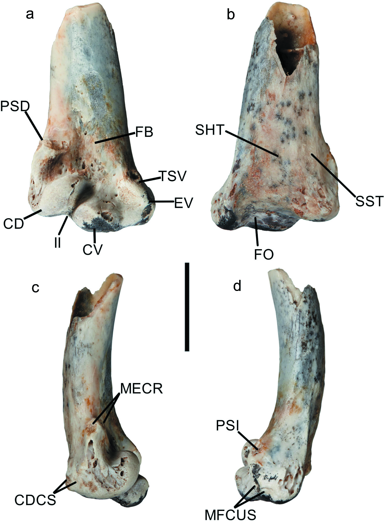
The fossil is broadly similar to accipitrids and displays the following features: (1) The tuberculum supracondylare dorsale ( Figure 11A View Figure 11 : PSD) is located well-proximal to the condylus dorsalis ( Figure 11A View Figure 11 : CD) and is small, barely projecting dorsally of the shaft, but projects slightly cranially as a proximodistally elongate rugosity; (2) the dorsal face/shaft margin between the tuberculum supracondylare dorsale and the epicondylus dorsalis is mildly inflated; Two shallow scars for the m. extensor carpi radialis are present on the tuberculum supracondylare dorsale ( Figure 11C View Figure 11 : MECR), (3) the larger palmar attachment scar on the cranial face adjacent to the dorsal margin is oval (4) and the smaller dorsal scar is located on the dorsal face of the processus. (5) In caudal view, the processus flexorius ( Figure 11 View Figure 11 : PF) terminates proximal to the condylus ventralis ( Figure 11A View Figure 11 : CV) but is prominent ventrally. (6) The sulcus scapulotricipitalis ( Figure 11B View Figure 11 : SST) forms a shallow but broad notch roughly 2 mm wide on the caudal face. (7) The fossa olecrani ( Figure 11B View Figure 11 : FO) is moderately deep, defining well the dorsal margin to the processus flexorius but does not create a discontinuity with the sulcus humerotricipitalis. (8) The sulcus humerotricipitalis ( Figure 11B View Figure 11 : SHT) is very shallow, and at 5.3 mm wide extends over half of shaft width of 9.7 mm at the same point. (9) The fossa m. brachialis ( Figure 11A View Figure 11 : FB) is shallow but distinct, with a proximodistal length of 13.8 mm extending well proximal to the tuberculum supracondylare dorsale, and a maximum dorsoventral width of 7.3 mm level with the proximal margin of the tuberculum supracondylare dorsale. In contrast, the shaft width measures 10.1 mm at the same point. Within the fossa, the impressio m. brachialis is slightly deeper. (10) The fossa is well separated (3 mm) from the dorsal margin of the shaft. (11) The epicondylus ventralis ( Figure 11A View Figure 11 : EV) is indistinct from the ventral margin and does not project ventrally past the processus flexorius. (12) Asingle distinct, shallow insertion scar is present on the ventrodistal section of the epicondylus ventralis, with a very faint and shallow second insertion ventrally adjacent to it. These insertions serve as the attachment point for the m. flexor carpi ulnaris. (13) The tuberculum supracondylare ventrale ( Figure 11A View Figure 11 : TSV) projects cranially but not ventrally from the shaft. (14) Ashallow insertion scar for the pronator superficialis is present just proximal to the tuberculum on the dorsal face. (15) The condylus dorsalis ( Figure 11A View Figure 11 : CD) is 5.9 mm proximodistally long, 4.7 mm dorsoventrally wide and 8.6 mm craniocaudally deep. (16) Two small, very shallow insertion scars are present on the caudal section of the dorsal face of the condylus dorsalis by the distal margin ( Figure 11C View Figure 11 : CDCS), directly craniocaudally adjacent to each other. (17) The condylus ventralis is 4.5 mm proximodistally long, 7.3 mm dorsoventrally wide and 5 mm craniocaudally deep. (18) The condylus dorsalis is separated by a distinct notch from and set well proximal to the distal margin of the condylus ventralis in cranial view. (19) The incisura intercondylaris ( Figure 11 View Figure 11 : II) is narrow, roughly 1.1 mm wide, but distinct. (20) The processus flexorius is surpassed distally by the condylus ventralis in caudal view, and strongly projects ventrally in caudal view. (21) The ventral margin of the condylus ventralis is not separated by a notch from the processus flexorius in cranial view.
Extant accipitrids differ as follows: (Trait 1) The tuberculum supracondylare dorsale projects much further dorsally in all subfamilies and species except Pernis apivorus (Perninae) , Polyboroides typus ( Gypaetinae , non-projecting), Aquilinae , Accipitrinae and species of Buteo (Buteoninae) . (16) The insertion scars towards the caudal margin of the condylus dorsalis are both deep in all subfamilies except Elaninae and Accipitrinae , with the latter having the cranial-most insertion being shallow and the caudal-most deep.
The fossil has the most similarities to species from the subfamily Elaninae (see SI.2 for more detailed differential comparisons), but differs markedly in regards to the inflation of the dorsal face between the tuberculum supracondylare dorsale and the epicondylus dorsalis, the size and shape of the palmar and dorsal attachment scars for the m. extensor carpi radialis, the distinct depression in the section of dorsal face caudal to the tuberculum supracondylaris and the epicondylus dorsalis, the sulcus humerotricipitalis width, the fossa m. brachialis length, the configuration of the insertion scars on the distal epicondylus ventralis, the position of the distal margin of the condylus dorsalis relative to that of the condylus ventralis in cranial view, the ventral projection of the processus flexorius, and the connectivity of the condylus ventralis and entepicondyle in cranial view.
As the Archaehierax sylvestris specimen SAMA P.54998 lacks a preserved distal humerus, it cannot be compared to SAMA P.58917. However, it is not believed to belong to the same species due to the significantly smaller size of SAMA P.58917 from the humerus size predicted for SAMA P.54998 (see comparative measurements below).
No known copyright restrictions apply. See Agosti, D., Egloff, W., 2009. Taxonomic information exchange and copyright: the Plazi approach. BMC Research Notes 2009, 2:53 for further explanation.