
Carinicateres merkli, Kolibáč, 2021
|
publication ID |
https://doi.org/10.11646/zootaxa.4985.4.3 |
|
publication LSID |
lsid:zoobank.org:pub:56896C86-CE69-4F3B-A887-3B1475A1D513 |
|
DOI |
https://doi.org/10.5281/zenodo.4964169 |
|
persistent identifier |
https://treatment.plazi.org/id/51C46532-413B-4452-9CC1-687C6898E4BA |
|
taxon LSID |
lsid:zoobank.org:act:51C46532-413B-4452-9CC1-687C6898E4BA |
|
treatment provided by |
Plazi |
|
scientific name |
Carinicateres merkli |
| status |
sp. nov. |
Carinicateres merkli sp. nov.
urn:lsid:zoobank.org:act:
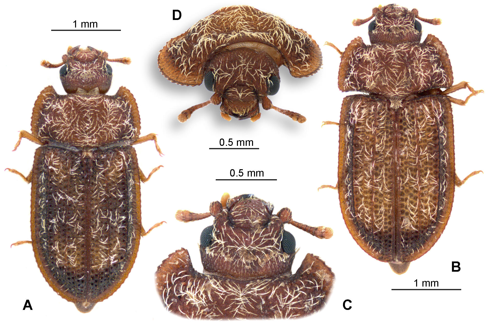
Type specimens: Holotype ( Fig. 1A View FIGURE 1 , sex unknown): “ Thailand / Khon Kaen / 10–15.ix.1978 ”, “ H.J. Bremer leg. / sub cortice arboris”. Nine paratypes ( 2 males, 1 females, 6 sex unknown): same label. The holotype and six paratypes are deposited in the collection of Hungarian Natural History Museum, Dept. of Zoology ( Budapest, Hungary); three paratypes are in the Moravian Museum , Dept. of Entomology ( Brno , Czechia).
Description: Measurements of figured paratype ( Fig. 1B View FIGURE 1 ) in millimetres: Body length (from abdominal apex to clypeus) 3.1, head dorsal length along middle 0.58, head maximum width behind eyes 0.77, head width through eyes 0.78, head width in front of eyes 0.55, head width between eyes 0.56, antenna length 0.46 (scape and pedicel 0.12, flagellum 0.19, club 0.15), pronotum maximum length 0.77, pronotum width at anterior part between corners 0.96, pronotum maximum width 1.7, elytral length along suture 2.34, elytral maximum width (at 2/3 length) 0.83, elytral width at humeral part 0.71, protarsus 0.3, mesotarsus 0.36, metatarsus 0.37 (all tarsi measurements including claws). Variability in the size among the type series specimens is minimal.
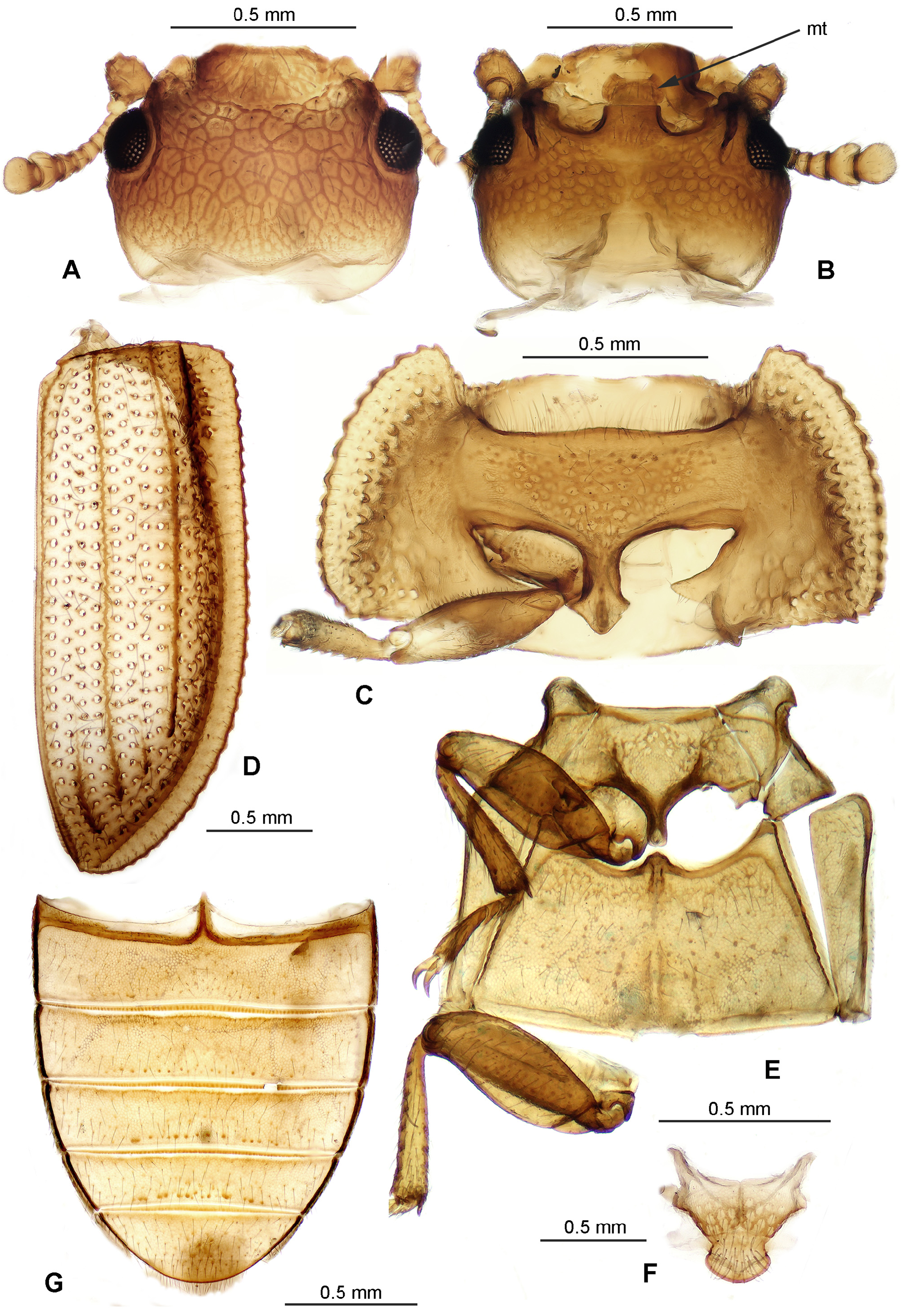
Coloration and sculpture ( Figs 1A–D View FIGURE 1 , 2A–G View FIGURE 2 ): Body broad oval, convex; all dorsal and ventral surfaces dull; head light brown, margins of pronotum and elytra black-brown with variable light brown pattern in centre; legs brown, become lighter from coxae to tarsi; antennae brown, club brown to light brown; ventral thoracic and abdominal sclerites dark brown.
Head dorsally with coarse sculpture composed of large angular impressions with thick white seta inside ( Figs 1 View FIGURE 1 CD, 2A); ventral surface with round punctures (interspaces smaller than diameter of punctures), short fine setae present in centre of each puncture. Pronotum also with large impressions and whitish decumbent or semierect setae, as same as those on dorsal surface of cranium; ventral surface with sparser punctation composed of round punctures in hypomeron and prosternum, surface around procoxae including postcoxal projections of hypomeron glabrous. Whitish pubescence of head and pronotum forms regular pattern. Elytron with regular dense punctation composed of round punctures, two rows of punctures always present between neighbour carinae. Pubescence of elytron sparser than that in head and pronotum but composed from the same whitish thick setae which form regular pattern.
Ventral surface of thorax and abdomen with fine sparse pubescence; meso- and metathorax sparsely punctate, abdominal ventrites with transverse rows of punctures along their apical margins. Legs without conspicuous sculpture, femora and tibiae sparsely pubescent. Antennae with short fine hairs.
Head ( Figs 1 View FIGURE 1 CD, 2AB, 3A–C): cranium transverse, almost orthognathous; frons flat, frontoclypeal suture distinct, deeply emarginate; eyes relatively small (space between them approximately 5–6 times as wide as eye diameter), moderately elevated (weakly exceeding contour of head), not emarginate, nearly rounded, situated laterally. Gular sutures relatively short, convergent; antennal grooves in ventral side of cranium short but conspicuous, running along eyes; antennal sockets not visible from above; epicranial acumination conspicuous; cervical sclerites welldeveloped. Ventral part of cranium without sensory setae at sides.
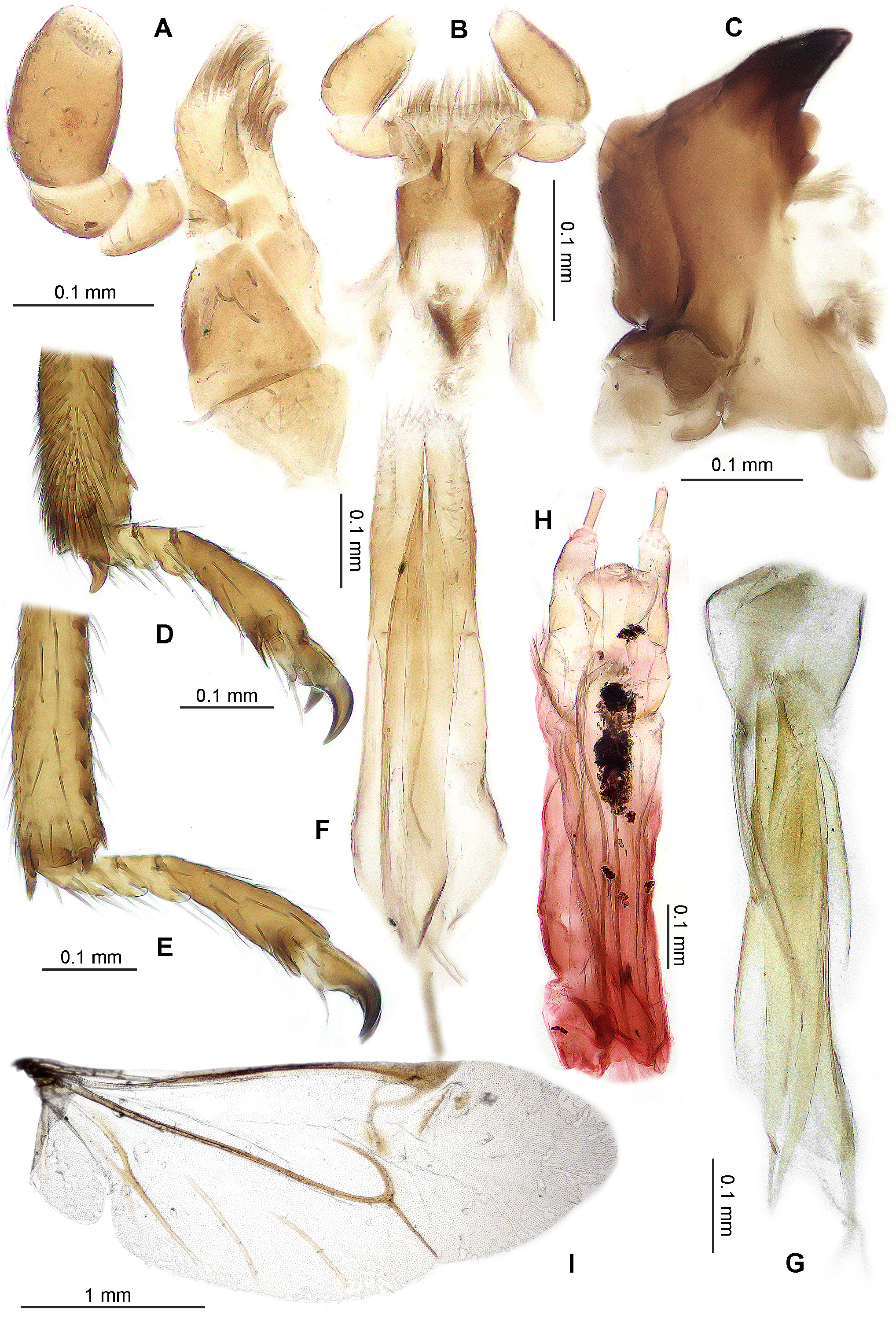
Maxilla ( Fig. 3A View FIGURE 3 ): lacinia with one or two dark spine-like setae and a number of thick setae along apical part of outer margin; galea with tuft of thick setae along apex; setae simple, non-ciliate (ciliate setae frequent in Trogossitidae ). Basigalea relatively large, feebly pigmented; galea elongate, not clavate; mediostipes relatively small, perfectly free, not fused with lacinia, nearly triangular in shape; basistipes distinct (pigmented) but probably fused with lacinia (weakly pigmented); palpifer relatively large, trapezoidal, not denticulate or crenulate along outer margin; palpifer rather narrow, triangular; maxillary palps 4-segmented, terminal joint cylindrical, robust; cardo, basistipes, mediostipes and palpifer finely and very sparsely ciliate.
Mandible ( Fig. 3C View FIGURE 3 ): bidentate, with two apical teeth situated side by side (in horizontal axis); two medial teeth distinct; mola present; basal notch weakly developed; prostheca composed of membranous appendage above mola (penicillus?) and tuft of rigid setae beneath medial teeth.
Labrum: labral sclerite oblong, its apical part nearly straight; dorsal surface sparse pubescent; tormal processes not observed.
Labium ( Figs 2B View FIGURE 2 , 3B View FIGURE 3 ): mentum deeply emarginate along anterior margin, strongly transverse, anterior corners extended, lateral margins obliquely sloping (general shape trapezoidal); prementum divided into two parts, with two spine-like processes; ligula weakly pigmented, shallowly emarginate, with numerous setae; “mental apodeme” (sclerites beneath mentum or between mentum and prementum) composed of two separated elongate sclerites; hypopharyngeal sclerite(s) (frequent cleroid structure on hypopharyngeal membrane between mentum and submentum) absent; hypopharynx with longitudinal strip of sensillae; labial palps 3-segmented; terminal joint of palps conical, as long as palpomeres 1+2 together.
Antenna ( Fig. 2A View FIGURE 2 ): 10-segmented; antennal club 2-segmented, compact but relatively small; scape robust, pedicel smaller than scape but wider than antennomere 3; 2–8 symmetrical, approximately the same in length; 9–10 (club) weakly asymmetrical, with conspicuous sensorial fields at apical portions; all antennomeres with sparse pubescence; antennae relatively short, extending backwards to base of cranium.
Prothorax ( Figs 1 View FIGURE 1 AB, 2C): pronotum transverse, width/length ratio about 2.2; anterior margin distinctly sinuate, anterior corners projecting, acuminate; lateral margins rounded, crenulate; lateral edge (carina) distinct, sharp; prosternal process distinctly dilated in front of apex; procoxal cavities transverse, narrowly separated, internally open, externally open to half (partially closed by postcoxal projections); protrochantin inconspicuous, fused with prosternum.
Mesothorax ( Figs 2 View FIGURE 2 EF): mesoventrite wide; prepectus present but narrow; mesocoxal cavities relatively large, rounded, narrowly separated or contiguous (meso- and metaventral processes conspicuously distanced), externally narrowly open; mesoventral process shorter than coxal diameter; projection of metaventrite short, not reaching apex of mesoventral process; mesotrochantin rectangular, conspicuous; scutum relatively wide, scutellum oval.
Wing ( Fig. 3I View FIGURE 3 ): well-developed and perfectly folded beneath elytra; lower margin of wing without conspicuous pubescence; radial cell complete; r4 present; pigmented fleck (below Rc) reduced in size but small pigmented spot along r4 present; medial field with all four veins: MP4 and MP3 not connected with other veins, veins AA3+4 and AA1+2 present.
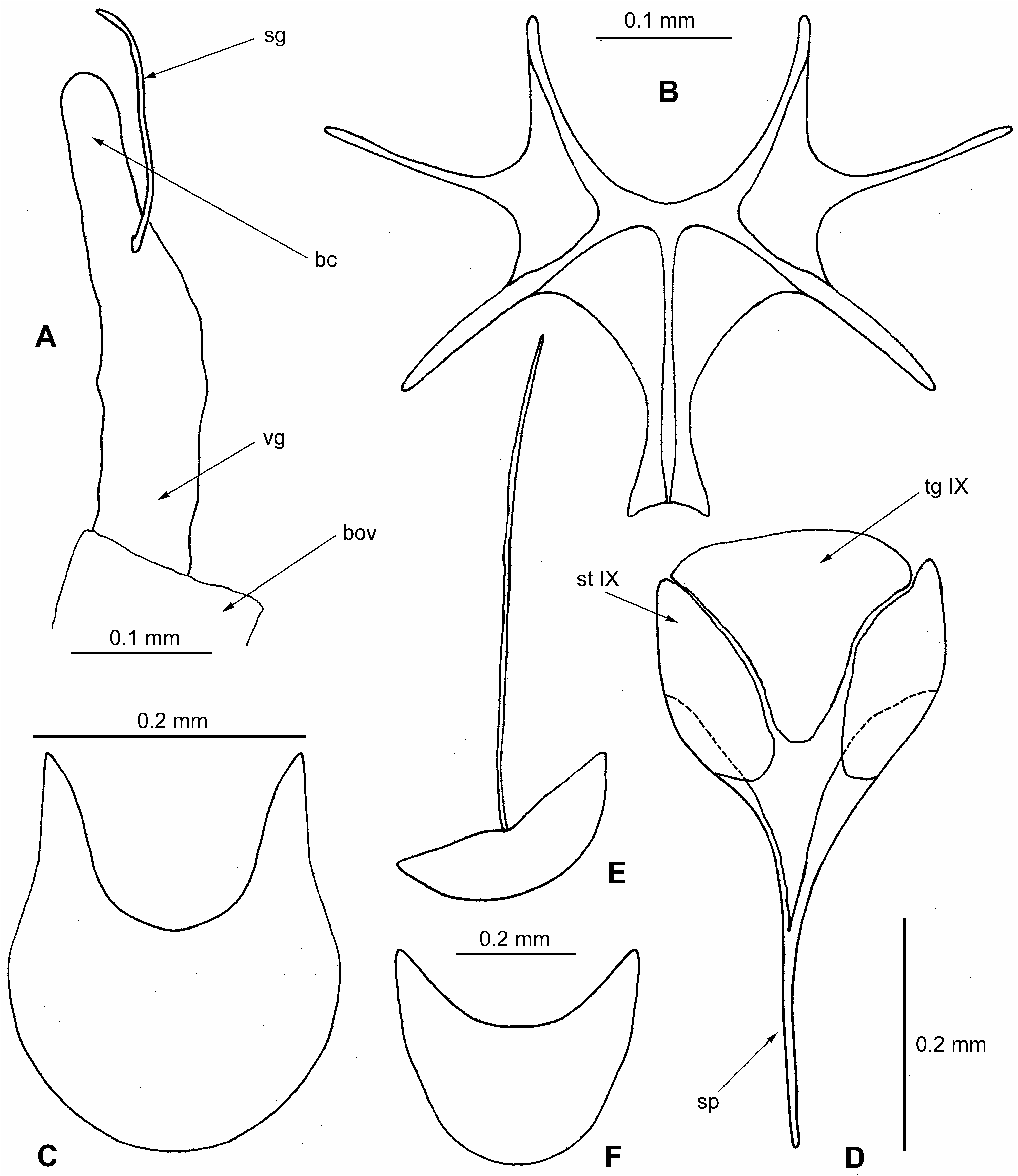
Metathorax ( Figs 2E View FIGURE 2 , 4B View FIGURE 4 ): metaventrite flat, transverse, slightly narrowed towards anterior portion; discriminal line (discrimen) runs to half of length of metaventrite; paracoxal sutures indistinct, closely separated from coxae; metepisternum narrowly triangular; metendosternite very wide, as in Fig. 4B View FIGURE 4 .
Elytron ( Figs 1 View FIGURE 1 AB, 2D): convex, in both sexes covering whole abdomen excepting pygidium; epipleuron wide, conspicuous along whole elytral length; interlocking mechanism absent; each elytron with five longitudinal carinae but carinae 1 and 3 distinct in apical quarter only, while carina 5 distinct only in basal third of elytron (counted from suture); lateral margins of elytra distinctly explanate.
Legs ( Figs 2 View FIGURE 2 CE; 3DE): procoxa very weakly projecting, transverse; mesocoxa oval, nearly spherical; metacoxa with longitudinal groove, extended to lateral margin of metathorax; trochanters relatively small, triangular; femora moderately clavate; protibia with six robust spines along outer margin; meso- and metatibia with about eight spines along outer margin; tibial apical spur pattern 2-2-2; one large protibial apical spur distinctly hooked; apices of all tibiae without (comb-like) row of spines; tarsomere 1 in all pairs of legs reduced in size and apparently conjoined with tarsomere 2 so that tarsal formula 4-4-4; tarsomere 4 longer than 1–3 combined; tarsal lobes absent but apices of tarsomeres 1–3 with long setae; claws without denticles; empodium large, projecting, bisetose.
Abdomen ( Figs 3F–H View FIGURE 3 ; 4A, C–F View FIGURE 4 ): all tergites but pygidium covered by elytra; five abdominal ventrites distinctly visible but sternite VIII may also be more or less partially visible; female sternite VIII weakly emarginate at apical margin, with setae along apical margin and in centre; female tergite VIII with long spiculum, base of sternite VIII deeply emarginate; ovipositor medium sized, membranous; coxital styli slender and relatively long. Bursa copulatrix well-developed; spermatheca inconspicuous but gland present. Male segment IX complete but pale and membranous, with divided sternite IX and its well-developed spiculum (“spicular fork”); tergite IX also conspicuous; aedeagus inverted (ventrally open); tegmen composed of three parts; parameres finely ciliate, distinctly separated each other and separated from phallobase; ventral piece of phallobase with pair of short lateral struts mutually conjoined with membrane; basal piece of phallobase with distinct median strut (apodeme); penis with pair of struts at base.
Biology: Collected under bark of an unidentified tree. The gut of three dissected specimens ( 2 males, 1 female) contains detritus.
Etymology: Named in honour of the late Ottó Merkl (Hungarian National Museum, Budapest) whose generosity made me study of an extensive cleroid material possible.
No known copyright restrictions apply. See Agosti, D., Egloff, W., 2009. Taxonomic information exchange and copyright: the Plazi approach. BMC Research Notes 2009, 2:53 for further explanation.
|
Kingdom |
|
|
Phylum |
|
|
Class |
|
|
Order |
|
|
SuperFamily |
Cleroidea |
|
Family |
|
|
Genus |