
Raymunida shraddhanandi, Tiwari & Padate & Cubelio & Osawa, 2022
|
publication ID |
https://doi.org/ 10.1080/00222933.2022.2138600 |
|
DOI |
https://doi.org/10.5281/zenodo.7383872 |
|
persistent identifier |
https://treatment.plazi.org/id/D23C87A0-FFBF-2E0F-FEF8-253AFCD2BB40 |
|
treatment provided by |
Plazi |
|
scientific name |
Raymunida shraddhanandi |
| status |
sp. nov. |
Raymunida shraddhanandi View in CoL sp. nov.
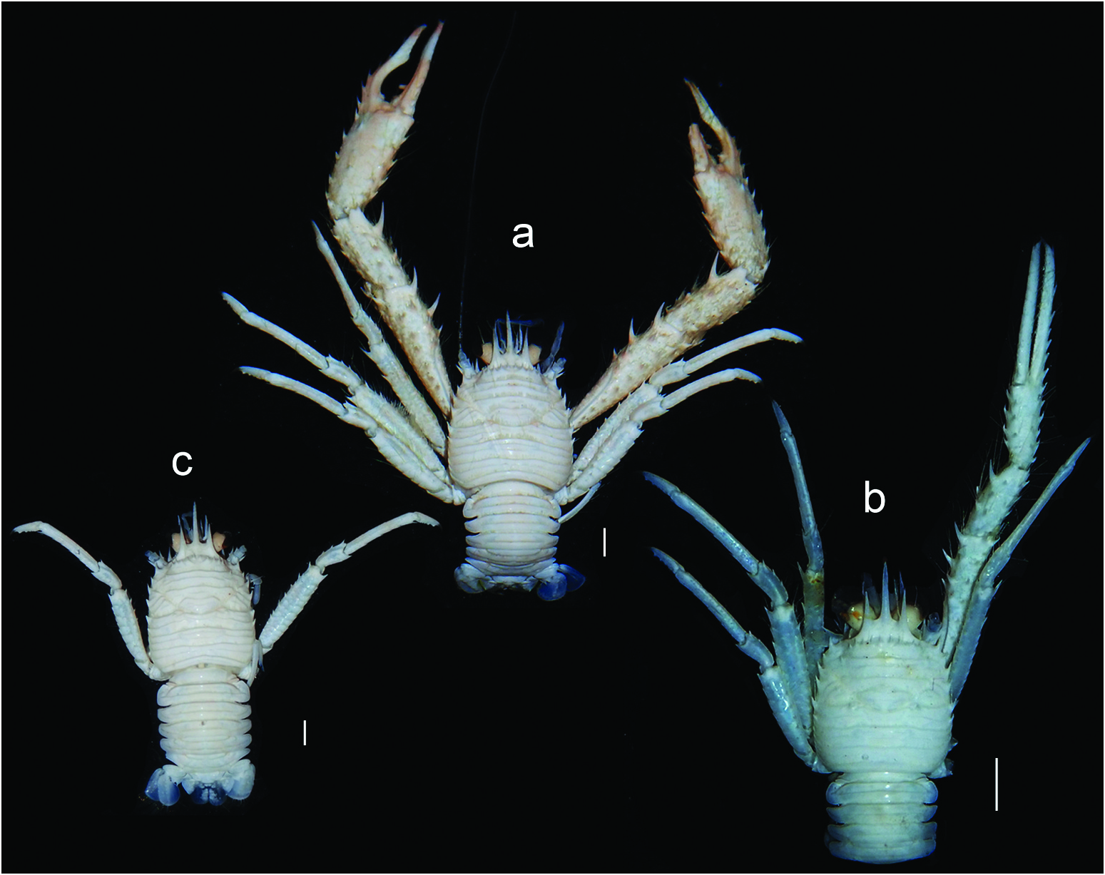
( Figures 2 View Figure 2 (a–), 3(a–3), Figure 4 View Figure 4 and Figure 5 View Figure 5 )
urn:lsid:zoobank.org:act:4A3E0887-2772-4221-9734-ADB4C4D9328D
Type material
Holotype. India • ♂ (PCL 9.7 mm, CW 8.2 mm); Andaman Sea , off Little Andaman Island; FORVSS stn. 38806; 10.72°N, 92.7°E; 53 m depth; Chain dredge; 10 August 2019; Vinay P. Padate leg.; CMLRE IO /SS/ANO/00141. GoogleMaps
Paratype. India • 1 ♀ (PCL 5.8 mm, CW 4.9 mm); same data as for holotype; CMLRE IO /SS/ ANO/00140 GoogleMaps .
Type locality
India: Andaman Sea , off Little Andaman Island; 10.72°N, 92.7°E; 53 m depth GoogleMaps .
Etymology
The species is named in honour of the Swami Shraddhanand College, University of Delhi, the alma mater of the first author.
Diagnosis
Carapace (exclusive of rostrum) 1.2 times as long as wide; dorsal surface with sparse long iridescent setae, 5 pairs of epigastric spines, 0–1 anterior branchial spine, 1 parahepatic spine and 1 post-cervical spine on either side; frontal margin with short spine mesial to anterolateral spine; branchial margin with 4 spines (2 on each of anterior and posterior margins). Rostrum 0.4 times PCL, 2 times longer than supraocular spines. Anterior margin of thoracic sternite 4 as wide as sternite 3, sternites 5–7 smooth or with few short oblique striae on lateral portions. Pleonal tergites 2 and 3 each with or without row of interrupted, short striae between 2 transverse ridges. Antennular peduncle article 1 (excluding distal spines) not reaching distal corneal margin. Antennal peduncle article 1 with distomesial spine slightly overreaching distal margin of article 3, not reaching distal margin of antennular peduncle article 1; article 2 with distomesial spine reaching distal margin of article 3, unarmed on mesial margin; article 3 with short distomesial spine. Mxp3 merus with dorsodistal spine, ventral margin with 2 slender spines;carpus with 1 or 2 distal spines on ventral margin. P1 3.1–3.6 times PCL,with numerous long, simple setae; chela 4.6 (paratype female) to 7.5 (holotype male) times as long as wide, with dorsolateral row of sparsely arranged spines; fingers each with distinct rounded crest along dorsal midline. P2–4 meri squamate on lateral surface, P4 mero-carpal articulation reaching level of lateral end of anterior cervical groove of carapace.
Description of holotype
Carapace ( Figures 2 View Figure 2 (a), 2(a), 2(a, 2)) (exclusive of rostrum) 1.2 times as long as wide, bearing a few long iridescent setae dorsally; transverse striae with dense short setae; gastric region with 6 transverse striae and 5 pairs of spines (epigastric spines); cardiac region with 2 complete transverse striae followed by 2 median striae and 1 pair of lateral striae; intestinal region with 3 transverse striae, first and third complete, second interrupted medially. One parahepatic and 1 postcervical spine present on each side; anterior branchial spines absent. Frontal margin moderately oblique, with 1 small spine between supraocular spine and anterolateral spine. Lateral margin gently convex. Anterolateral spine distinct, but falling far short of level of sinus between rostrum and supraocular spine, followed by small spine anterior to cervical groove. Anterior and posterior branchial margins each with 2 spines, posteriormost spine smallest. Rostrum spiniform, 0.4 times PCL, 2 times longer than supraocular spines, directed slightly downward. Supraocular spines horizontal, subparallel, overreaching distal margin of cornea.
Thoracic sternite 3 ( Figure 4 View Figure 4 (c)) 3.3 times as wide as long, with 2 flattened lobes separated by shallow V-shaped notch on anterior margin, anterior margins of lobes faintly granulate, lateral angle with distinct acute projection. Sternite 4 with anterior margin as wide as sternite 3, with 2 transverse striae, anterior stria medially interrupted, bearing sparse setae, posterior stria uninterrupted. Sternites 5 and 6 with short oblique stria on each lateral portion. Sternite 7 with few short striae laterally.
Pleonal tergites ( Figure 4 View Figure 4 (d)) with few moderately long iridescent setae and row of short plumose setae on transverse ridges; tergites 2 and 3 each with row of short, interrupted striae between 2 transverse ridges, tergite 4 without striae between 2 transverse ridges; tergites 5 and 6 with 2 medially interrupted ridges, those on tergite 5 longer and nearly straight, those on tergite 6 somewhat squamiform.
Eye ( Figure 4 View Figure 4 (a)) moderately large, corneal diameter 0.3 times distance between mesial bases of anterolateral spines, eye lashes simple and relatively short, long stout seta on rounded dorsodistal margin of peduncle.
Antennular peduncle article 1 ( Figure 4 View Figure 4 (f)) (distal spines excluded) only reaching proximal margin of cornea; distomesial spine distinctly shorter than distolateral spine; 2 lateral spines, anterior spine distinctly overreaching tip of distolateral spine, posterior spine much shorter than anterior spine.
Antennal peduncle article 1 ( Figure 4 View Figure 4 (f)) with long distomesial spine slightly overreaching distal margin of article 3, not reaching distal margin of antennular article 1; article 2 with distomesial spine reaching distal margin of article 3, distolateral spine nearly reaching distal margin of article 3, mesial margin unarmed; article 3 with short distomesial spine; article 4 unarmed.
Mxp3 ( Figure 4 View Figure 4 (g)) ischium subequal in length to merus measured along dorsal margin, with strong spine each at dorsodistal and ventrodistal angles; crista dentata consisting of 33 denticles. Merus with small dorsodistal spine; ventral margin with 3 spines, second spine much smaller than other 2 slender main prominent spines. Carpus with 2 spines distally on ventral margin. Epipod present.
P1 ( Figure 4 View Figure 4 (h,)) subequal from right to left, massive, 3.6 times PCL, moderately depressed, with numerous long, iridescent setae; surfaces of merus, carpus and palm of chela squamate. Merus 1.1 times PCL; dorsal and ventral surfaces each with 2 irregular rows of spines, distomesial spine strongest; mesial surface with 2 spines distal to mid length, distal spine much larger than proximal spine; lateral surface with 1 subdistal spine, ventrolateral margin with 1 small distal spine. Carpus with 2 irregular rows of spines on dorsal surface and with 1 spine each on ventral surface, mesial margin and lateral margin; mesial margin also with elongated spine on distal one-third. Chela 4.6 (left) or 4.8 (right) times as wide as long. Palm 1.3 times longer than carpus, 2.1 (right) or 2.3 (left) times as long as wide; dorsal surface with 2 irregular rows of spines and small spine at dactylar articulation; ventral surface with 1 mesial spine at mid length; mesial surface with longitudinal row of 4 spines; lateral margin with row of sparsely arranged spines extending to entire length of fixed finger. Fingers distally broken, but distinctly longer than palm at least, each with rounded longitudinal crest on dorsal surface; fixed finger curved on proximal half; dactylus with 3 spines on proximal one-third portion, 1 median spine and small, unevenly spaced spinules on distal portion of mesial margin; occlusal margins minutely dentate, dactylus with blunt teeth on proximal one-third fitting into large concavity on proximal half of fixed finger.
P2–4 ( Figure 4 View Figure 4 (j–)) somewhat compressed laterally, 2.1 (P2, P3) and 1.7 (P4) times PCL; P4 mero-carpal articulation reaching only level of first branchial spine (lateral end of anterior cervical groove of carapace) ( Figure 2 View Figure 2 (a)); meri 6.3 (P2), 4.5 (P3) and 3.5 (P4) times as long as wide, 0.9 (P2), 0.8 (P3) and 0.6 (P4) times PCL, 3.0 (P2), 2.6 (P3) and 2.2 (P4) times longer than carpi; propodi 7.5 (P2), 6.7 (P3) and 5.7 (P4) times as long as wide, 2.1 (P2), 2.0 (P3) and 1.9 (P4) times longer than carpi, 2.3 (P2), 2.2 (P3) and 2.0 (P4) times longer than dactyli. Meri covered with distinct squamiform ridges on lateral and mesial surfaces; dorsal margins each with row of 10 (P2), 6 (P3) and 3 (P4) spines randomly arranged and increasing in size distally; lateral surfaces each with dorsal row of 3 (P2), 9 (P3) and 8 (P4) spines; ventral margins with 4 (P2), 3 (P3) and 2 (P4) spines on distal one-third. Carpi with 5 (P2), 4 (P3) and 1 (P4) dorsal spines; ventral margins each with 1 distal spine. Propodi dorsally unarmed; ventral margins with 7 (P2), 4 (P3) and 4 (P4) slender corneous spines. Dactyli with 6 (P2), 5 (P3) and 4 (P4) slender corneous spines on ventral margins. Epipods present on P1–3.
Male with G1 ( Figure 4 View Figure 4 (n)) and G2 ( Figure 4 View Figure 4 (o)) as illustrated.
Uropodal protopod ( Figure 4 View Figure 4 (e)) with 1 distal spine and proximal produced portion on lateral margin; endopod broader than exopod, lateral and distal margins each with row of spines (lateral spines damaged); exopod also spinose on lateral and distal margins.
Variations
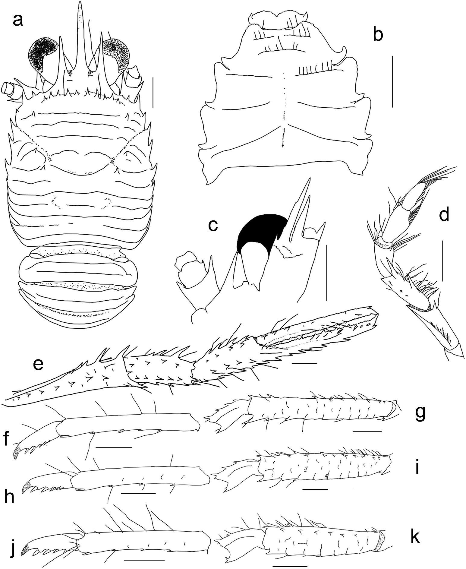
The female paratype differs from the male holotype in the following points, although the difference in the P1 represents sexual dimorphism. Carapace ( Figure 5 View Figure 5 (a)) bearing 1 anterior branchial spine on dorsal surface. Thoracic sternites 5–7 ( Figure 5 View Figure 5 (b)) smooth. Pleonal tergites 2 and 3 ( Figure 5 View Figure 5 (a)) without striae. Antennal peduncle article 2 with distomesial spine feebly overreaching distal margin of article 3 ( Figure 5 View Figure 5 (c)). Mxp3 ( Figure 5 View Figure 5 (d)) merus with only 2 main spines on ventral margin, carpus with 1 distal spine on ventral margin. P1 slender, 3.1 times PCL, chela 7.5 times as long as wide; palm subequal in length to carpus, ventral surface with 2 irregular longitudinal rows of spines, lateral margin with row of spines extending to distal one-fourth of fixed finger; fingers about 1.9 times as long as palm, parallel to each other, occlusal margins minutely dentate, each with slightly larger acute teeth at regular intervals. P2–4 ( Figure 5 View Figure 5 (f–i)) 1.9 (P2), 2.2 (P3) and 1.7 (P4) times PCL; meri with 4 (P2), 4 (P3) and 2 (P4) spines on dorsal margins, lateral surfaces with dorsal row of 6 (P2) and 6 (P4) spines, P4 ventral margin with only 1 distal spine; carpi with 4 (P2), 3 (P3) and 2 (P4) spines on dorsal margins; P2 propodus and dactylus each with 5 ventral spines.
Remarks
Among the known congeners, R. shraddhanandi sp. nov. is morphologically closest to R. formasanus (known from Taiwan and the south-eastern Australia, 104–300 m depths) in having the carapace with some long setae on the dorsal surface, the frontal carapace margin with a small spine mesial to the anterolateral spine, the antennal peduncle article 2 without a small spine on the mesial margin, the P1 bearing numerous long simple setae, the P1 fixed finger with a row of relatively sparse spines on the dorsolateral margin, and the P1 dactylus with a distinct longitudinal crest along the dorsal midline.
However, the new species differs from R. formasanus ) in the following characters (see Ahyong and Poore 2004; Lin et al. 2004).
(1) The anterior branchial carapace region is unarmed or has only one spine, instead of three to five spines as in R. formasanus .
(2) The distomesial spine of the antennal peduncle article 2 reaches only or feebly overreaches the distal margin of the article 3, while it clearly overreaches that margin in R. formasanus based on the re-examination of the holotype.
(3) The antennal peduncle article 3 has a short distomesial spine, which is absent in R. formasanus .
(4) The Mxp3 merus has a small but distinct dorsodistal spine, which is absent in R. formasanus .
(5) The P4 mero-carpal articulation reaches only the lateral end of the anterior cervical groove of the carapace, whereas it overreaches the frontal margin of the carapace in R. formasanus .
Munida alcocki Southwell,1906 was described from the Gulf of Mannar (Dutch Modragam Paar and Aripu Reef,off western coast of Sri Lanka),in shallow water to a depth of 66 m. The identity of the taxon is not clear, although it has been questionably included in the synonymy of Raymunida elegantissima (see Baba et al. 2008). Southwell (1906) described Munida alcocki as having seven spines on the carapace lateral margin; the number agrees only with that of R. iranica ;in the other known Raymunida species,only five or six spines are present ( Osawa and Safaie 2014). It is unlikely that Munida alcocki is conspecific with the present new taxon.
Geographical distribution
So far known only from the Andaman Sea, 53 m depth.
| IO |
Instituto de Oceanografia da Universidade de Lisboa |
No known copyright restrictions apply. See Agosti, D., Egloff, W., 2009. Taxonomic information exchange and copyright: the Plazi approach. BMC Research Notes 2009, 2:53 for further explanation.
|
Kingdom |
|
|
Phylum |
|
|
Class |
|
|
Order |
|
|
Family |
|
|
Genus |