
Pachylus crassus ( Roewer, 1943 )
|
publication ID |
https://doi.org/ 10.11646/zootaxa.4984.1.13 |
|
publication LSID |
lsid:zoobank.org:pub:BA9DD175-7CFA-4D80-9250-16EE6C8CE0C4 |
|
DOI |
https://doi.org/10.5281/zenodo.5191757 |
|
persistent identifier |
https://treatment.plazi.org/id/D25087B9-FFEF-EA4C-09D6-F8DED82D3BE7 |
|
treatment provided by |
Plazi |
|
scientific name |
Pachylus crassus ( Roewer, 1943 ) |
| status |
|
Pachylus crassus ( Roewer, 1943) View in CoL
Figs 1–4A, C, E View FIGURES 1 View FIGURES 2 View FIGURES 3 View FIGURES 4 , 5 View FIGURE 5
urn:lsid:zoobank.org:act:6B8D2AA6-EFB4-4DC7-AD8F-4C06C5C993AE
Pachyloidellus crassus Roewer 1943: 17 View in CoL , pl. 1, figs 5, 5a–b; Ringuelet 1959: 281 (generic placement uncertain).
Progyndes crassus: Soares & Soares 1954: 292 (synonymized Pachyloidellus View in CoL with Progyndes View in CoL ); Cekalovic 1968: 9; 1985: 23; Soares & Bauab Vianna 1972: 213 (provisional combination; generic placement uncertain).
Acanthopachylus crassus: Capocasale & Bruno Trezza 1964: 20 .
Pachylus crassus: Acosta 1993: 3 View in CoL ; 1996a: 220 (implied combination); 2020: 131; Kury 2003: 184.
Type series. SMF RII 1382 /75, 3 ♂, 2 ♀ syntypes (poorly preserved and very brittle, several legs detached and lying at the bottom of the vial), examined. The largest male is hereby designated as the lectotype (left leg IV detached, presumably by Roewer [1943] to draw fig. 5b); the remaining specimens become paralectotypes.
Type locality. “ Chile: Santiago ”, probably inaccurate.
Other material examined. CHILE: Maule Region: Curicó Province, Los Queñes , alt. 700 m, 17.10.1992 (leg. N. Platnick, P. Goloboff, K. Catley), 1 ♂, 1 ♀ ( AMNH); [Estero] Las Tablas, 27/ 29.9.1983 (leg. L. Peña), 2 ♂, 5 ♀ (MACN-Ar 28799) , 1 ♂, 1 ♀ ( CDA 000.910); Talca Province, [Alto de] Tonlemo , 14.– 21.12.1984 (leg. L. Irarrazával), 2 ♂ ( AMNH).
Redescription: Measurements: DS length in males 7.7–8.7 (mean 8.3, n = 9), females 7.5–8.6 (mean 8.0, n = 9). Femur length of males in % of DS length 77–92. Measurements of lectotype ♂ and one female: Table 1 View TABLE 1 . Tarsal formula 5:7:6:6.
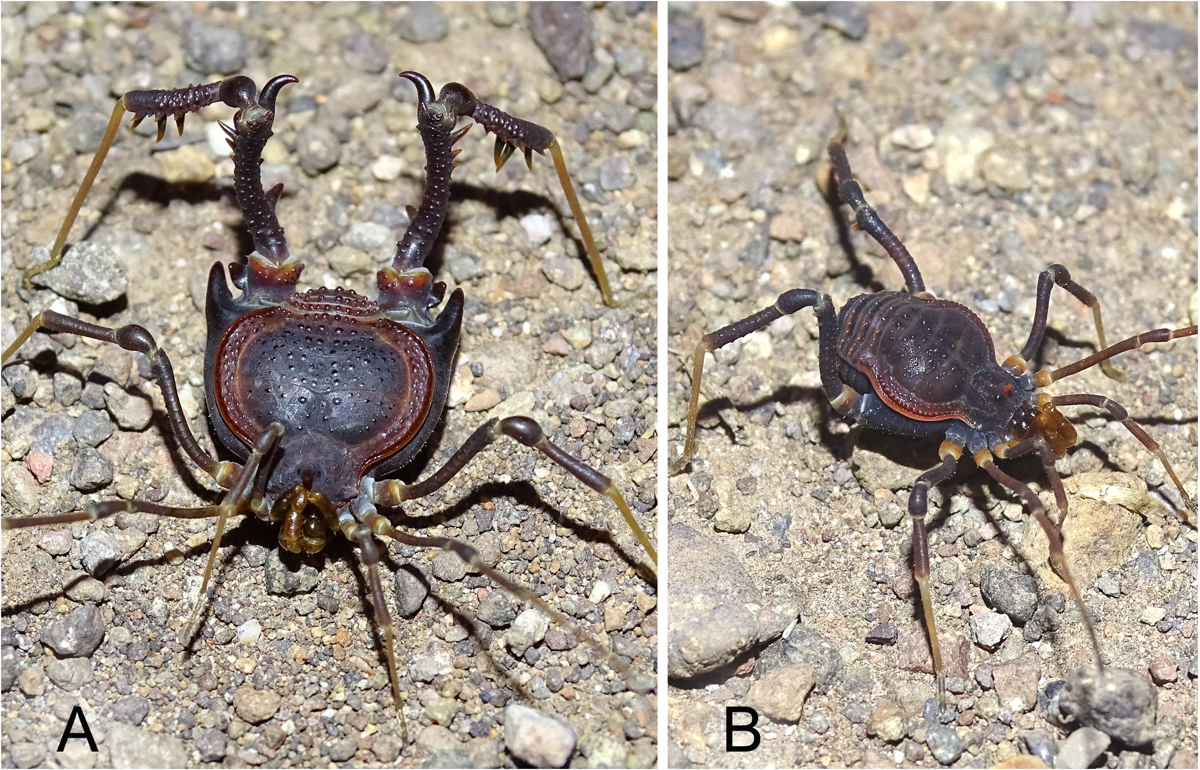
Color in ethanol 70%: General color dark reddish brown, with lateral and posterior areas and free tergites lighter, often orangish or yellowish; granules of DS normally standing out darker. Ventral and lateral sides of coxae IV reticulate; cheliceral bulla, pedipalps and legs I–III lighter, with fine pigment reticulation; metatarsi and tarsi of all legs yellowish. Coxa to tibia IV of males with the general color. Some preserved specimens overall light yellowish hazel, with lateral areas even lighter. These color contrasts are more vivid in living animals ( Fig. 1 View FIGURES 1 ).
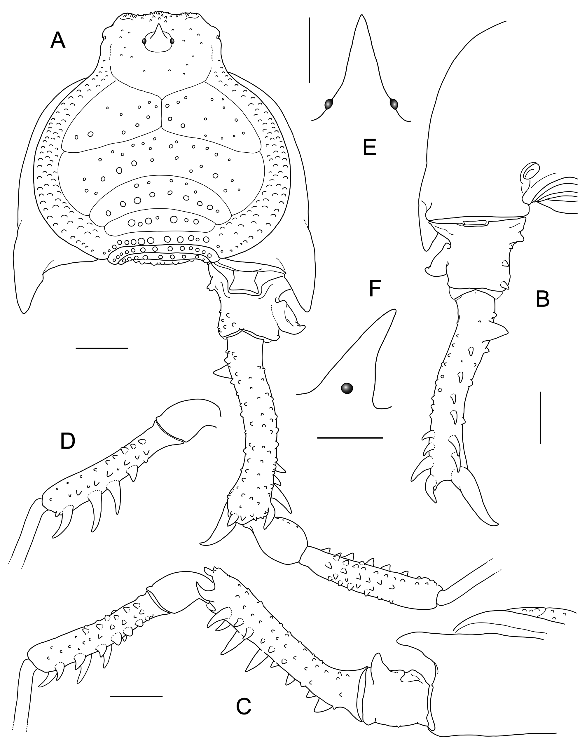
Male. Dorsum: DS of type γR (gamma rotund), its posterior margin concave (in a few specimens in the form of an open square bracket: Fig. 2A View FIGURES 2 ). Granulation in general conspicuous, with granules in paramedian position larger than others on each scutal area, also increasing in size from area I to V, often looking like small rounded tubercles. Prosomal part of DS: anterior margin bordered by a row of conical grains, these also sparsely covering low frontal hump, rest of carapace region covered by scattered granules. Ocular mound situated in anterior half of carapace region, without granules, bearing a large spine-like apophysis inclined frontwards ( Figs 2E–F View FIGURES 2 ). DS raised in lateral areas and somewhat depressed toward the posterior margin. Sulci delimiting scutal areas complete and well defined; area I divided; areas I–IV entire. Areas I–III with irregular granulation, roughly forming two rows, the posterior one more regular and/or with larger grains. Areas IV–V and free tergites each with a regular row of pearl-like grains or small tubercles, the two paramedian ones rarely slightly larger or distinct from the rest (most commonly so on free tergite III). Dorsal anal plate with an anterior row of similar pearl-like granules, plus isolated or aligned ones posteriorly. Lateral areas with two rows of flat granules: outer row of rounded granules reaching primary constriction of scutum; inner row of scattered, less well-defined granules (giving the sector a rugulose appearance) continuous with a well-marked ridge on each side of prosoma, almost reaching ozopores.
Venter: Posterior margin of stigmatic segment bordered by a gentle tegumentary thickening delimiting a narrow and deep concavity, in most specimens trapezoid-shaped (in lectotype in the shape of a catenary arch), with tracheal stigmata on both sides ( Fig. 4A View FIGURES 4 ).
Chelicerae: Developed as usual in the genus; bulla with 1–2 posterolateral granules.
Pedipalps: Weak, from trochanter to patella smooth; minute ventral setigerous tubercles on trochanter and femur (one basal and one in the middle); true medial subapical spine on femur lacking, instead a vestigial setigerous tubercle present close to patellar joint. Pedipalp spination: tibia i[Ii] (lateral), Iiii (medial); tarsus IiIi(i) (lateral), IIi (medial).
Legs I–III: Unarmed, sparsely granulous on all segments; proventral edge of distal half of femur III with a row of conical grains of increasing size.
Leg IV: Coxa IV with smooth ventral and lateral sides; prolateral apophysis stout and conical, almost horizontal and directed backwards, bearing a faint retroventral tuberosity (resembling the vestige of a presumed bifid condition; Fig. 4C View FIGURES 4 ); retrolateral apophysis represented by a minute process, best viewed from ventral, often hidden between tegumentary borders; coxa-trochanter joint transverse in ventral view, articulating in longitudinal axis of body.
Trochanter IV ( Figs 2A–B View FIGURES 2 , 4C View FIGURES 4 ) sub-trapezoidal, armed with several distinctive apophyses: a large, ear-like prolateral one; a proximal truncate apophysis and two retroventral conical ones (on distal end and in the middle) on retrolateral side. Retrodorsal distal sector covered with a cluster of grains.
Femur IV ( Figs 2A–C View FIGURES 2 ) slightly curved in lateral view, with very short pre- and long post-flexure sections, forming an angle of about 150°, in dorsal view looking gently arched; dorsal surface with coarse granulation forming irregular longitudinal rows; strong armature visible in ventral view; a large sub-basal retroventral, blunt to acute apophysis directed downwards and medially; a very conspicuous, hook-like and sigmoid apical-retroventral apophysis first directed backwards, then curved medially (these curvatures attenuated in lectotype and paralectotypes); a row of less strongly developed (though still large) conical apophyses on retroventral edge, the subapical ones larger than the rest; distally on lateroventral edge one distal hook-like curved apophysis, followed by a large acute subdistal apophysis pointing sideways, and a row of smaller apophyses of decreasing size turning into tubercles towards base of femur.
Patella IV pyriform, unarmed and conspicuously smooth ( Figs 1 View FIGURES 1 , 4E View FIGURES 4 ).
Tibia IV club-shaped, basal two-thirds granulous on dorsal side, the rest smooth; ventral side armed with pro- and retroventral rows of strong apophyses, the three distal ones on retroventral side remarkably large ( Figs 1A View FIGURES 1 , 2D View FIGURES 2 ).
Male genitalia ( Fig. 3 View FIGURES 3 ): VP sub-trapezoidal, longer than wide, narrower at base; distal margin slightly concave; in lateral view VP gently inclined dorsad with respect to truncus axis, with a distinct ventral hump indicating truncus-VP transition ( Fig. 3B View FIGURES 3 ). Four strong macrosetae on each side in anterior half of VP, the three distal ones corresponding to C1–C2 (transverse) and C3 (diagonal); basal to them a strong diagonal macroseta, which might be interpreted as a fourth C (R. Pinto-da-Rocha, in litt.) or, following Kury & Villarreal M. (2015: 28), as a displaced A macroseta; basal group of two transverse A macrosetae situated close to each other (B likely absent); in hiatus between distal and basal groups of macrosetae a short D1; minute E1–E3 ventrally to row of four distal macrosetae. Truncus noticeably extended distad beyond base of VP, with truncus-glans articulation displaced to mid-level of VP (at level of D1); widened subapical portion of truncus hiding base of VP in dorsal view ( Fig. 3A View FIGURES 3 ). Glans short, globose, smoothly continuing into a thick stem, then bent dorsad into a cylindrical, slightly sigmoid unarmed stylus; at point of flexure of stylus a sessile, spiny VPS directed opposite to stylus ( Fig. 3B View FIGURES 3 ).
Female. Different from male as follows ( Fig. 1B View FIGURES 1 ): DS type αK (alpha keyhole), not depressed posteriorly; posterior margin straight to gently convex. Granules on area V pearl-like, gradually becoming higher and more conical towards dorsal anal plate; paramedian conical grains distinct from rest of same row on free tergite III, seldom also on other segments. Posterior margin of stigmatic segment gently arched, concave in ventral view.
Legs I–III unarmed; proventral row of grains in distal half of femur III present but less conspicuous than in males. Leg IV: Coxa with a small conical prolateral apophysis, lateral sides granulous; a minute retrolateral apophysis also present but very often difficult to see. Trochanter articulated diagonally with coxa IV; with an acute small apophysis on the retroapical margin, two retrolateral conical grains and scattered grains on rest of article. Femur curved, granulous throughout; small acute apophyses standing out distally, with increasing size on lateroventral margin; largest apophysis in retrodistal position, first directed backwards, then curved medially. Patella unarmed as in males but with retrolateral side somewhat rugulose instead of smooth. Tibia covered by conical grains all around, retro- and proventral rows larger than others.
Comparisons. Males of P. crassus are easily distinguishable from all congeners. First, in this species femur IV is the least curved: in lateral view pre- and post-flexure sections form an angle of ~150° ( Fig. 2C View FIGURES 2 ), whereas in other congeners it is 120° to 90° (see Roewer 1913: figs 12–13; Muñoz 1969, 1970). In addition, in P. crassus all granules are mostly equal-sized, the paramedian ones on free tergite III only subtly larger than the rest, whereas in other Pachylus species the paramedian granules/small tubercles on area V and free tergites are, as a rule, larger than the rest on the same row (very often on other mesotergal areas too). Moreover, the ear-shaped lateral apophysis on trochanter IV is unique to this species; in other congeners this apophysis is either triangular or truncate. While the sigmoid retroapical apophysis of femur IV is characteristic for the whole genus, in P. crassus it is remarkably large and conspicuous, giving the apical end of femur IV a hooked appearance ( Fig. 1A View FIGURES 1 ). Finally, the trapezoid-shaped concavity of the stigmatic segment ( Fig. 4A View FIGURES 4 ) is seemingly unique to P. crassus , even if a few specimens may show some variability (e.g., in the lectotype it is more like a catenary arch).
Species of Pachylus can be roughly sorted into two informal species-groups: (1) the chilensis -group, comprising P. chilensis , P. paessleri and P. vachoni , with femur IV bent between 90° and 110°, and prolateral apophysis of coxa IV bifid, its dorsal branch often bent medially; and (2) the crassus -group, which includes P. crassus and P. quinamavidensis , with femur IV bent between 120° and 150°, and prolateral apophysis of coxa IV shorter and not clearly bifid. The geographical ranges of these tentative groups also seem to be distinct ( Fig. 5 View FIGURE 5 ): the chilensis -group occupies the northern part of the generic range (records from 31.9°S to 33.8°S in the Metropolitan, Valparaíso and Coquimbo Regions); the crassus -group occurs in the southern part (34.8°S to 36.9°S, in the O’Higgins, Maule and Bio-Bio Regions). At a quick glance males of P. crassus and P. quinamavidensis can be confused because of their similar appearance (with apophyses on coxa IV short and stout), however, they show many clear differences, as given in Table 2 View TABLE 2 .
Distribution. Central Chile: Maule and O’Higgins Regions ( Fig. 5 View FIGURE 5 ). Includes three additional records from O’Higgins Region (J. Pérez-Schultheiss, in litt.): Colchagua Province: Quebrada el Sauce, Chimbarongo; Apalta, Millahue. Cardenal Caro Province: Tanumé, Pichilemu.


No known copyright restrictions apply. See Agosti, D., Egloff, W., 2009. Taxonomic information exchange and copyright: the Plazi approach. BMC Research Notes 2009, 2:53 for further explanation.
|
Kingdom |
|
|
Phylum |
|
|
Class |
|
|
Order |
|
|
Family |
|
|
Genus |
Pachylus crassus ( Roewer, 1943 )
| Acosta, Luis E. 2021 |
Pachylus crassus:
| Kury, A. B. 2003: 184 |
| Acosta, L. E. 1996: 220 |
| Acosta, L. E. 1993: 3 |
Acanthopachylus crassus:
| Capocasale, R. & Bruno Trezza, L. 1964: 20 |
Progyndes crassus:
| Cekalovic K. 1985: 23 |
| Soares, H. E. M. & Bauab Vianna, M. J. 1972: 213 |
| Cekalovic K. 1968: 9 |
| Soares, B. A. M. & Soares, H. E. M. 1954: 292 |
Pachyloidellus crassus
| Ringuelet, R. A. 1959: 281 |
| Roewer, C. F. 1943: 17 |