
Pannychia rinkaimaruae, Ogawa & Kobayashi & Kohtsuka & Fujita, 2023
|
publication ID |
https://doi.org/10.11646/zootaxa.5323.1.6 |
|
publication LSID |
lsid:zoobank.org:pub:4F1F4092-5A70-4BE7-B361-C4978C847899 |
|
DOI |
https://doi.org/10.5281/zenodo.8203978 |
|
persistent identifier |
https://treatment.plazi.org/id/D2570167-CA67-FFDB-FF33-FF10D1F6FA6D |
|
treatment provided by |
Plazi |
|
scientific name |
Pannychia rinkaimaruae |
| status |
sp. nov. |
Pannychia rinkaimaruae View in CoL sp. nov.
( Figs. 6–8 View FIGURE 6 View FIGURE 7 View FIGURE 8 )
[New Japanese name: Misaki-hage-namako]
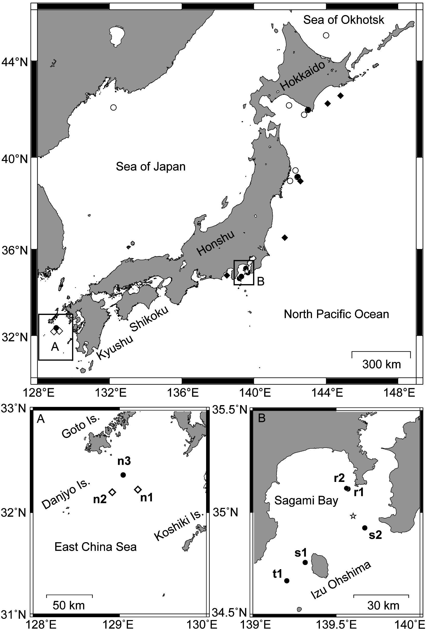
Type materials. Holotype: NSMT E-13404 ( INSD accession number LC750305, 629 bps), off Misaki , Miura Peninsula, Kanagawa, Sagami Bay, Japan, 35°6.998′– 35°7.291′N 139°34.158′– 139°33.36′E, depth 267–276 m, R / V Rinkai-maru, St. 2, 15 May 2013 (r1 in Fig. 1 View FIGURE 1 ) GoogleMaps . Paratypes: NSMT E-13416 ( 1 specimen, INSD accession number LC750306, 641 bps), west of Johgashima Island , Miura Peninsula , Kanawaga, Sagami Bay, Japan, 35°7.186′– 35°7.275′N 139°33.618– 139°33.272′E, depth 267–276 m, R / V Rinkai-maru St. 2, 20 Oct. 2014 (r 2 in Fig. 1 View FIGURE 1 ) GoogleMaps ; NSMT E-9439 ( 1 specimen), Sagami Bay , Japan, 34°39.889′– 34°40.03′N 139°12.191– 139°13.866′E, depth 504–551 m, R / V Tansei-maru KT 07-31 cruise St. L-3-500, 27 Nov. 2007 (t 1 in Fig. 1 View FIGURE 1 ) GoogleMaps ; NSMT E-9440 ( 1 specimen), northwest of Izu-Ohshima , Sagami Bay, Japan, 34°45.3′– 34°45.4′N 139°18.8– 139°19.5′E, depth 406– 371 m, T/V Shin’yo-maru St. 40, 24 Oct. 2002 (s 1 in Fig. 1 View FIGURE 1 ) GoogleMaps ; NSMT E-9441 ( 4 specimens), Okinose, Sagami Bay , Japan, 34°55.5′– 34°55.4′N, 139°40.2– 139°40.5′E, depth 375– 275 m, T/V Shin’yo-maru St. 9, 18 Oct. 2003 (s 2 in Fig. 1 View FIGURE 1 ) GoogleMaps ; NSMT E-13417 ( 1 specimen, INSD accession number LC750307, 644 bps), east of Kasayama Bank , off Goto Islands , the East China Sea, Japan, 32°12.013′N, 128°54.825′E, depth 403–413 m, T / V Nagasaki-maru N 365 cruise St. 6, 19 Nov. 2012 (n 3 in Fig. 1 View FIGURE 1 ) GoogleMaps .
Description of holotype. Body sub-cylindrical with flat ventrum, 101 mm long, 31 mm wide, 16 mm high ( Fig. 6 View FIGURE 6 ). Body wall thick, soft. Mouth subventral. Anus at posterior end of body. Dorsal papillae conical, distributed on both sides of ventral-lateral and dorsal radii. Ventral-lateral papillae, up to 3 mm long, count 22 and 23 in number on left and right ventral-lateral radius, arranged in single row on each radius. Dorsal papillae up to 4 mm, mixing several larger ones, up to 12 mm long, count 70 and 65 in number on left and right dorsal radii, arranged in double rows on each radius. Tube feet cylindrical, slightly tapering toward tips on both sides of ventral-lateral and mid ventral radii. Ventral-lateral tube feet count 26 and 29 in number on left and right ventral radii, arranged in a single straight or zigzag row in each radius. Mid-ventral tube feet count 21 in number, arranged in a zigzag row, more crowded posteriorly. Tentacles 19, with cylindrical stems, and circle terminal discs. Papillae, tube feet, and tentacles nonretractile.
Calcareous ring a rudimentary fragile continuous ring, consists of 5 radial and 5 interradial elements, surrounding the pharynx, but outline of each element not clear ( Fig. 7A View FIGURE 7 ). Each element of the calcareous ring incompletely calcified, tilted so that the anterior opens outward. Radial elements (R in Fig. 7A View FIGURE 7 ) with 3 short anterior processes (Rap in Fig. 7A View FIGURE 7 ) and a large opening at base of anterior processes (Ro in Fig. 7A View FIGURE 7 ). Posterior edge of radial elements slightly depressed. Interradial elements (IR in Fig. 7A View FIGURE 7 ) with single anterior process (IRap in Fig. 7A View FIGURE 7 ), without posterior process. Gonads slightly yellow paired tuft, comprising multi-branched genital vesicles, connected into central genital duct opened at anterior mid dorsal body wall with single genital papilla. Polian vesicle single, elongated tube, 19.3 mm long, derived from left ventral side of pharynx, white color. Stone canal single cylindrical tube with a hemispherical end, 0.26 mm long, derived from right ventral side of pharynx, tube white colored, brown color at the ends.
Dorsal body skin pink to reddish brown in alcohol ( Fig. 6A View FIGURE 6 ), light pink in living specimens ( Fig. 6C View FIGURE 6 ). Ventral body skin yellowish white in alcohol ( Fig. 6B View FIGURE 6 ), transparent white in living specimens ( Fig. 6D View FIGURE 6 ). Dorsal papillae dark violet in living state ( Fig. 6C View FIGURE 6 ), its color well preserved in alcohol ( Fig. 6A View FIGURE 6 ). Discs of tube feet white in living state ( Fig. 6D View FIGURE 6 ). Discs of tentacles light yellow in living state ( Fig. 6D View FIGURE 6 ). After preservation, both changed from off white to yellowish white in alcohol ( Fig. 6B View FIGURE 6 ).
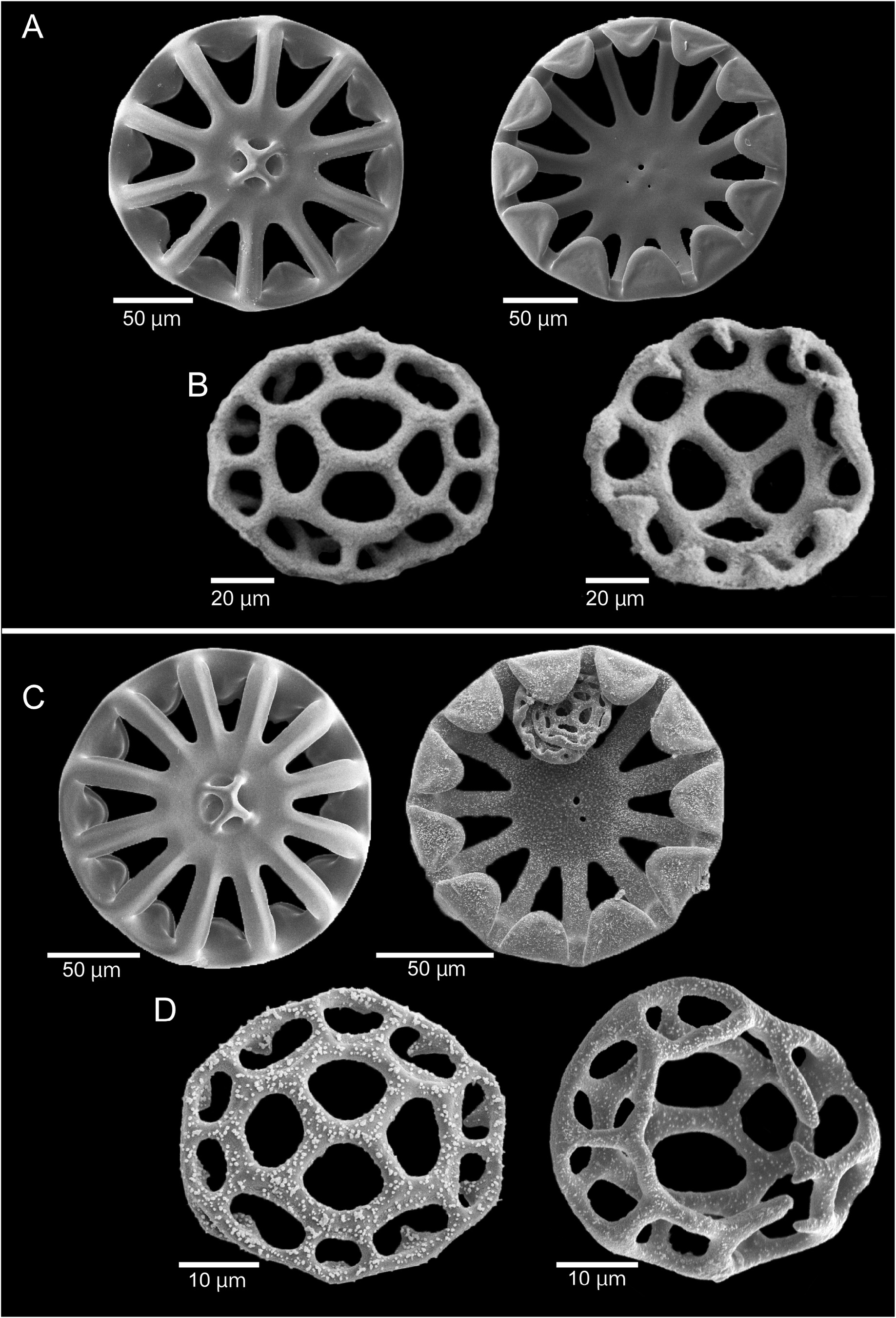
Ossicles. Ossicles in dorsal body wall are two forms of wheels: large wheels having both triangular rim teeth and a calcareous membrane, and small wheels lacking calcareous membrane, but having cylindrical rim teeth ( Fig. 8A, 8B View FIGURE 8 ). Large wheels ( Fig. 8A View FIGURE 8 ) 141–214 µm in diameter (mean 198 µm, N = 30), with 10–12 spokes (N = 30), 4 central rays (N = 14), triangular rim teeth on inner rim, 21–41 µm high (mean 33 µm, N = 30). Calcareous membranes derived from inner edge of connecting central portion. Small wheels ( Fig. 8B View FIGURE 8 ) 40–73 µm in diameter (mean 48 µm, N = 10), with 6–10 spokes (N = 10), usually 4, rarely 3 central rays (N = 10), mostly with cylindrical rim teeth derived inward from inter-spokes part of rim. Tips of rim teeth sometimes T-shaped branched and bridged with neighbor ones. Ossicles distributed high densely, omnipresently in dorsal body wall.
Ossicles in ventral body wall usually consist of two forms of wheels: large wheels and small wheels ( Fig. 8C, 8D View FIGURE 8 ). Large wheels slightly smaller than dorsal ones ( Fig. 8C View FIGURE 8 ), 109–209 µm in diameter (mean 163 µm, N = 23), with usually 11 or 12 spokes, rarely 8 or 10 (N = 23), usually 4 central rays (N = 11), triangular rim teeth on inner rim 17–31 µm long (mean 26 µm, N = 23). Calcareous membranes derived from inner edge of connecting central portion, sometimes. Small wheels ( Fig. 8D View FIGURE 8 ) 34–62 µm in diameter (mean 40 µm, N = 27), with mostly 8–10, rarely 6 or 7 spokes (N = 26), usually 4, rarely 3 central rays (N = 73), always with cylindrical rim teeth like dorsal ones. Ossicles distributed high densely, omnipresently in ventral body wall.
Ossicles in tentacles only rods ( Fig. 7B View FIGURE 7 ). Rod ossicles distinctly arched, 91–583 µm long (mean 351 µm, N = 25), 5–36 µm wide (mean 18 µm, N = 25), usually with minute conical spines scattered on whole ossicles and clouded on both ends.
Ossicles in tube feet rods and end plates ( Fig. 7C, 7D View FIGURE 7 ). Rod ossicles nearly straight, 209–364 µm long (mean 282 µm, N = 24), 10–24 µm wide (mean 15 µm, N = 24), usually with minute conical spines restricted on ends or sometimes completely lacking spines.
Variations of paratypes. Body sizes of the 8 paratypes range between 32–96 mm in length, 11–19 mm in width, and 12–16 mm in height, obviously smaller than holotype. Numbers of ventral-lateral papillae are 5–23, and numbers of dorsal papillae 23–81. Papillae arrange in a single row on each ventrolateral radius, and a zigzag row or double rows on each dorsal radius. Numbers of tube feet are 19–29 in ventral-lateral radii and 20–35 in mid radius. The number of mid tube feet dose not correlate with the body length. Tentacle numbers are 15–20, but some paratypes loose several tentacles during sampling.
Dorsal body skin is pink to reddish brown in alcohol, sometimes decolorized and becoming transparent (NSMT E-9439–9441). Ventral body skin is white to yellowish white in alcohol. Dorsal papillae are reddish brown to dark violet, well preserved in alcohol. Discs of tube feet and tentacles are off white to yellowish white in alcohol, sometimes pigmented with reddish or violet particles (NSMT E-13416 and E-13417).
Ossicles. Most of paratypes possess two types of large wheel ossicles in dorsal body wall while a paratype, NSMT E-9439, comprises large wheels and small wheels. Large wheel ossicles of paratypes show larger ranges of ossicle diameters than holotype: 142–269 μm in diameter of ossicles (mean 250 µm, N = 7 in NSMT E-9440; mean 232 µm, N = 28 in NSMT E-9441-A3 individuals), 7–13 spokes (N = 35 from NSMT E-9440 and E-9441-A), and usually 4 but rarely 5 central rays (N = 26 from NSMT E-9440 and E-9441-A). Small wheels of the dorsal body wall show similar ranges of ossicle diameters, 39–67 µm in diameter (mean 50 µm, N = 44 in NSMT E-9440; mean 49 µm, N = 25 in NSMT E-9441-A), and the other morphological characters are consistent with those of holotype. Ossicles of paratypes are also distributed in high density and are omnipresent in their dorsal body walls.
Morphologies of ossicles in ventral body wall are similar between holotype and a paratype NSMT E-9439.
Distribution. Known from the upper continental slopes located in the warm temperate zone of Japanese waters: 251–551 m deep in Sagami Bay and 304–310 m deep off Goto Islands (closed circles in Fig.1 View FIGURE 1 , present study).
Etymology. The species is named after the R/V Rinkai -maru collected holotype.
Remarks. Pannychia rinkaimaruae sp. nov. is characterized by the following 5 morphological characters ( Table 1 View TABLE 1 ): i) colorings of the body skin, pink to reddish brown on the dorsum, white on the venter, and reddish brown to dark violet on the dorsal papillae, ii) dorsal papillae arranged in two rows on each radius, iii) ventral papillae absent, iv) mid-sized wheel ossicles absent, and v) small wheel ossicles in the dorsal and ventral body walls with rim teeth.
Compared to all the eight nominal species/subspecies of this genus, P.rinkaimaruae sp. nov. shows morphological differences. The present new species differs from P. henrici , P. virgulifera , and P. moseleyi mollis in the arrangement of dorsal papillae in double rows on each radius and in the absence of rod ossicles in dorsal and ventral body walls. This new species is distinguished from P. taylorae and P. multiradiata by the maximum diameter of large wheel ossicles in dorsal body walls not exceeding 300 µm, and from P. moseleyi and P. pallida by small wheel ossicles in dorsal and ventral body walls having rim teeth, and from L. fecundum by the absence of ventral papillae. The other congeners and some species of Laetmogone Théel, 1879 also have small wheel ossicles, but the small wheel ossicles of P. rinkaimaruae sp. nov. have rim teeth, and this microstructure is unique to this new species. This microstructure shows that this species is clearly distinct from congeners including synonymous taxa of P. moseleyi and P. virgulifera in terms of morphological characters, and genetic differences between the present species and congeners in our molecular phylogenetic analysis ( Fig. 9 View FIGURE 9 ) also support this morphological character reflecting interspecific differences between the present species and congeners.


| NSMT |
National Science Museum (Natural History) |
| R |
Departamento de Geologia, Universidad de Chile |
| V |
Royal British Columbia Museum - Herbarium |
| T |
Tavera, Department of Geology and Geophysics |
No known copyright restrictions apply. See Agosti, D., Egloff, W., 2009. Taxonomic information exchange and copyright: the Plazi approach. BMC Research Notes 2009, 2:53 for further explanation.
|
Kingdom |
|
|
Phylum |
|
|
Class |
|
|
Order |
|
|
Family |
|
|
Genus |