
Diaphanosoma turkanae, Korovchinsky, Nikolai M., Walsh, Elizabeth J. & Smolak, Radoslav, 2017
|
publication ID |
https://doi.org/10.11646/zootaxa.4250.1.6 |
|
publication LSID |
lsid:zoobank.org:pub:6DAAA28B-A425-4A3F-BBD2-188EAA8B3BE |
|
DOI |
https://doi.org/10.5281/zenodo.5618206 |
|
persistent identifier |
https://treatment.plazi.org/id/D25B879B-890A-FF81-0DDA-F990FEB3B74E |
|
treatment provided by |
Plazi |
|
scientific name |
Diaphanosoma turkanae |
| status |
sp. nov. |
Diaphanosoma turkanae sp. nov.
( Figs. 2–3 View FIGURE 2 View FIGURE 3 )
Etymology. The species name is derived from the name of lake in which the species is found.
Type locality. Lake Turkana, Kenya.
Type material. Holotype: a female preserved in 4 % buffered formalin deposited in Zoological Museum of Moscow State University (Ml 154).
Paratypes: four females preserved in 4 % buffered formalin deposited in the same Museum (Ml 155).
Description. Parthenogenetic female. Body measurements of specimens are presented in Table 1 View TABLE 1 . Habitus. Body rectangular-elongated, head of moderate size (length is 38–41 % of body length), with more or less developed dorsal part, the shape of which varies to some degree ( Fig. 2 View FIGURE 2 A–D).
Eye of medium size (5.7–7.7 % of body length) and situated close to antero-ventral corner of head.
Antennule small and situated ventrally, with nine aesthetascs and rather long sensory seta ( Fig. 2 View FIGURE 2 E).
Swimming antennae comparatively short (62.0–68.8 % of body length), with upper branch not far from reaching the posterior valve margin ( Fig. 2 View FIGURE 2 A). Basipodite massive, with a small, sharp spine on the outer side of its apical end close to the base of the lower branch ( Fig. 2 View FIGURE 2 F). Proximal segment of upper two-segmented antennal branch (exopodite) (12.6–15.6 % of body length) with small apical spine ( Fig. 2 View FIGURE 2 G), while that of the distal segment of the branch (15.8–19.2 % of body length) is somewhat larger ( Fig. 2 View FIGURE 2 H). Proximal segment of upper antennal branch always bearing three swimming setae, while the distal one has six or seven setae (of six specimens examined, two had six setae and four had seven setae) ( Fig. 2 View FIGURE 2 I, 2J). All antennal setae are uniformly armed by rough setulae of the “swimming” type. Formula for antennal setae: 3–(6–7) / 0–1–4.
Shell with slightly arched dorsal side and inconspicuous or conspicuous dorso-posterior angle ( Fig. 2 View FIGURE 2 A, 2L, 2M). Dorsally, it has a structure consisting of more or less pronounced longitudinal ribs ( Fig. 2 View FIGURE 2 K). Valves with posterior margin of moderate length smoothly connected with ventral margin ( Fig. 2 View FIGURE 2 L) and forming a comparatively narrow, short inflexion shifted anteriorly and bearing 5–6 long, finely setulated setae, the proximal one somewhat diminished and implanted slightly submarginally; thin marginal setulae sometimes present between these setae ( Fig. 3 View FIGURE 3 D). Postero-ventral valve margin armed with a row of 11–19 uniform small denticles with thin setulae between each two to five of them ( Fig. 2 View FIGURE 2 L, 2M, 2N, 2O, 2P). A few similar setulae and rows of marginal and submarginal spinulae along posterior valve margin ( Fig. 2 View FIGURE 2 L, 2M).
Six pairs of thoracic limbs (tl I–tl VI), all with epipodites. Structure and armament of limbs are schematically summarized in Table 2 View TABLE 2 . Exopodite of tl I is comparatively narrow at its end ( Fig. 3 View FIGURE 3 E, 3F), while the exopodite widens terminally in limbs tl II–tl VI ( Fig. 3 View FIGURE 3 G, 3H). Those of tl I–tl V bear 5–6 terminal and 3–5 lateral, unsegmented, thick and long, densely setulated setae. Endopodites of tl I–tl V are inconspicuously subdivided into four segments and bear 22–38 long, two-segmented, setulated filtering setae and one (tl I) or two (tl II–tl V) outer setae, similar to those of the exopodite ( Fig. 3 View FIGURE 3 E, 3G: arrow). Among the latter, the subterminal one is conspicuously shorter than the other ( Fig. 3 View FIGURE 3 G). Small, thorn-like, naked seta on the end of proximal segment above the row of filtering setae of tl I ( Fig. 3 View FIGURE 3 E: ns). Gnathobase of tl I with a row of seven two-segmented, distally finely setulated setae, six of which inclined down to the food groove and one inclined in opposite direction; a group of small, curved spines near its base ( Fig. 3 View FIGURE 3 E). Gnathobases of tl II–tl V are larger, bearing 16–26 filtering setae, one naked seta (p) proximally and one long, two-segmented seta (I) distally with rough setulae ( Fig. 3 View FIGURE 3 G); an additional modified naked, hooked seta (J) with few lateral denticles near the previous one is also present in tl III– tl V. Tl VI is small and strongly modified ( Fig. 3 View FIGURE 3 H, 3I); its exopodite is reduced up to terminal plate and is armed with six terminal and one lateral setae; endopodite with seven similar setae and rounded outgrowth; gnathobase with two long setae and two thorns of different sizes.
Postabdomen cone-shaped with rather long postabdominal setae (43–56 % of body length), groups of spinulae on its lateral and dorsal sides, terminal claws with three basal spines, the distal one longest ( Fig. 3 View FIGURE 3 A, 3B). Groups and short rows of spinulae are situated above the basal spines and distally along the outer lateral side of claws.
Size. Body length: 0.62–0.80 mm.
Gamogenetic females and males unknown.
Differential diagnosis. The new species markedly differs from all known Diaphanosoma species in presence of a low number of swimming setae on both segments of upper two-segmented antennal branch ( Fig. 2 View FIGURE 2 I, 2J). In most other features, this species is most similar to D. orghidani , especially to its subspecies D. orghidani orghidani , having similar body structure and armament, and in particular the shell valve margins. D. turkanae sp. nov. differs from the latter species in armament of swimming antennae, lower number of marginal setae on the ventral valve inflexion ( Fig. 3 View FIGURE 3 D), and presence of smaller and thinner denticles on postero-ventral valve margins ( Fig. 2 View FIGURE 2 N, 2O, 2P).

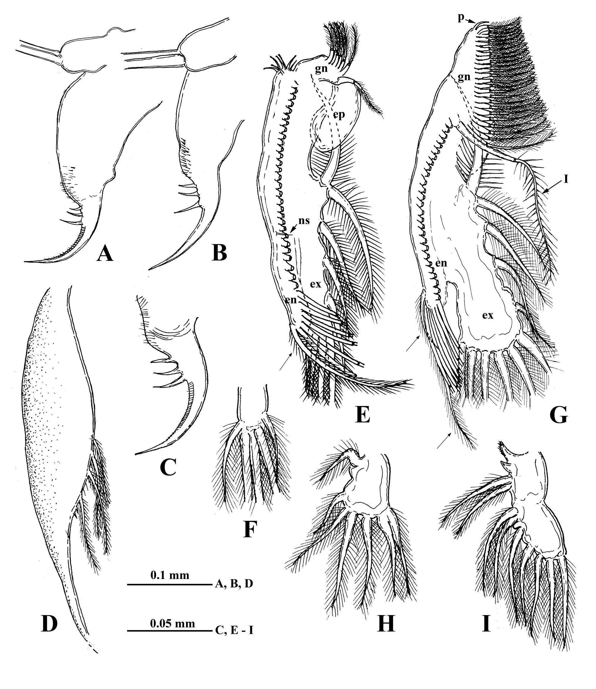
TABLE 1. Body measurements of representatives of three species of Diaphanosoma from Lake Turkana (in columns for D. excisum from top to bottom: range, M, SD, CV; for other two species represented by small number of individuals, only range and M) (for abbreviations see text above)
| BL, mm HL:BL,% | DE:BL, % | AL:BL,% | PSL:BL,% | DSL:BL,% | UBL:BL, % | LBL:BL,% | ND |
|---|---|---|---|---|---|---|---|
| 1) D. turkanae sp. nov. (n | = 6) | ||||||
| 0.62– 0.80 37.6–41.0 | 5.7–7.7 | 62.0–68.8 | 12.6–15.6 | 15.8–19.2 | 28.4–34.6 | 19.3–24.4 | 11–19 |
| 0.73 38.8 | 6.8 | 65.2 | 14.2 | 18.0 | 32.2 | 21.9 | 15 |
| 2) D. lacustris (n = 11) | |||||||
| 0.78–0.98 34.5–38.8 | 5.2–6.8 | 60.0–73.8 | 12.2–13.9 | 19.0–23.4 | 32.4–36.4 | 20.9–24.8 | 18–37 |
| 0.87 35.9 | 6.3 | 67.1 | 13.1 | 21.1 | 34.3 | 22.8 | 26 |
| 3) D. excisum (n = 15) | |||||||
| 0.79– 0.90 35.3–38.9 | 7.1–8.9 | 65.0–81.8 | 13.6–15.9 | 20.8–26.0 | 34.9–41.1 | 23.0–28.0 | 3–12 |
| 0.85 37.0 | 8.0 | 73.1 | 14.6 | 22.9 | 37.4 | 24.3 | 8 |
| 1.1 | 0.6 | 5.0 | 0.7 | 1.4 | 1.9 | 1.6 | |
| 3.0 | 6.9 | 6.8 | 5.0 | 6.1 | 5.1 | 6.5 |
TABLE 2. Structure and armament of thoracic limbs of Diaphanosoma turcanae sp. nov. from Lake Turkana (Kenya, East Africa) (for explanation and abbreviations see text above and Fig. 3).
| Limb pairs | Exopodite (apical +lateral setae) | Endopodite | Gnathobase | Epipodite |
|---|---|---|---|---|
| I | 5 + 5 | (n6 + 1) + (n3) + (n3) + (n26) | n7 | + |
| II | 6 + 5 | (n7 + 1) + (n3 + 1) + (n3) + (n23) | (n26 + p) + I | + |
| III | 6 + 5 | (n7 + 1) + (n3 + 1) + (n3) + (n23) | (n25 + p) + I + J | + |
| IV | 6 + 5 | (n7 + 1) + (n3 + 1) + (n3) + (n23) | (n17 + p) + I + J | + |
| V | 5 + 3 | (n5 + 1) + (n3 + 1) (n14) | (n16 + p) + I + J | + |
| VI | 6 + 1 | 7 + one outgrowth | 2 + two thorns | + |
No known copyright restrictions apply. See Agosti, D., Egloff, W., 2009. Taxonomic information exchange and copyright: the Plazi approach. BMC Research Notes 2009, 2:53 for further explanation.
|
Kingdom |
|
|
Phylum |
|
|
Class |
|
|
SuperOrder |
Cladocera |
|
Order |
|
|
Family |
|
|
Genus |