
Indopamphantus, Malipatil, M. B., 2017
|
publication ID |
https://doi.org/ 10.11646/zootaxa.4242.2.4 |
|
publication LSID |
lsid:zoobank.org:pub:E76C1E6E-AC88-4B65-BEEB-FA432799B56E |
|
DOI |
https://doi.org/10.5281/zenodo.6002002 |
|
persistent identifier |
https://treatment.plazi.org/id/D2654E77-E917-F60D-87A8-FB754CE9FB53 |
|
treatment provided by |
Plazi |
|
scientific name |
Indopamphantus |
| status |
gen. nov. |
Genus Indopamphantus View in CoL gen. nov.
Type-species: Indopamphantus makutaensis sp. nov.
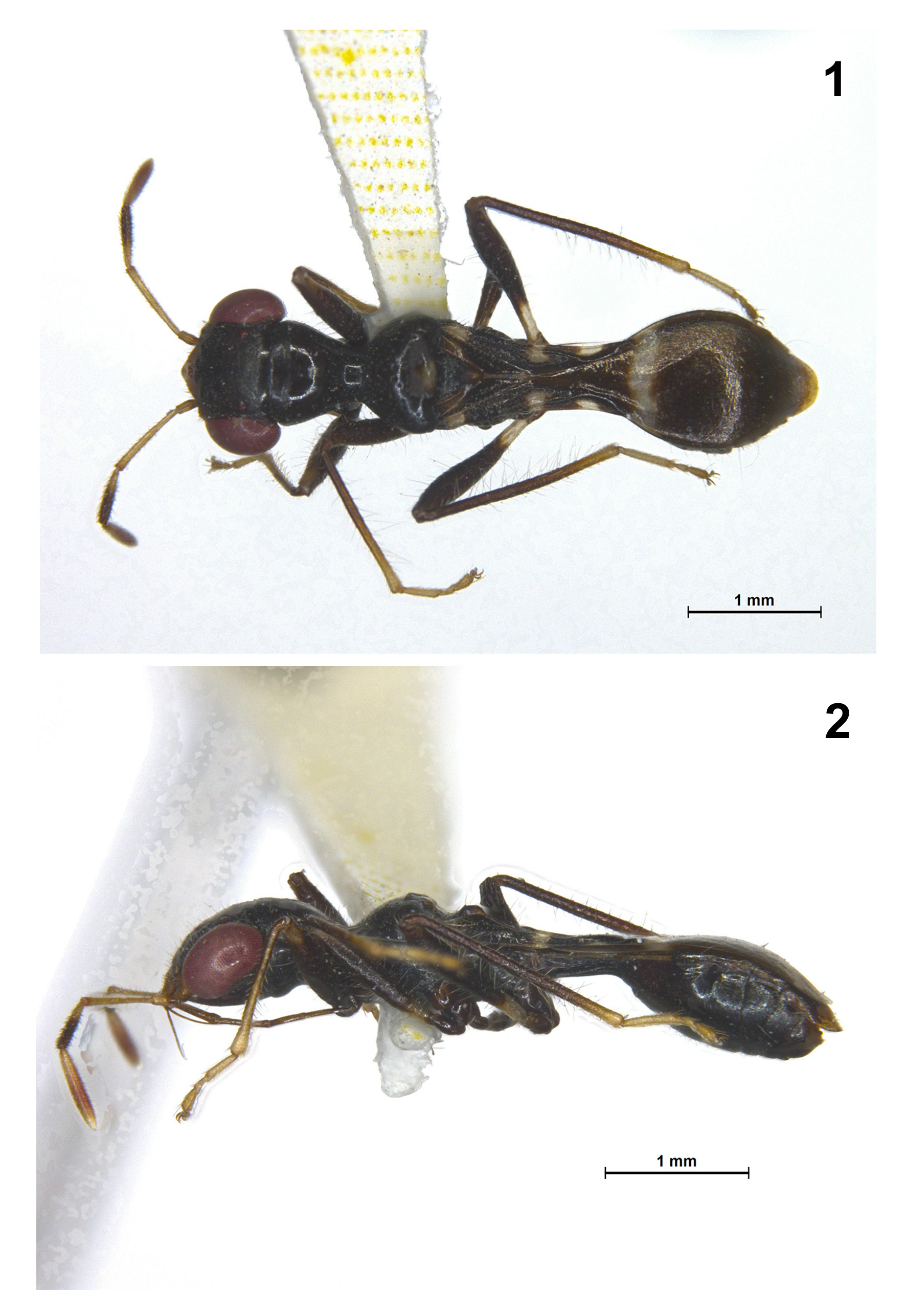
Body elongate, slender, ant mimetic ( Figs. 1, 2 View FIGURES 1 − 2 ).
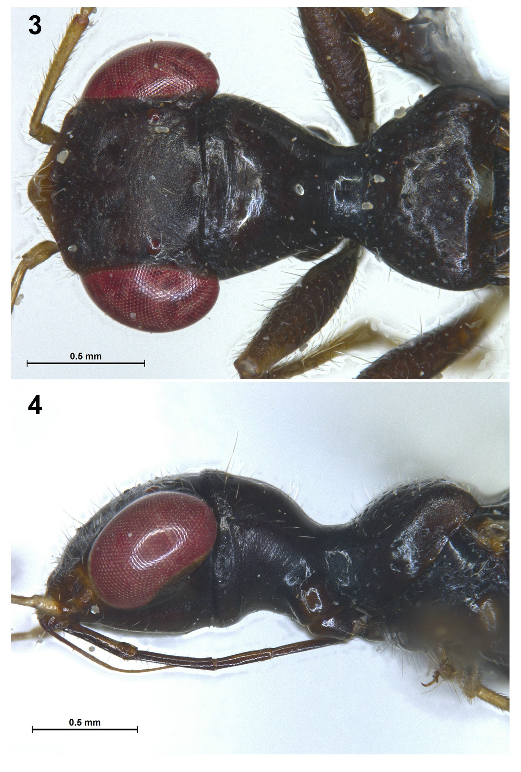
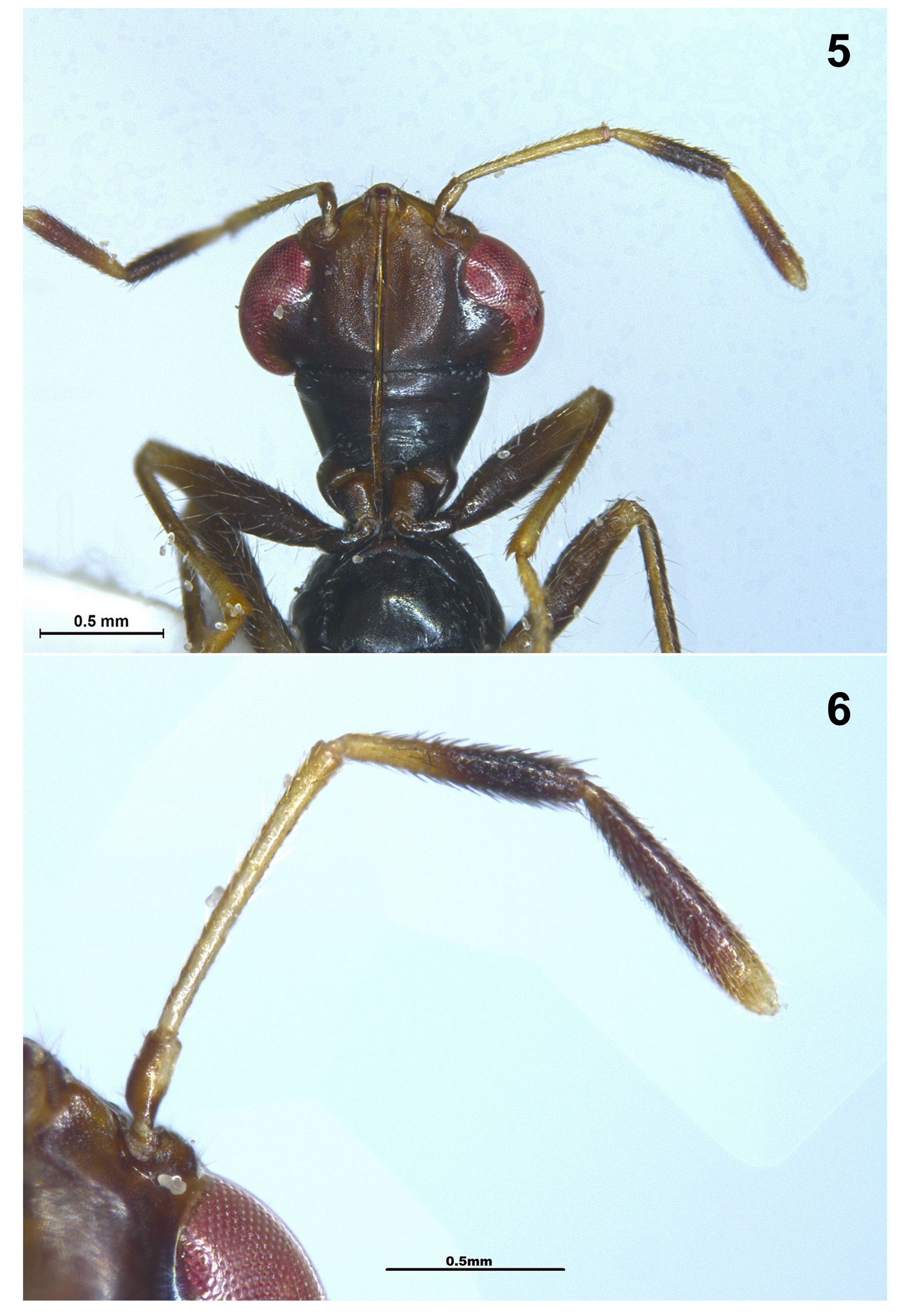
Head: Evenly sloping downward from posterior margin ( Fig. 2 View FIGURES 1 − 2 ), vertex flat, lacking any indication of lateral or median carinae ( Fig. 3 View FIGURES 3 – 4 ); juga faintly carinate ( Fig. 3 View FIGURES 3 – 4 ); bucculae small, produced near base as flaps ( Fig. 4 View FIGURES 3 – 4 ), then gradually diminishing posteriorly to almost to surface of head near base of antenna; head below medially broadly shallowly concave and with a narrow linear median groove for receipt of labium ( Fig. 5 View FIGURES 5 – 6 ); eyes somewhat reniform and curving backward over anterolateral corners of pronotum; slightly stylate, touching pronotal margin (e.g., Fig. 3 View FIGURES 3 – 4 ). Labium with 3rd segment shortest. Labrum slender, about as long as 1st labial segment ( Fig. 4 View FIGURES 3 – 4 ). Antenna with 1st segment shortest, 2nd and 4th subequal ( Figs. 5, 6 View FIGURES 5 – 6 ).
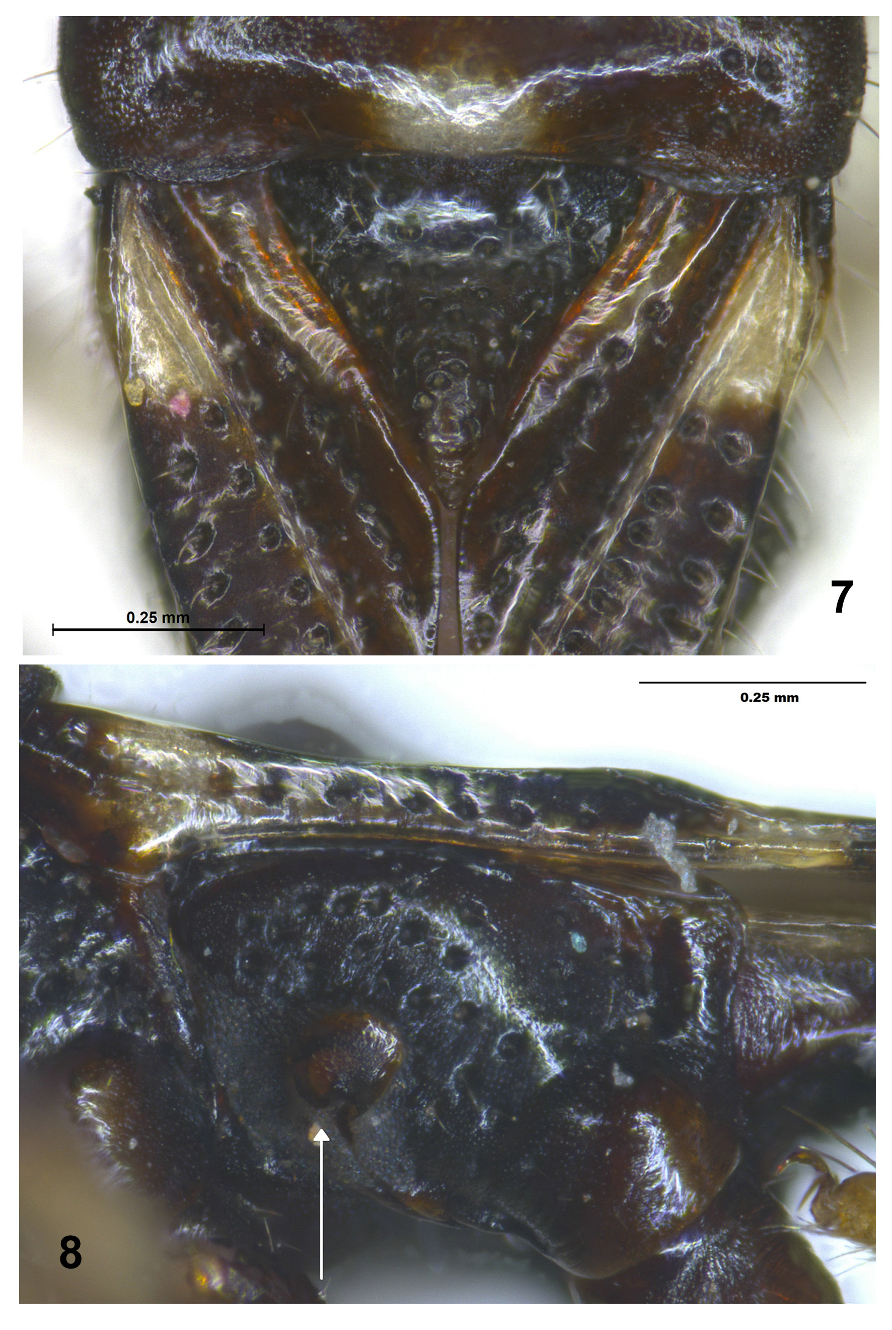
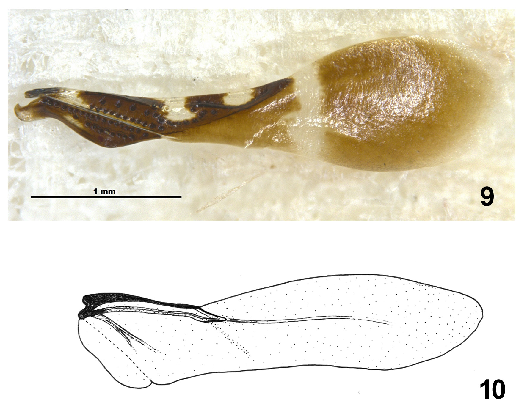
Thorax: Pronotum with broad deep median transverse impression at about half length, lateral margins sinuate, evenly rounded and non-carinate ( Fig. 3 View FIGURES 3 – 4 ). Thoracic pleura with sparse, coarse punctures ( Fig. 8 View FIGURES 7 – 8 ), metathoracic scent gland auricle short and rounded projecting above surface of pleuron with spout turning upwards, opening narrow and directed posterior; evaporative area dull and distinct ( Fig. 8 View FIGURES 7 – 8 ). Scutellum with median impression on basal 1/3 area, coarse deep punctures on sides and on posterior area, the latter slightly projecting above surface (e.g., Fig. 7 View FIGURES 7 – 8 ). Hemelytra hyaline ( Fig. 9 View FIGURES 9 – 10 ), reaching but not exceeding abdomen ( Fig. 1 View FIGURES 1 − 2 ); corium with more or less three rows of thick-set brown punctures, first row on basal third running near costal margin, then somewhat deviating from it, second row in the middle, third near claval suture and indistinctly continuing from apex along apical margin of corium to apex of first row; clavus with two rows of coarse punctures, one complete row along claval suture, 2nd row complete on inside apical, inner margin impunctate ( Fig. 9 View FIGURES 9 – 10 ). Hind wing ( Fig. 10 View FIGURES 9 – 10 ) narrowed, venation reduced, hamus and intervannals absent, vannal folds indistinct, radius (? or R+M) long, extending to distal half of wing, anterior and posterior vannals distinct but fused on basal area, jugal lobe small and jugal vein absent, cubitus indistinct, the leading margin (subcostal or? Sc+R) sclerotized and greatly narrowed towards distal end of discal cell, corresponding to the basal constriction of abdomen ( Fig. 1 View FIGURES 1 − 2 ). All femora almost similarly uniformly incrassate, unarmed ( Figs. 1, 2 View FIGURES 1 − 2 , 5 View FIGURES 5 – 6 ).
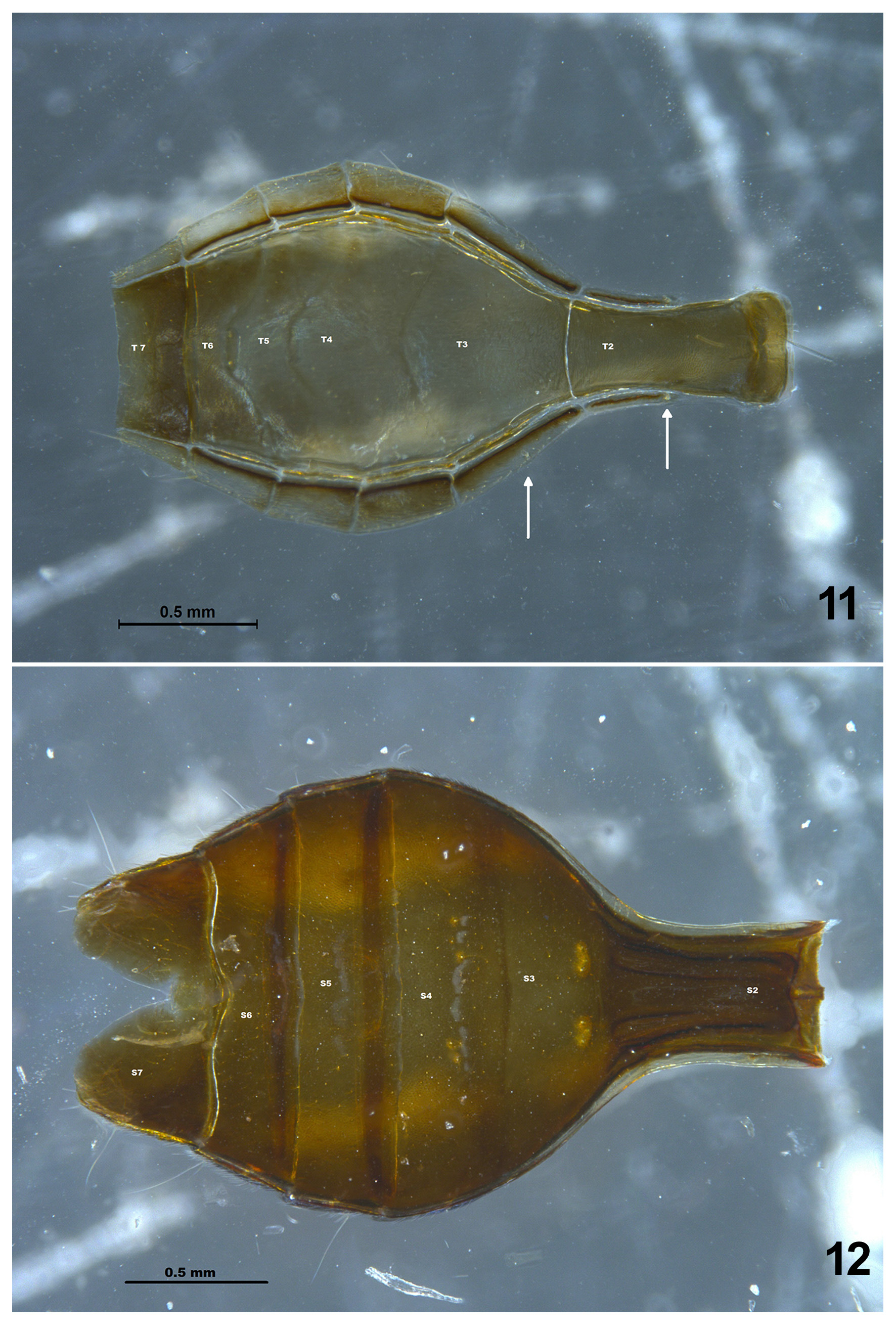
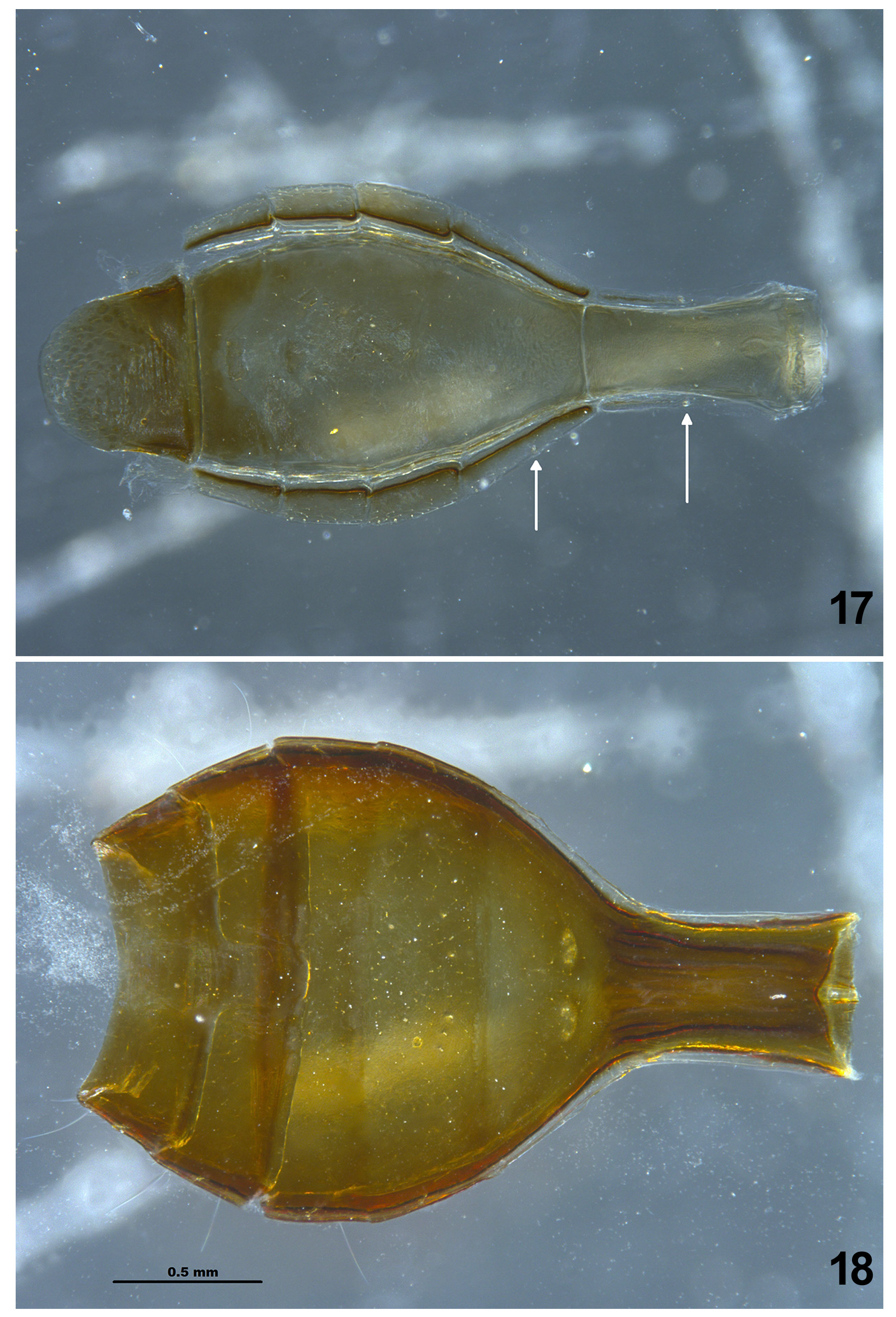
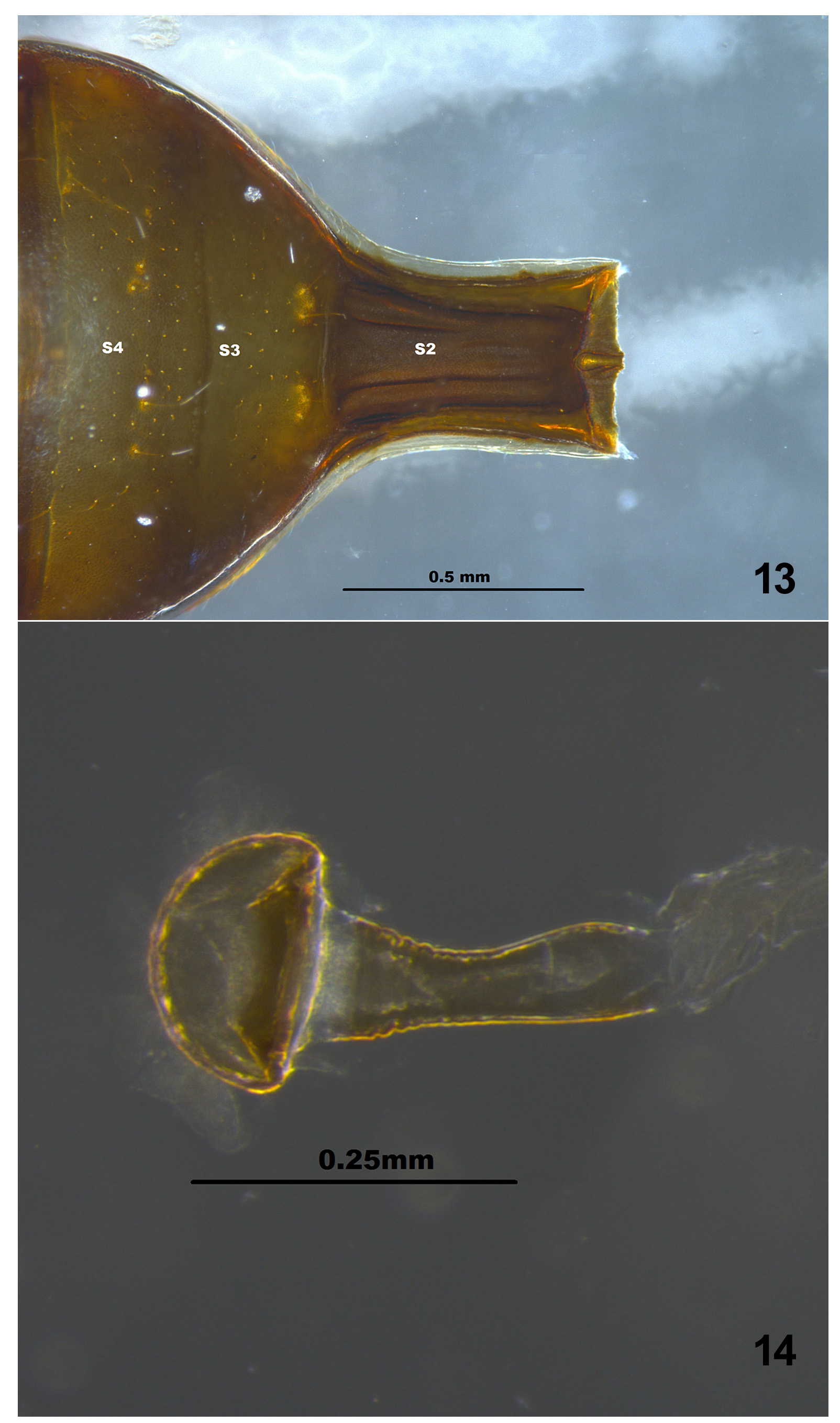
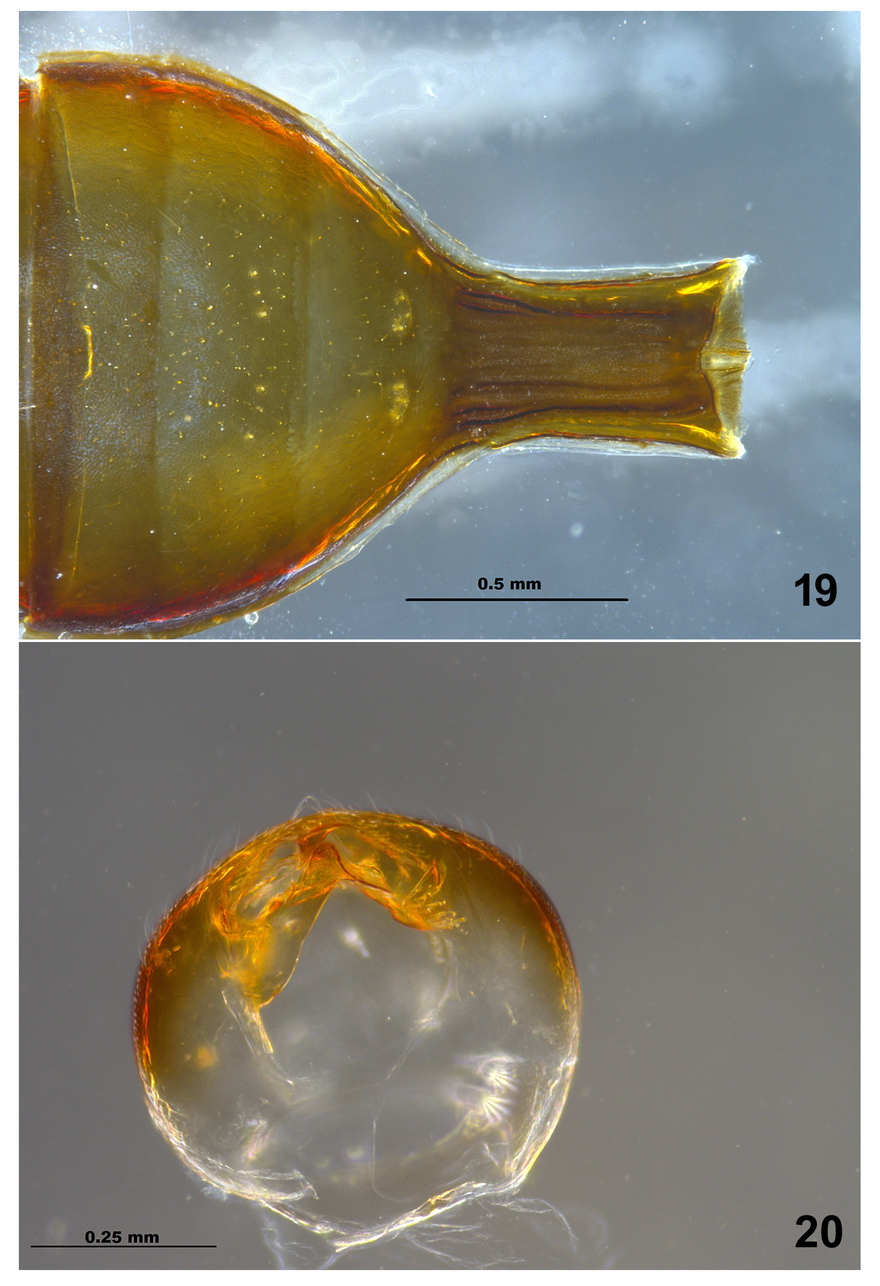
Abdomen: Vasiform ( Figs.1 View FIGURES 1 − 2 , 11, 12 View FIGURES 11 − 12 , 17, 18 View FIGURES 17 − 18 ). First visible (i.e., second) segment strongly constricted to form a parallel-sided tube, and strengthened with longitudinal ridges on ventral surface, appearing like a node as in ants ( Figs. 1, 2 View FIGURES 1 − 2 , 12 View FIGURES 11 − 12 ). Spiracles II and III dorsal on connexiva ( Figs. 11 View FIGURES 11 − 12 , 17 View FIGURES 17 − 18 ), IV, V, VI and VII ventral ( Figs. 11 View FIGURES 11 − 12 , 17 View FIGURES 17 − 18 ). Sutures between all tergal segments distinct, those between terga III–IV only slightly and those between IV–V and V–VI strongly curved caudad from margin to meson and with distinct scent gland scars which are subequal (the latter fractionally wider) in width, scar between III–IV indistinct (e.g., Figs. 11 View FIGURES 11 − 12 , 17 View FIGURES 17 − 18 ). Inner laterotergites present, very narrow particularly adjoining terga III to VI ( Figs. 11 View FIGURES 11 − 12 , 17 View FIGURES 17 − 18 ). Sterna II, III and IV with intersegmental sutures faint and not fused, sutures reaching abdominal margin ( Figs. 12 View FIGURES 11 − 12 , 18 View FIGURES 17 − 18 ). Trichobothria on sternum III in loose triangle and those on IV almost linear to open triangle ( Figs. 12 View FIGURES 11 − 12 , 13 View FIGURES 13 – 14 ), variable in spacing and degree of development, sometimes one trichobothrium reduced or absent (e.g., Fig. 13 View FIGURES 13 – 14 ); trichobothria on V, VI and VII are sublateral, those posterior to spiracle one above the other, spiracle of V and VI situated almost equidistant between anterior and posterior trichobothria, all trichobothrial setae very long.
In male, anterior margin of VII sternum internally with median broad process extending almost to entire length of VI ( Fig. 18 View FIGURES 17 − 18 ). In female, suture between IV–V and V–VI broadly overlapping, suture between III–IV slightly corrugated in middle; seventh sternum cleft with ovipositor ( Fig. 12 View FIGURES 11 − 12 ).
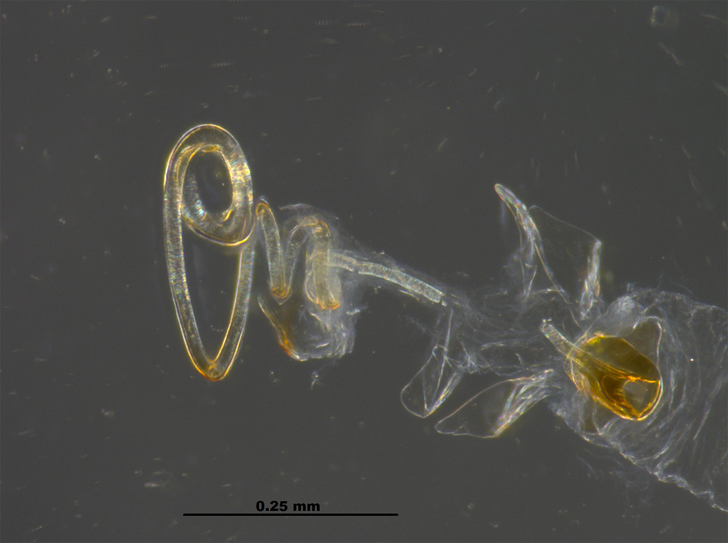
Female genitalia. Spermatheca with apical bulb heavily sclerotized ( Fig. 14 View FIGURES 13 – 14 ), almost spherical, lacking flanges, duct near base of bulb short, saccoid, uniformly moderately sclerotized and tubular gradually narrowing to join a basal unevenly expanded clear membranous duct that is short and slightly wider than previous duct ( Fig. 14 View FIGURES 13 – 14 ).
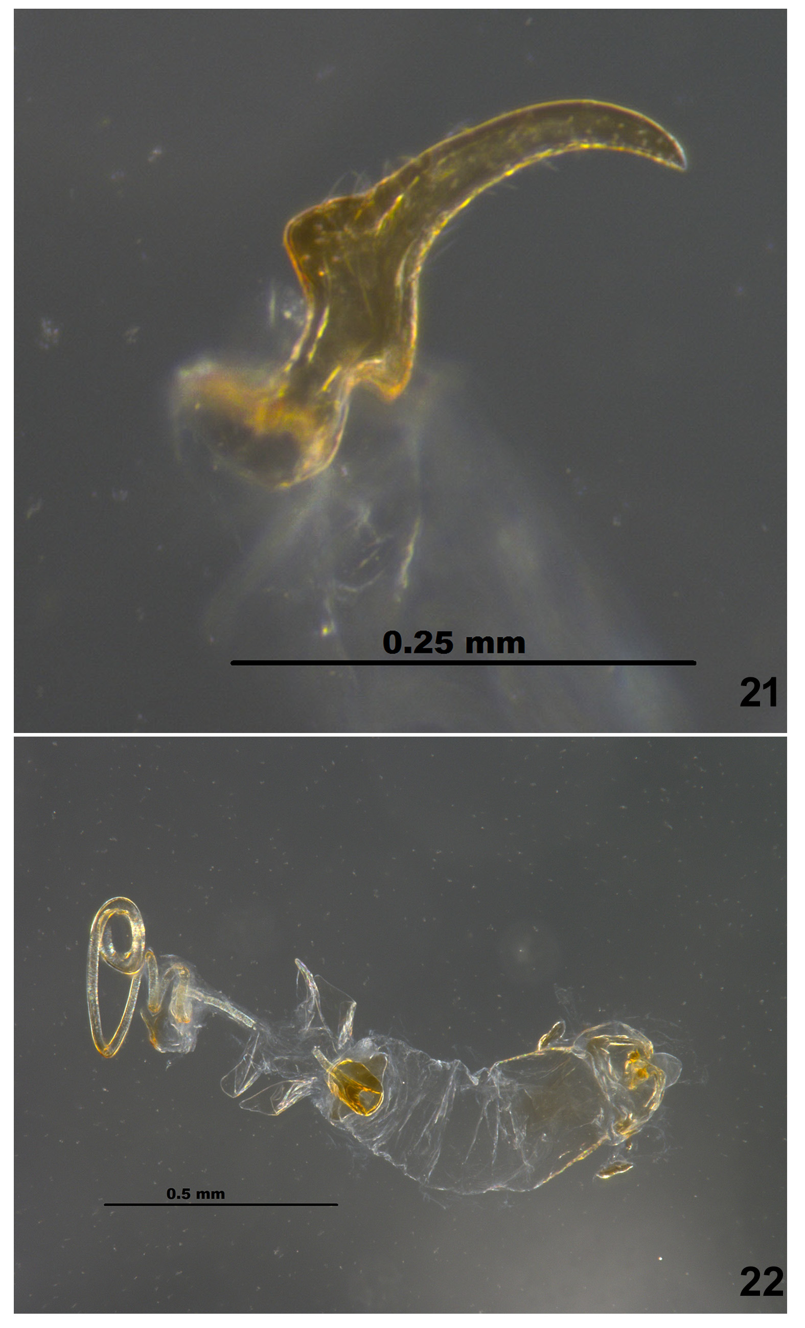
Male genitalia. Pygophore sclerotized anteriorly, gradually rounded posteriorly, lacking processes ( Fig. 20 View FIGURES 19 – 20 ). Paramere ( Fig. 21 View FIGURES 21 – 22 ) slender, with dorsal flange more prominent than ventral lobe, which has long setae, blade narrowly pointed, sickle-shaped. Aedeagus ( Figs. 22 View FIGURES 21 – 22 ) with phallotheca moderately sclerotized on basal area, body and wings of ejaculatory reservoir well developed, neck well developed, no holding sclerites present, conjunctiva membranous with vesica adjoining helicoid process with two pairs of leaf-like processes, helicoid process with about 2 coils, gonoporal process beyond helicoid process with 2–3 large coils, secondary gonopore simple, only slightly flaring ( Fig. 23 View FIGURES 23 ).
No known copyright restrictions apply. See Agosti, D., Egloff, W., 2009. Taxonomic information exchange and copyright: the Plazi approach. BMC Research Notes 2009, 2:53 for further explanation.