
Nesodiprion biremis ( Konow, 1899 )
|
publication ID |
https://doi.org/10.5281/zenodo.209562 |
|
DOI |
https://doi.org/10.5281/zenodo.6178611 |
|
persistent identifier |
https://treatment.plazi.org/id/D26C4539-FFB6-FFA1-7AEE-590BFE5D4D5C |
|
treatment provided by |
Plazi (2016-04-13 09:12:23, last updated 2024-11-27 18:56:44) |
|
scientific name |
Nesodiprion biremis ( Konow, 1899 ) |
| status |
|
Nesodiprion biremis ( Konow, 1899)
Figs. 2A–F View FIGURES 2 A – L ; 3E–H; 4 E–H; 5D–F; 6C, D; 7C, D; 8G–L; 9C; 11A–L; 13F–I; 14E–G
Lophyrus biremis Konow, 1899: 43 . Oehlke and Wudowenz (1984: 370).
Nesodiprion biremis: Rohwer (1910: 104) , Wei et al. (2006: 551), Taeger et al. (2010: 209).
Female [condition of lectotype in brackets]. Length 7.5–8.5 [8.5] mm. Black, shiny without metallic reflection [faintly violet on abdomen] ( Figs. 2A–D View FIGURES 2 A – L ). Clypeus brown to black [brown], ventrally becoming paler. Labrum dark brown to black [dark brown]. Mandible apically reddish. Basal two antennomeres faintly or distinctly yellow to brown [distinctly yellow] ( Figs. 2A–D View FIGURES 2 A – L , 6C, D View FIGURES 6 A – E ). Palpi yellow to pale brown. Pronotum widely or narrowly yellowish white on posterior corner [widely] ( Figs. 2A–D View FIGURES 2 A – L ). Postspiracular sclerite slightly pale or not [slightly pale]. Median mesoscutal lobe brownish or not [brownish]. Mesoscutellum mostly yellowish white or black and centrally slightly brownish [mostly yellowish white]. Mesepisternum centrally slightly brownish ( Fig. 2A View FIGURES 2 A – L ) or not ( Fig. 2D View FIGURES 2 A – L ) [slightly brownish]. Legs white to yellow on apices of coxae to trochantelli, apices of femora, fore and middle tibiae, wide basal part of hind tibia and tarsi; hind tibia dark brown to black on apical third to fourth [third], but narrowly brown at apex; fore and middle tibiae and tarsi each apically faintly brownish; spurs brown. Wings hyaline, apically very faintly blackish; veins largely brown to black; in fore wing, vein C except for apical part yellow, vein R1 basal to stigma partly yellowish, and stigma pale apically. Sixth abdominal tergum laterally narrowly whitish or not [whitish]. Seventh and eighth abdominal terga each laterally with yellowish white spot. Cercus black. Setae largely whitish.
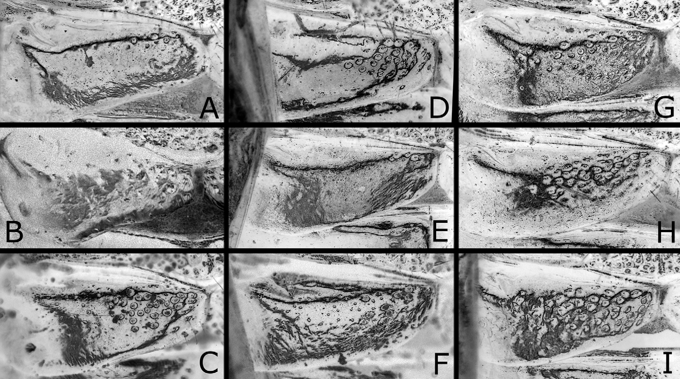
Head and thorax weakly shiny, with punctures mostly dense and distinct ( Figs. 3F View FIGURES 3 A – L , 4E, F View FIGURES 4 A – L ); dorsum of head with punctures somewhat small, moderately or predominantly contiguous [predominantly], and interspaces mostly narrower than punctures; mesoscutum with punctures on posterior part of median lobe not so small, distinct or somewhat vague [distinct], largely contiguous, and larger than those on lateral mesoscutal lobe, and interspaces on posterior part of median lobe predominantly narrower than punctures; interspaces on mesoscutellum moderately or predominantly linear-shaped [moderately]; on mesepisternum, punctures mostly contiguous, and interspaces predominantly linear-shaped ( Fig. 4F View FIGURES 4 A – L ). Clypeus with wide ventromedial part smooth or weakly punctured [smooth]. Labrum smooth. Abdomen shiny ( Figs. 2A, D View FIGURES 2 A – L ); first tergum punctured on narrow medial part to medial third [medial third] ( Figs. 5D, E View FIGURES 5 A – I ); second to fifth terga dorsally nearly smooth; sixth tergum to apex faintly punctured; ventral surface somewhat dull and weakly punctured.
Postocellar area weakly convex ( Figs. 3E, F View FIGURES 3 A – L ); lateral furrow distinct or weak, present on anterior third to half [distinct, present on anterior half]; anterior furrow rather sharp, medially narrowly inconspicuous; median furrow absent. Distances between eye and hind ocellus, between hind ocelli, and between hind ocellus and posterior margin of head 1.0–1.2: 1.0: 1.0–1.1 [1.0: 1.0: 1.0]; distances between eye and hind ocellus and between hind ocellus and posterior margin of head 1.0: 1.0 [1.0: 1.0]. Distance between torulus and eye 1.4–1.7 [1.7] × distance between toruli. Width of malar space 0.3–0.4 [0.3] × width of front ocellus, 0.3–0.4 [0.3] × length of second antennomere. Clypeus with ventral margin roundly concave. Antenna ( Figs. 6C, D View FIGURES 6 A – E ) with 20–22 [22] antennomeres; length of second antennomere 0.9–1.1 [1.0] × width of front ocellus; length of ramus of third antennomere 1.5–2.2 [2.2] × length of third antennomere. Mesoscutellum dorsally slightly or distinctly convex roundly [slightly so], without median furrow ( Fig. 4E View FIGURES 4 A – L ). Hind leg ( Fig. 7C View FIGURES 7 A – F ) with length of inner tibial spur 1.2–1.4 [1.2] × length of first tarsomere (exclusive of pulvillar pad), 1.4–1.7 [1.4] × breadth of tibia; length of first tarsomere 1.1–1.3 [1.1] × breadth of tibia; second and third tarsomeres combined 1.0 [1.0] × first in length.
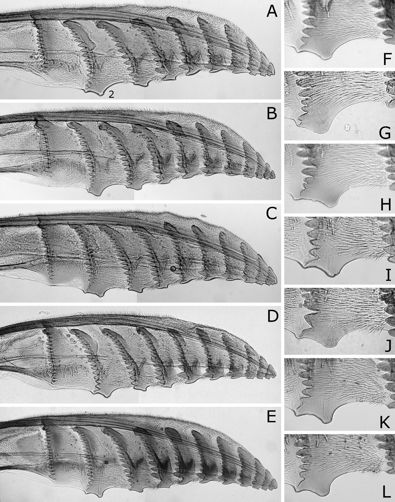
Sawsheath in dorsal view narrow, not tapering apically, with inner margin concave and apex much wider than cercus (Figs. 8G, J), in lateral view slightly roundly convex apically (Figs. 8H, K), in posterior view with scopa vertically elongate (Figs. 8I, L). Lance in lateral view with dorsal margin slightly concave at middle ( Fig. 9C View FIGURES 9 A – D ); apices of lances asymmetrical, either left or right one longer than the other as in Fig. 9A View FIGURES 9 A – D [right one longer]. Lancet ( Figs. 11A–E View FIGURES 11 A – L ) with 10–12 [10] annuli, widest at second annulus, and length from apex to ventral end of basal row of spines 2.5–2.6 [2.6] × maximum width; spines relatively long; border of first and second annuli ventrally very slightly convex; serrula of second annulus ( Figs. 11F–L View FIGURES 11 A – L ) apically narrowly or widely truncate, or nearly rounded [nearly narrowly truncate], with anterior slope shorter than posterior slope; serrula of third annulus with anterior slope nearly straight.
Male. Length 6.5–7.5 mm. Coloration as in female ( Figs. 2E, F View FIGURES 2 A – L ), but clypeus at most slightly pale ventrally, labrum black, sometimes widely brown ventrally, basal two antennomeres scarcely or slightly pale, thorax black except for posterior corner of pronotum narrowly white or brown, trochantelli widely brownish, hind tibia brown on apical third, and abdomen entirely black.
Structure as in female except for usual sexual differences. Punctures more distinct ( Figs. 3H View FIGURES 3 A – L , 4G, H View FIGURES 4 A – L ); first abdominal tergum punctured on medial third to half ( Fig. 5F View FIGURES 5 A – I ). Distances between eye and hind ocellus, between hind ocelli, and between hind ocellus and posterior margin of head 1.0: 1.0: 0.8–0.9; distances between eye and hind ocellus and between hind ocellus and posterior margin of head 1.1–1.2: 1.0. Distance between torulus and eye 1.2–1.6 × distance between toruli. Width of malar space 0.5–0.6 × width of front ocellus, 0.9–2.0 × length of second antennomere. Antenna with 20–26 antennomeres, 1.1–1.2 × as long as head width; length of second antennomere 0.3–0.6 × width of front ocellus; ramus of third antennomere very long. In hind leg ( Fig. 7D View FIGURES 7 A – F ), length of inner tibial spur 1.2 × length of first tarsomere (exclusive of pulvillar pad), 1.4–1.7 × breadth of tibia; length of first tarsomere 1.2–1.3 × breadth of tibia; second and third tarsomeres combined 1.0–1.1 × first tarsomere in length.
FIGURES 8A–R. Sawsheath of Nesodiprion japonicus (A–F), N. biremis (G–L) and N. orientalis (M–R). A, D, G, J, M, P, Sawsheath and cercus, dorsal view; B, E, H, K, N, Q, sawsheath, lateral view; C, F, I, L, O, R, do., posterior view. A, C, Right sawsheath is removed. H, N, Q, Reversed images. N. japonicus : A–C, Amami-oshima; D–F, Honshu, Zu. N. biremis : G–I, Lectotype; J–L, Guizhou Prov., Chishui. N. orientalis : M–O, Paratype, Thailand, Bo Luang; P–R, do.
Genitalia with valviceps in dorsal view laterally strongly convex on apical half, with sharp dorsal ridge basally connected with sharp lateral ridge ( Figs. 13F–I View FIGURES 13 A – M ), in lateral view distinctly widened on apical half, with dorsal margin rather angularly convex and apex ventrally pointed ( Fig. 14E–G View FIGURES 14 A – J ).
Type material examined. Lectotype (here designated): Ƥ, labeled “ Lophyrus biremis Kon. , China ”, “ Hong Kong, China ”, “Coll. Konow”, “ Type ” and “ Holotypus ” ( SDEI).
Konow (1899) included information on intraspecific variation indicating the presence of more than one type specimen, but he did not state how many specimens he had. Only one type specimen is preserved in SDEI, and there is no information on other type specimens (S. M. Blank, 2011, personal communication). Oehlke and Wudowenz (1984) stated “(?) Holotypus: Ƥ, China, Hongkong. DEI: Wahrscheinlich lag dem Autor nur ein Exemplar vor. Dieses ist vorhanden.” Because Konow (1899) possibly had more than one specimen, and because he did not designate a holotype, we here designate the type specimen, labeled as above, in SDEI as the lectotype.
Other material examined. CHINA ―Shandong Prov.: 1Ƥ13, Taian, 28. V. 1992, Host: Pinus sp., J. Hua ( USNM). Zhejiang Prov.: 1Ƥ13, Anji, VIII. 1991, Host: Pinus sp., J.-j. Zheng ( USNM). Jiangxi Prov.: 1Ƥ, “Kuling, Musée Heude” and “ 24. 8. 35, O. PIEL, coll.” ( OPU). Guizhou Prov.: 1Ƥ, Chishui, Jinsha, 20-23. IX. 2000, C.-r. Li ( SDEI). Guangdong Prov.: 13, Ruyuan, 15. V. 2007, Z.-j., Li ( SDEI). Hong Kong: 13, “ Hong Kong: N.T., Sai Kung Station, 25. XI. 1964 ” ( BPBM); 13, do., but 7. V. 1965 ( BPBM); 13, do., but 12. V. 1965 ( BPBM).
Distribution. China: Shandong Prov., Zhejiang Prov., Jiangxi Prov., Guizhou Prov., Guangdong Prov. and Hong Kong; Korea ( Zombori 1978) (?).
The record from Korea by Zombori (1978) needs confirmation; the characters he gave are not sufficient for species recognition.
Host plant. Pinaceae : Pinus sp.
Life history. Adults have been collected from early May to late November.
Comparative notes. This species is similar to N. japonicus , N. orientalis and N. kagaensis , and probably to N. yananicus , N. zhejiangensis , N. huanglongshanicus and N. degenicus , as discussed under Comparative notes of N. japonicus , and is distinguished from them except for N. zhejiangensis and N. huanglongshanicus as follows.
From N. japonicus and N. kagaensis [characters given in brackets]: On mesoscutum, punctures on posterior part of median lobe large, largely contiguous and larger than those on lateral lobe, and interspaces on posterior part of median lobe mostly narrower than punctures ( Figs. 4E, G View FIGURES 4 A – L ) [punctures fine, mostly separated and about as large as those on lateral lobe, and interspaces mostly wider than punctures ( Figs. 4A, C View FIGURES 4 A – L )]; on hind leg, first tarsomere length (exclusive of pulvillar pad) 1.1–1.3 × tibia breadth ( Figs. 7C, D View FIGURES 7 A – F ) [1.3–1.8 × ( Figs. 7A, B View FIGURES 7 A – F ), 1.5–1.9 × respectively]; in female, ramus of third antennomere 1.5–2.1 × length of the third antennomere ( Figs. 6C, D View FIGURES 6 A – E ) [2.3–4.1 × ( Fig. 6A View FIGURES 6 A – E ), 1.9–2.9 × respectively]; in lancet, border of first and second annuli ventrally very slightly convex ( Figs. 11A–L View FIGURES 11 A – L ) [distinctly and angularly convex ( Figs. 10A–L View FIGURES 10 A – L )]; in male, valviceps in lateral view not sinuate and apically suddenly widened ( Figs. 14E–G View FIGURES 14 A – J ) [sinuate and apically gradually widened ( Figs. 14A–D View FIGURES 14 A – J , 16G View FIGURES 16 A – G )].
From N. orientalis: Interspaces on mesoscutellum and mesepisternum moderately or predominantly linear-shaped ( Figs. 4E–H View FIGURES 4 A – L ) [predominantly not linear-shaped ( Figs. 4I –L View FIGURES 4 A – L )]; in female, sawsheath in dorsal view relatively wide, with inner margin concave and apex wider than cercus (Figs. 8G, J) [narrow, with inner margin not concave and apex about as wide as cercus (Figs. 8M, P)]; in male, trochanters yellowish white ( Fig. 2F View FIGURES 2 A – L ), and valviceps in dorsal view laterally strongly convex apically ( Figs. 13F–I View FIGURES 13 A – M ) and in lateral view with dorsal convexity relatively obtuse ( Figs. 14E–G View FIGURES 14 A – J ) [fore and middle or all trochanters widely brown ( Fig. 2K View FIGURES 2 A – L ), and valviceps in dorsal view laterally weakly convex apically ( Figs. 13J–M View FIGURES 13 A – M ) and in lateral view with dorsal convexity relatively acute ( Figs. 14H–J View FIGURES 14 A – J )].
From N. yananicus: Punctures on mesoscutum large ( Figs. 4E, G View FIGURES 4 A – L ) [small ( Xiao et al. 1981)]; in female, lancet wide, with length from apex to ventral end of basal row of spines 2.5–2.6 × maximum width ( Figs. 11A–E View FIGURES 11 A – L ) [narrow, 3.5–3.7 × ( Xiao et al. 1981: fig. 4; Xiao et al. 1985: fig. 35)]; in male, valviceps in lateral view not constricted, and apically strongly widened ( Figs. 14E–G View FIGURES 14 A – J ) [constricted at apical third, with apex not so wide as in Fig. 16E View FIGURES 16 A – G ( Xiao et al. 1981: fig. 1; Xiao et al. 1985: fig. 15)].
From N. degenicus : Head and thorax without metallic reflection ( Figs. 2A–F View FIGURES 2 A – L ) [with bluish purple metallic reflection ( Xiao et al. 1984, 1985)]; in female lancet, serrula of third annulus with anterior slope nearly straight ( Figs. 11A–E View FIGURES 11 A – L ) [distinctly concave ( Fig. 15A View FIGURES 15 A – C ; Xiao et al. 1985: fig. 30)]; in male, valviceps in lateral view not constricted, and apically strongly widened ( Figs. 14E–G View FIGURES 14 A – J ) [strongly constricted at apical third, with apex not so wide ( Fig. 15C View FIGURES 15 A – C ; Xiao et al. 1981: fig. 1, Xiao et al. 1985: fig. 12)].
The differences of N. biremis from N. zhejiangensis and N. huanglongshanicus are not clear. At least the male of N. huanglongshanicus has the penis valve as in Fig. 16E View FIGURES 16 A – G , quite similar to that of N. yananicus ( Xiao et al. 1981: fig. 2; Xiao et al. 1985: fig. 14), and it is distinguishable from the male of N. biremis . The penis valve of N. zhejiangensis is apically distinctly widened ( Xiao et al. 1981: fig. 3; Xiao et al. 1985: fig. 13) and similar to that of N. biremis ( Figs. 14E–G View FIGURES 14 A – J ). The lancet is variable in outline, wide or narrow in both N. zhejiangensis ( Xiao et al. 1981: fig. 6; Xiao et al. 1985: fig. 29) and N. huanglongshanicus ( Xiao et al. 1981: fig. 5; Xiao et al. 1985: fig. 28). Detail studies on the holotypes of N. zhejiangensis and N. huanglongshanicus (both females) are necessary to clarify their identities.
Konow, F. W. (1899) Ueber einige neue Chalastogastra. Wiener entomologische Zeitung, 18 (2 - 3), 41 - 46.
Oehlke, J. & Wudowenz, J. (1984) Katalog der in den Sammlungen der Abteilung Taxonomie der Insekten des Institutes fur Pflanzenschutzforschung, Bereich Eberswalde (ehemals Deutsches Entomologisches Institut), aufbewahrten Typen - XXII (Hymenoptera: Symphyta). Beitrage zur Entomologie, 34 (2), 363 - 420.
Rohwer, S. A. (1910) Japanese sawflies in the collection of the United States National Museum. Proceedings of the United States National Museum, 39 (1777), 99 - 120.
Taeger, A., Blank, S. M. & Liston, A. D. (2010) World catalog of Symphyta (Hymenoptera). Zootaxa, 2580, 1 - 1064.
Wei, M., Nie, H. & Taeger, A. (2006) Sawflies (Hymenoptera: Symphyta) of China - Checklist and review of research. In: Blank, S. M, Schmidt, S. & Taeger, A. (Eds.), Recent Sawfly Research: Synthesis and Prospects. Goecke & Evers, Keltern, pp. 505 - 574.
Xiao, G. - r., Huang, X. - y. & Zhou, S. - z. (1981) Three new species of Nesodiprion from China (Hymenoptera, Symphyta, Diprionidae). Scientia Silvae Sinicae, 17, 247 - 249, pl. 1. (In Chinese, with English abstract.)
Xiao, G. - r., Huang, X. - y. & Zhou, S. - z. (1984) The Chinese sawflies of the family Diprionidae (Hymenoptera, Symphyta). Scientia Silvae Sinicae, 20, 366 - 371. (In Chinese; English abstract in Xiao et al. 1985.)
Xiao, G. - r., Huang, X. - y. & Zhou, S. - z. (1985) The Chinese sawflies of the family Diprionidae (Hymenoptera, Symphyta) (continuation). Scientia Silvae Sinicae, 21, 30 - 43. (In Chinese, with English abstract.)
Zombori, L. (1978) New sawfly species from Korea (Hymenoptera: Symphyta). Acta Zoologica Academiae Scientiarum Hungaricae, 24 (1 - 2), 253 - 268.
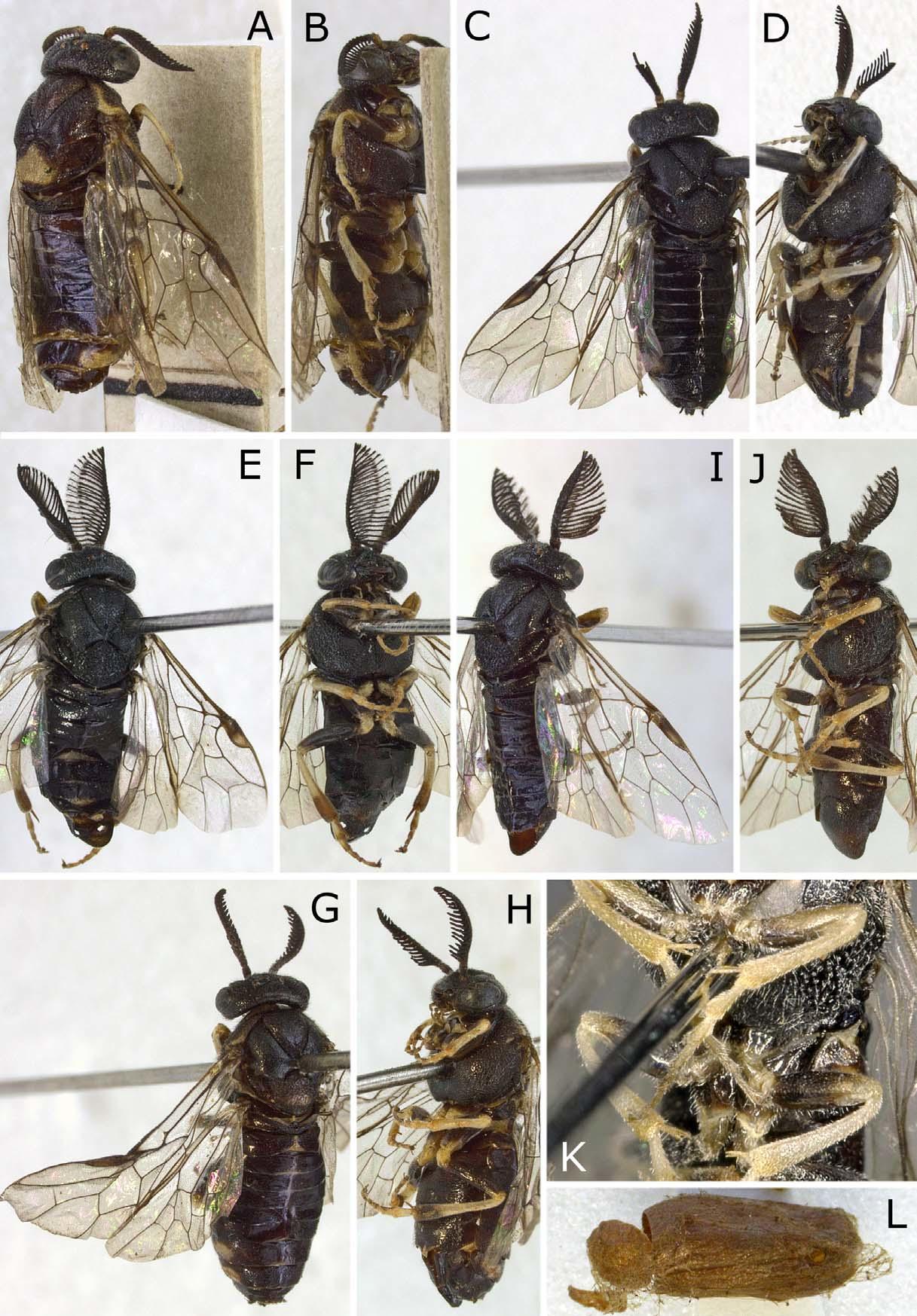
FIGURES 2 A – L. Nesodiprion biremis (A – F) and N. orientalis (G – L). N. biremis: A, B, Female, lectotype, dorsolateral and ventrolateral views; C, D, female, Guizhou Prov., Chishui, dorsal and ventral views; E, F, male, Guangdong Prov., Ruyuan, dorsal and ventral views. N. orientalis: G, H, Female, paratype, Thailand, Bo Luang, dorsal and ventrolateral views; I – L, male, holotype; I, J, dorsolateral and ventrolateral views; K, center of J; L, cocoon.
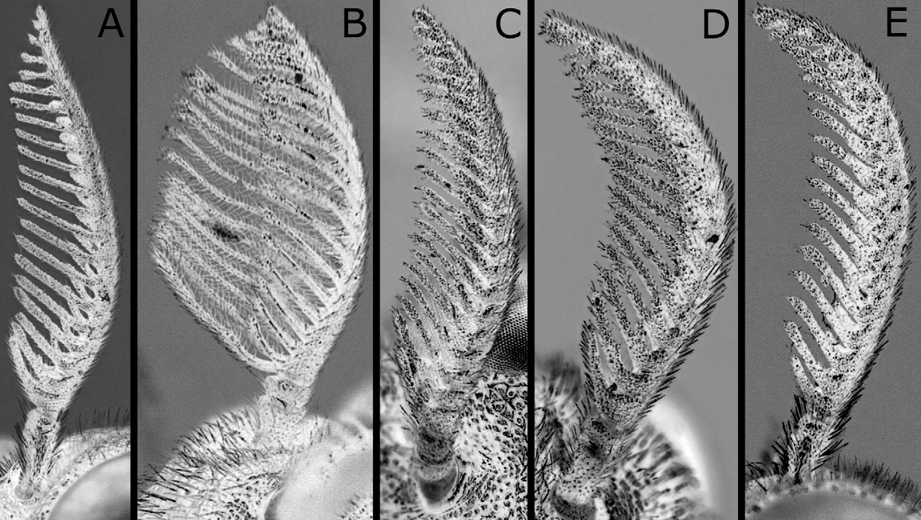
FIGURES 6 A – E. Antenna of Nesodiprion japonicus (A, B), N. biremis (C, D) and N. orientalis (E). A, C – E, Female; B, male. A, E, Outer lateral view; B – D, inner lateral view. D, E, Reversed images. N. japonicus: A, Amami-oshima; B, lectotype. N. biremis: C, Lectotype; D, Zhejiang Prov., Anji. N. orientalis: E, paratype, Thailand, Bo Luang.
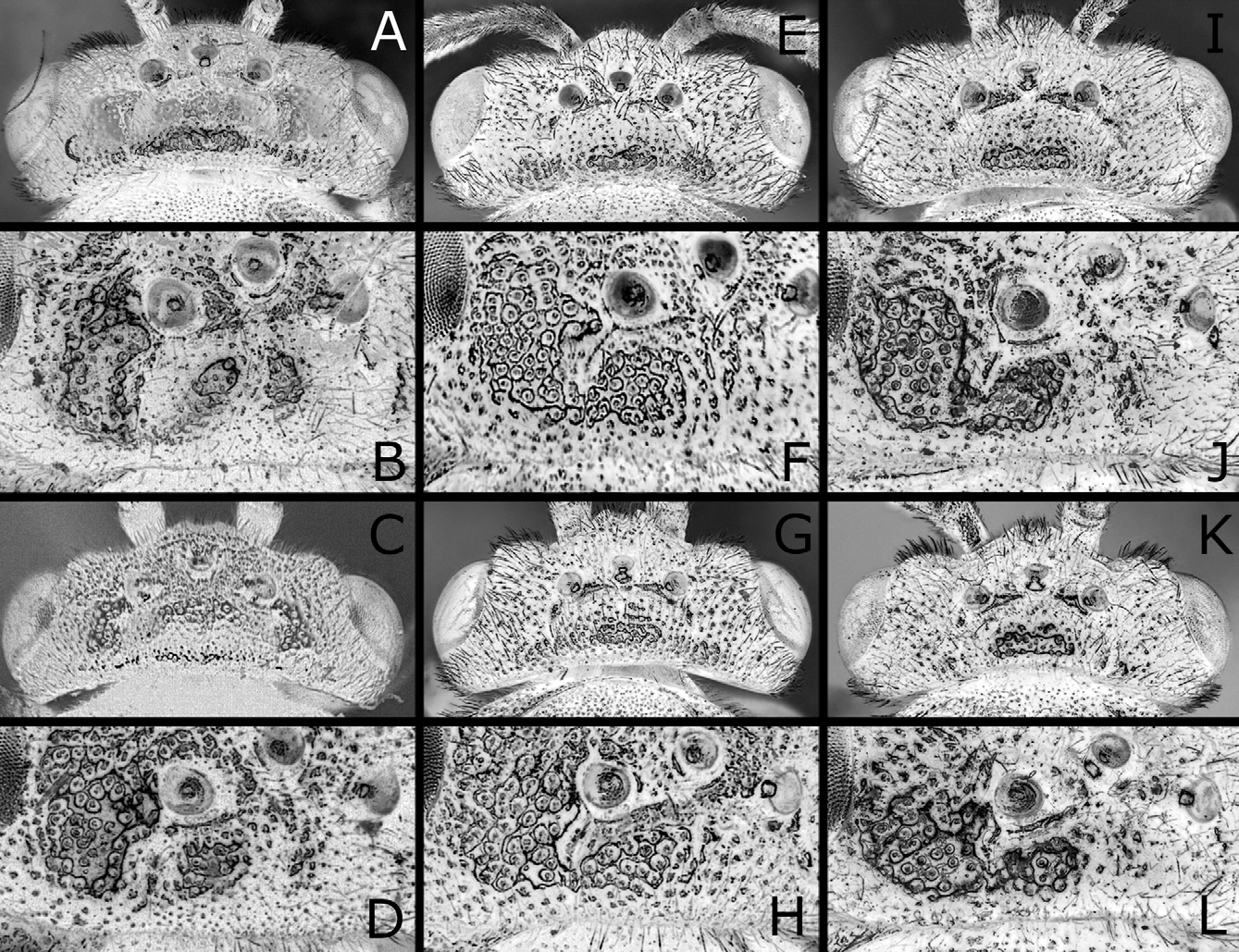
FIGURES 3 A – L. Head of Nesodiprion japonicus (A – D), N. biremis (E – H) and N. orientalis (I – L). A, E, I, Head, female, dorsal view; C, G, K, do., male; B, F, J, dorsum of head, female, dorsolateral view; D, H, L, do., male. N. japonicus: A, Amami-oshima; B, paralectotype, Honshu, Gifu; C, D, lectotype. N. biremis: E, F, Lectotype; G, H, Guangdong Prov., Ruyuan. N. orientalis: I, J, Paratype, Thailand, Bo Luang; K, L, holotype.
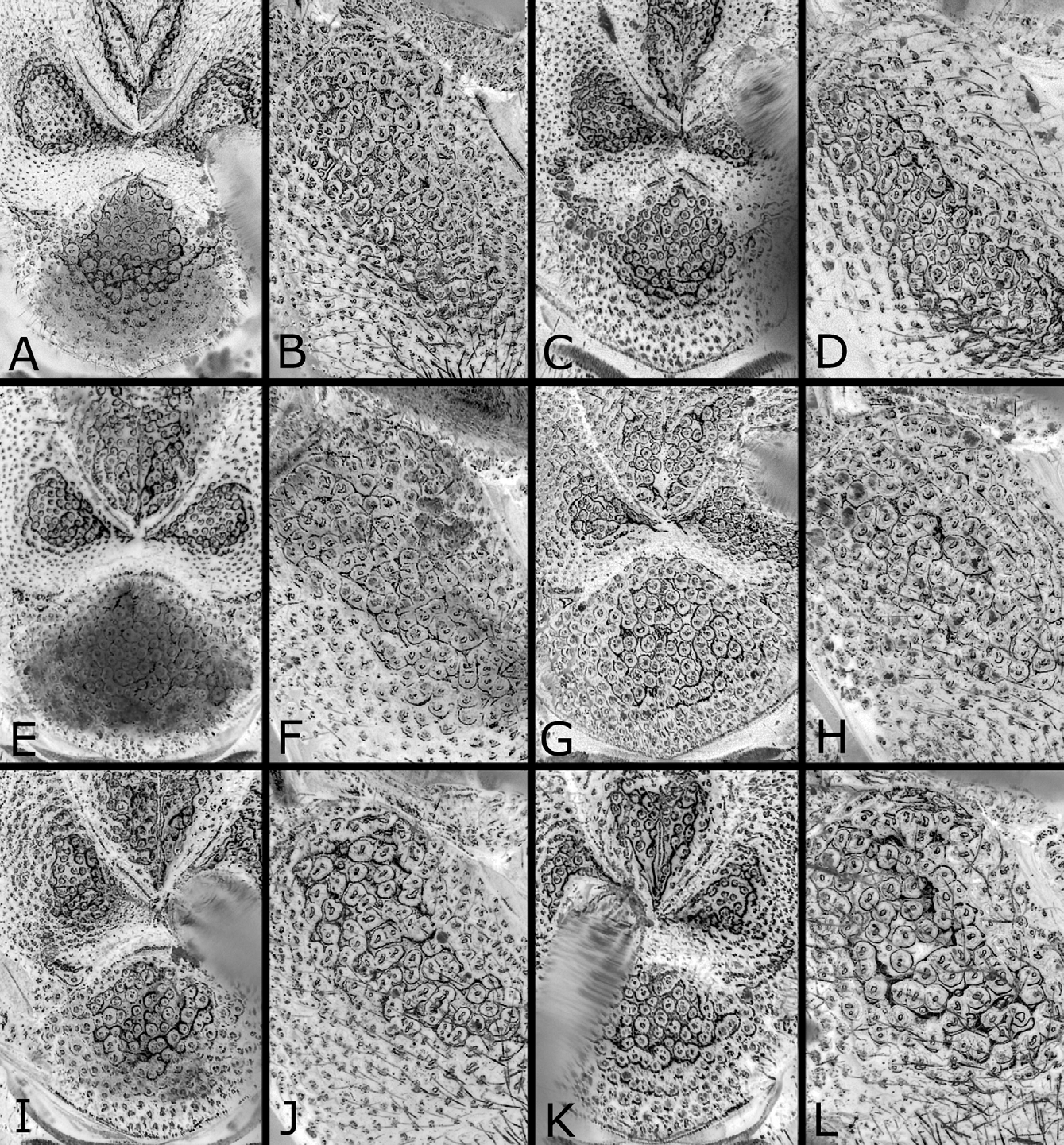
FIGURES 4 A – L. Thorax of Nesodiprion japonicus (A – D), N. biremis (E – H) and N. orientalis (I – L). A, E, I, Posterior part of mesoscutum and mesoscutellum, female, dorsal view; C, G, K, do., male; B, F, J, mesepisternum, female, lateral view; D, H, L, do., male. N. japonicus: A, B, Amami-oshima; C, lectotype; D, Honshu, Fukushima Pref. N. biremis: E, F, Lectotype; G, H, Guangdong prov., Ruyuan. N. orientalis: I, J, Paratype, Thailand, Bo Luang; K, L, holotype.
FIGURES 5 A – I. First abdominal tergum of Nesodiprion japonicus (A – C), N. biremis (D – F) and N. orientalis (G – I). A, D, E, G, H, Female; B, C, F, I, male. A, G, H, I, Reversed images. N. japonicus: A, Honshu, Zu; B, lectotype; C, Korea, Tokchomkogae. N. biremis: D, Lectotype; E, Zhejiang prov., Anji; F, Guangdong Prov., Ruyuan. N. orientalis: G, H, Paratypes, Thailand, Bo Luang; I, holotype.
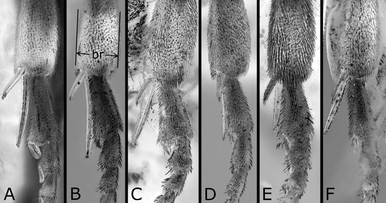
FIGURES 7 A – F. Apex of tibia and basal tarsomeres of hind leg of Nesodiprion japonicus (A, B), N. biremis (C, D) and N. orientalis (E, F), anterior view. A, C, E, Female; B, D, F, male. Small br in B indicates breadth. C, D, Reversed images. N. japonicus: A, Amami-oshima; B, Okinawa-jima. N. biremis: C, Lectotype; D, Guangdong Prov., Ruyuan. N. orientalis: E, Paratype, Thailand, Bo Luang; F, paratype, Thailand, “ Pine. Res. C. ”.

FIGURES 9 A – D. Saw of Nesodiprion japonicus (A, B), N. biremis (C) and N. orientalis (D). A, B, Lance, dorsal and lateral views; C, D, saw, lateral view. B – D, Reversed images. N. japonicus: A, B, Amami-oshima. N. biremis: C, Zhejiang Prov., Anji. N. orientalis: D, Paratype, Thailand, Bo Luang.
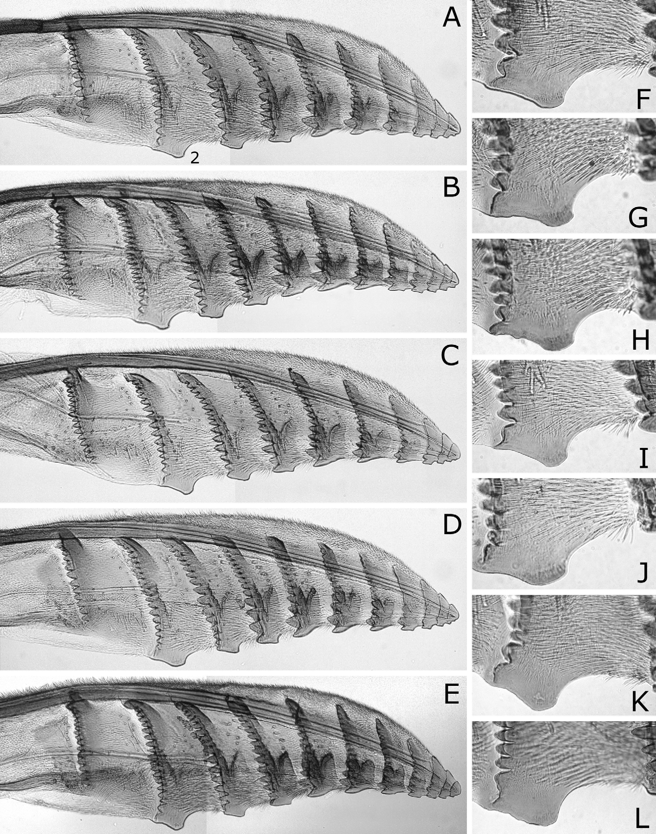
FIGURES 11 A – L. Lancet of Nesodiprion biremis. A – E, Lancet; F – L, serrula of second annulus. Small 2 in A indicates the second annulus. B, H, E, K, Reversed images. A, F, Lectotype; B, G, H, Shandong Prov., Taian; C, I, Zhejiang Prov., Anji; D. J, Guizhou Prov., Chishui; E, K, L, Jiangxi Prov., “ Ku-ling ”.
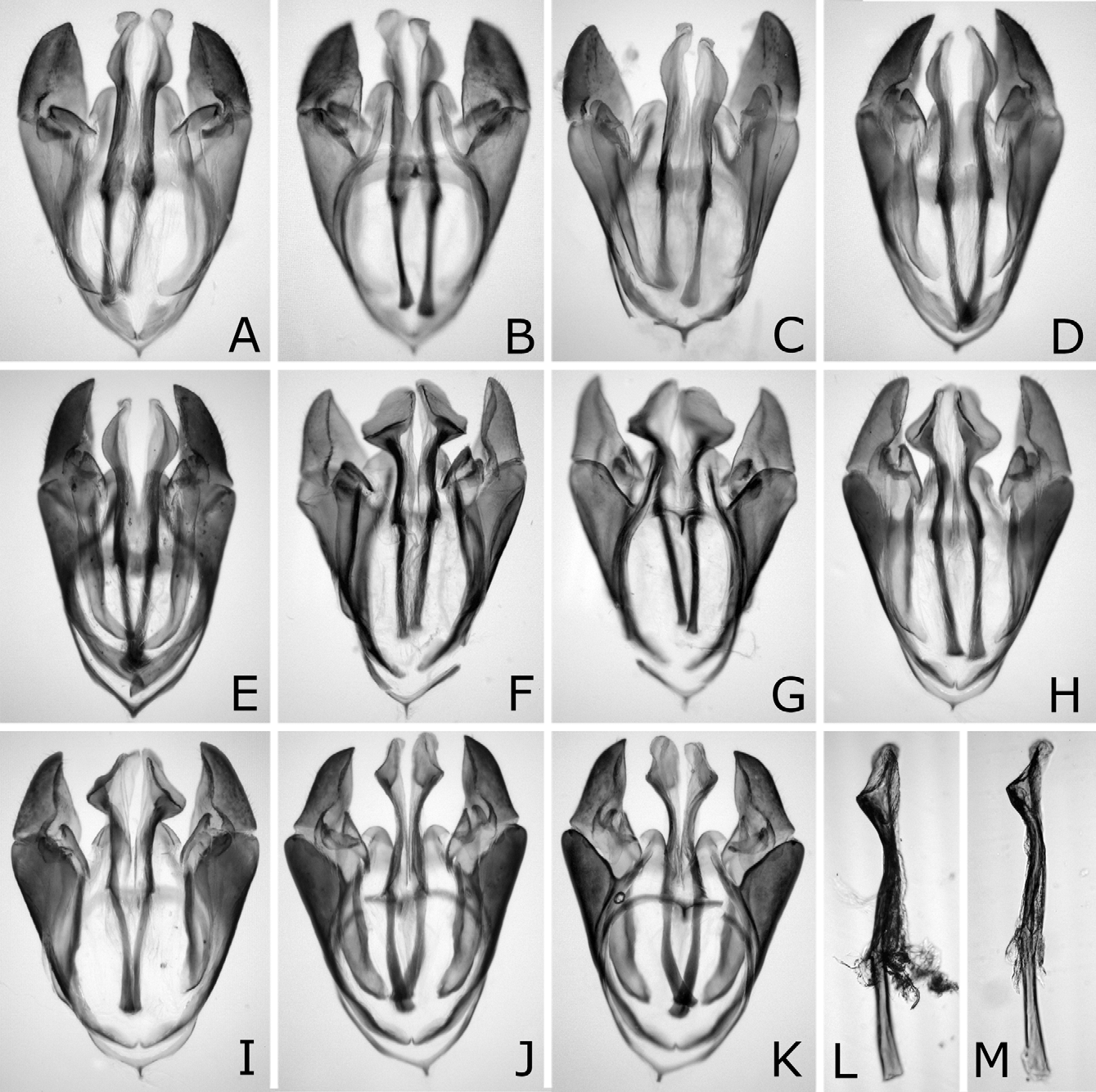
FIGURES 13 A – M. Male genitalia of Nesodiprion japonicus (A – E), N. biremis (F – I) and N. orientalis (J – M). A, C – F, H – J, Male genitalia, dorsal view; B, G, K, do., ventral view; L, M, right penis valve, dorsal view. N. japonicus: A, B, Lectotype; C, Amamioshima; D, Korea, Iksan; E, Taiwan. N. biremis: F, G, Guangdong prov., Ruyuan; H, Zhejiang Prov., Anji; I, Shandong Prov., Taian. N. orientalis: J, K, Holotype; L, paratype, Thailand, “ Pine Res. C. ”; M, Yunnan Prov., Anning.
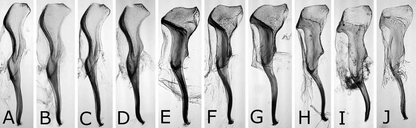
FIGURES 14 A – J. Penis valve of Nesodiprion japonicus (A – D), N. biremis (E – G) and N. orientalis (H – J), lateral view. I, J, Reversed images. N. japonicus: A, Lectotype; B, Amami-oshima; C, Korea, Iksan; D, Taiwan. N. biremis: E, Guangdong prov., Ruyuan; F, Zhejiang Prov., Anji; G, Shandong Prov., Taian. N. orientalis: H, Holotype; I, paratype, Thailand, “ Pine Res. C. ”; J, Yunnan Prov., Anning.
FIGURES 10 A – L. Lancet of Nesodiprion japonicus. A – E, Lancet; F – L, serrula of second annulus. Small 2 in A indicates second annulus. D, I, L, Reversed images. A, F, Hokkaido, Mori; B, G, Honshu, Kyoto; C, H, Amami-oshima; D, I, J, Korea, Chuncheon; E, K, L, Taiwan.
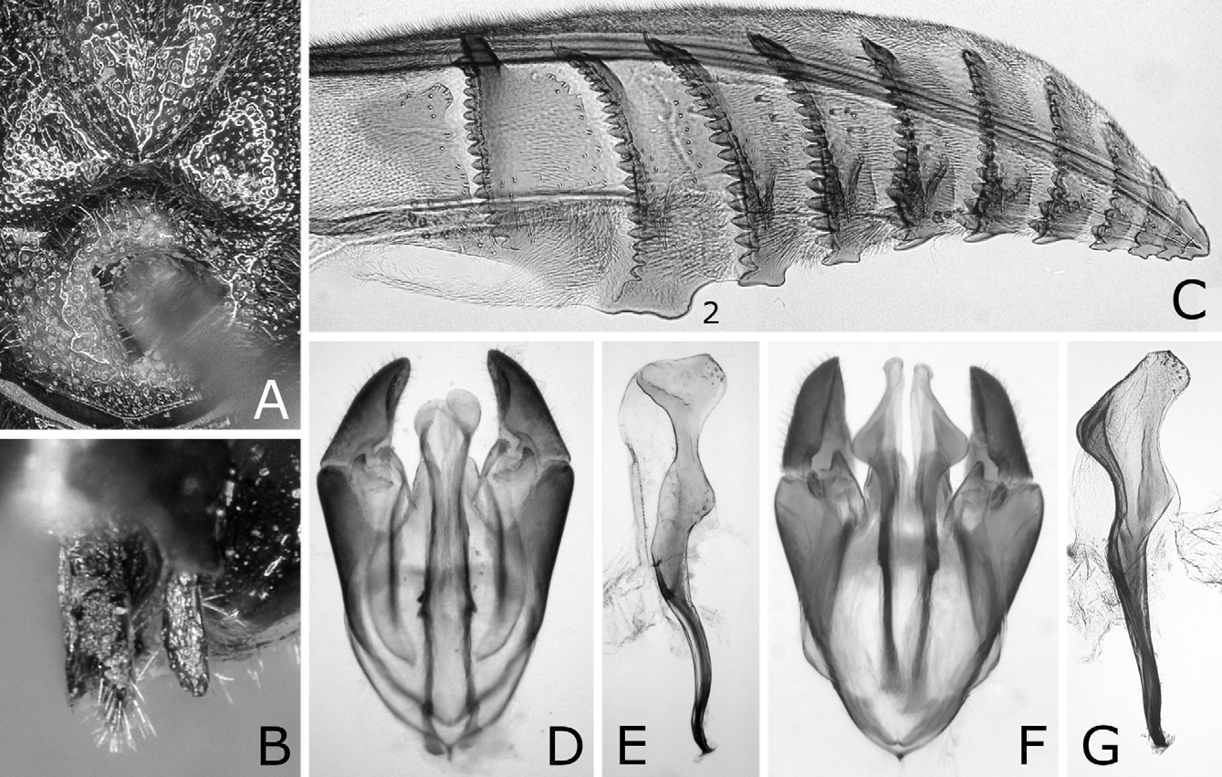
FIGURES 16 A – G. Nesodiprion sp. (? huanglongshanicus) (A – E) and N. kagaensis (F, G) A, Posterior part of mesoscutum and mesoscutellum, female, dorsal view; B, sawsheath and cercus, dorsal view (left sawsheath removed); C, lancet; D, F, male genitalia, dorsal view; E, G, penis valve, lateral view (left dorsal). Small 2 in C indicates the second annulus. N. sp.: A – C, Taiwan, Riyuetan; D, E, do. N. kagaensis: F, G, Honshu, Morioka.

No known copyright restrictions apply. See Agosti, D., Egloff, W., 2009. Taxonomic information exchange and copyright: the Plazi approach. BMC Research Notes 2009, 2:53 for further explanation.
|
Kingdom |
|
|
Phylum |
|
|
Class |
|
|
Order |
|
|
Family |
|
|
Genus |
Nesodiprion biremis ( Konow, 1899 )
| Hara, Hideho & Smith, David R. 2012 |
Nesodiprion biremis:
| Taeger 2010: 209 |
| Wei 2006: 551 |
| Rohwer 1910: 104 |
Lophyrus biremis
| Oehlke 1984: 370 |
| Konow 1899: 43 |
1 (by plazi, 2016-04-13 09:12:23)
2 (by ImsDioSync, 2016-12-19 15:40:21)
3 (by ImsDioSync, 2016-12-19 15:42:23)
4 (by ImsDioSync, 2017-06-17 14:19:02)
5 (by ImsDioSync, 2017-06-17 19:17:39)
6 (by ExternalLinkService, 2019-09-26 18:55:20)
7 (by ExternalLinkService, 2019-10-18 14:08:54)
8 (by ExternalLinkService, 2022-01-30 12:43:58)
9 (by ExternalLinkService, 2022-02-20 05:03:51)
10 (by plazi, 2023-10-26 10:39:04)