
Piromis suni, Salazar-Vallejo, Sergio I., 2011
|
publication ID |
https://doi.org/ 10.5281/zenodo.277211 |
|
DOI |
https://doi.org/10.5281/zenodo.6183597 |
|
persistent identifier |
https://treatment.plazi.org/id/D34C87B8-4D24-2636-FF44-FC7D6511FE7E |
|
treatment provided by |
Plazi |
|
scientific name |
Piromis suni |
| status |
sp. nov. |
Piromis suni View in CoL n. sp.
Figure 9 View FIGURE 9
Stylarioides eruca: Okuda 1937a:52 View in CoL , Text fig. 2–3 (non Claparède, 1868). Pherusa eruca: Imajima & Hartman 1964:302 View in CoL –303 (non Claparède, 1868).
Type material. South China Sea. Holotype (SMF-15348) and paratypes ( SMF), Senckenberg Museum Hainan 1992 Expedition, Sta. B92-17B-13, Sanya Bay (37°07ʹ0 3ʺ N, 122°30ʹ30ʺ E), Shandong Sheng, China, 6 m, 25 Mar. 1992, D. Fiege, coll.
Additional material. Japan. An anterior fragment (CMNH-1051), damaged, off Shimoda, Japan, 40 m (about 20 chaetigers, cephalic cage chaetae broken; sediment grains completely cover the body, dorsally and ventrally; neurochaetae all bidentate, with short articles).
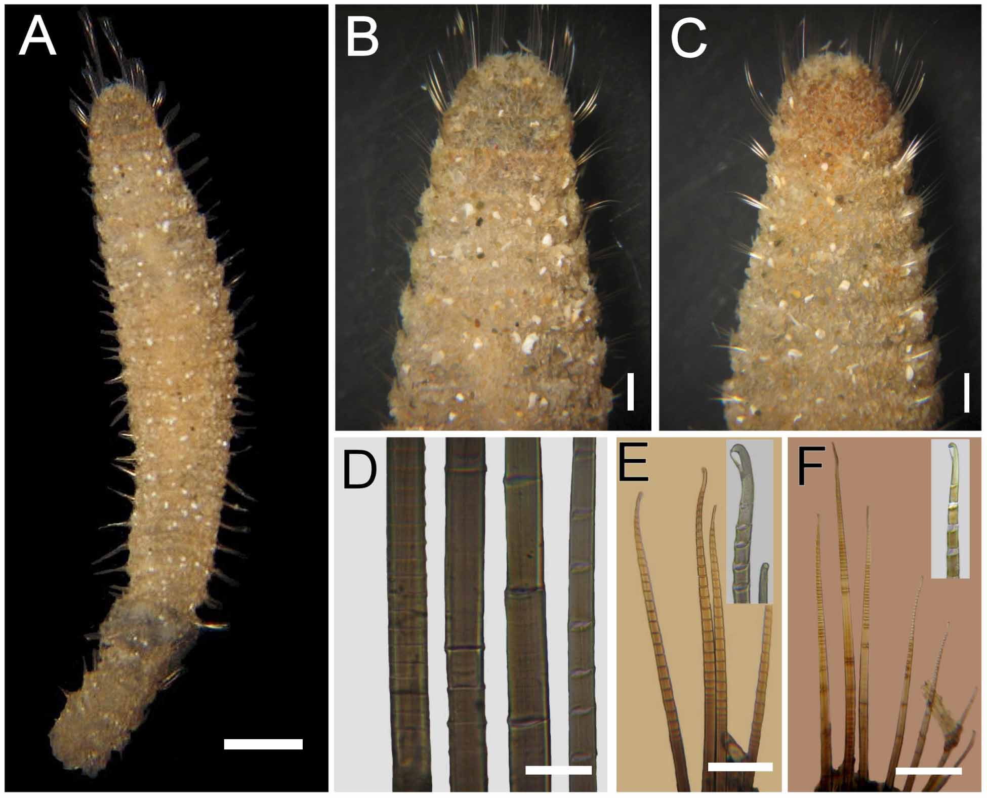
Description. Holotype (SMF-15348) anterior fragment ( Fig. 9 View FIGURE 9 A); body tapering posteriorly to blunt cone in paratypes, pale brown; tunic papillated with sediment particles abundant, densely packed, completely covering body wall; anterior region not bent ventrally. Larger capitate papillae arranged in transverse rows, one per segment dorsally ( Fig. 9 View FIGURE 9 B) and ventrally ( Fig. 9 View FIGURE 9 C), each with about ten papillae per row. Holotype 22 mm long, 3.8 mm wide, cephalic cage 4 mm long, 30 chaetigers.
Cephalic hood not exposed. Holotype not dissected to avoid further damage; paratype transparent, without eversible anterior end, probably exposed and broken during sampling. Anterior end details unknown.
Cephalic cage chaetae about as long as body width. Chaetigers 1-3 involved in the cephalic cage; chaetae arranged in short rows, dorsolateral in chaetiger 1, lateral in chaetigers 2-3; chaetiger 1 with seven notochaetae and six neurochaetae per side; chaetigers 2–3 with 5–6 notochaetae and 4 neurochaetae per side.
Anterior dorsal margin of chaetiger 1 with a trifid swollen bulb, median projection broken, tip lost, laterals about 1.5 times longer than median bulb. Anterior chaetigers with long papillae, chaetigers 2–5 with notopodial lobes. Chaetigers 1–3 progressively larger. Chaetal transition from cephalic cage chaetae to body chaetae abrupt; shorter multiarticulate bidentate neurohooks start in chaetiger 4. Gonopodial lobes not seen.
Parapodia well developed, forming notopodial lobes in anterior chaetigers (1–5). Parapodia lateral, median neuropodia ventrolateral. Noto- and neuropodia with long chaetal lobes and long digitate papillae, two pre- and two postchaetal papillae per ramus.
Median notochaetae arranged in short transverse rows, 7–8 per bundle, about 1/3 as long as body width; all notochaetae multiarticulated capillaries ( Fig. 9 View FIGURE 9 D), short articles basally, medium-sized then long medially, becoming slightly shorter distally. Neurochaetae multiarticulated capillaries in chaetigers 1–3; multiarticulated bidentate neurohooks from chaetiger 4, with articles medium-sized medially, becoming long by chaetiger 10 ( Fig. 9 View FIGURE 9 E). Each median neurohook with short articles basally, long articles medially, then progressively decreasing to mediumsized articles almost to the distal article (3–4 times longer than wide). Median and posterior neurochaetae with anchylosed articles basally, 2–3 long articles medially, and then short to medium-sized articles to the distal article. ( Fig. 9 View FIGURE 9 F). Tip hooked, bidentate; accessory tooth flat, wide, as long as the fang.
Posterior end blunt (observed in paratype), last 20 chaetigers without sediment particles; pygidium conical, anus dorso-terminal, without anal cirri.
Etymology. This species is being named after Dr. Ruiping Sun, from the Institute of Oceanology, Academia Sinica, in recognition of his many publications on the taxonomy of Chinese polychaetes, and because he was involved in the field and lab work during the Senckenberg Museum Hainan Expedition.
Type locality. Sanya Bay, South China Sea.
Variation. Paratypes were an anterior and a posterior fragments; anterior fragment damaged, 32.5 mm long, 4.0 mm wide, cephalic cage 2 mm long, 52 chaetigers; whereas the posterior fragment had 48 chaetigers; without sediment particles over the last 20 chaetigers.
Remarks. Piromis suni n. sp. is closely allied to P. e r u c a ( Claparède, 1868) in the general body pattern. They clearly differ in the type of neurohooks, however. In P. suni n. sp. the median and posterior chaetigers have neurohooks with a single or a few long articles medially, while the distal part is multiarticulated and continuing as such almost to the tip. In contrast, in P. eruca , median and posterior neurospines have few long articles while the distal one is often the longest. The previous records of P. e r u c a from Japan do not agree with the Mediterranean species, and are herein regarded as likely conspecific with P. suni .
Distribution. The species is widespread in the Western North and Central Pacific Ocean, from Japan to the South China Sea, in shallow water.
| SMF |
Forschungsinstitut und Natur-Museum Senckenberg |
No known copyright restrictions apply. See Agosti, D., Egloff, W., 2009. Taxonomic information exchange and copyright: the Plazi approach. BMC Research Notes 2009, 2:53 for further explanation.
|
Kingdom |
|
|
Phylum |
|
|
Class |
|
|
Order |
|
|
Family |
|
|
Genus |
Piromis suni
| Salazar-Vallejo, Sergio I. 2011 |
Stylarioides eruca:
| Imajima 1964: 302 |
| Okuda 1937: 52 |