
Leptoplectus kijimunaa Jałoszyński & Nomura, 2021
|
publication ID |
https://doi.org/ 10.11646/zootaxa.4915.3.9 |
|
publication LSID |
lsid:zoobank.org:pub:45463ABA-2457-416F-B9A5-47A26AFF0ACA |
|
DOI |
https://doi.org/10.5281/zenodo.4495399 |
|
persistent identifier |
https://treatment.plazi.org/id/D36187C2-FF84-895C-42B8-F9EAFEBDFDB4 |
|
treatment provided by |
Plazi |
|
scientific name |
Leptoplectus kijimunaa Jałoszyński & Nomura |
| status |
sp. nov. |
Leptoplectus kijimunaa Jałoszyński & Nomura View in CoL sp. n.
( Figs 1–27 View FIGURES 1–4 View FIGURES 5–11 View FIGURES 12–18 View FIGURES 19–27 )
Type material. Holotype: JAPAN (Okinawa Pref.): ♁, two labels: „JAPAN, OKINAWA Island / Rd. 58 Todoroki waterfall / - Michi-no-eki Kyoda , / ~ 10 m, roadside, 08.02.2019 / under bark of dead Vernicia / tree, leg. P. JAŁOSZYŃSKI” [white, printed], “ LEPTOPLECTUS / kijimunaa / Jałoszyński & Nomura / HOLOTYPUS ” [red, printed] ( NSMT) . Paratypes (12 exx.): 1 ♁, 3 ♀♀ (one disarticulated and mounted in Canada balsam), same data as for holotype; 2 ♁♁, same data except for 17.02.2019 ; 1 ♁, Yona, Kunigami-son, Okinawa Is. , 18. ix. 2017, N. Tokushige leg. ; 2 ♁♁, 1 ♀, same data except 7. x. 2017 ; 2 ♁♁, Mt. Nago-dake, Nago-shi, Okinawa Is. , 22. xii. 2018, N. Tokushige leg. (paratypes in NSMT, MNHW, cPJ) .
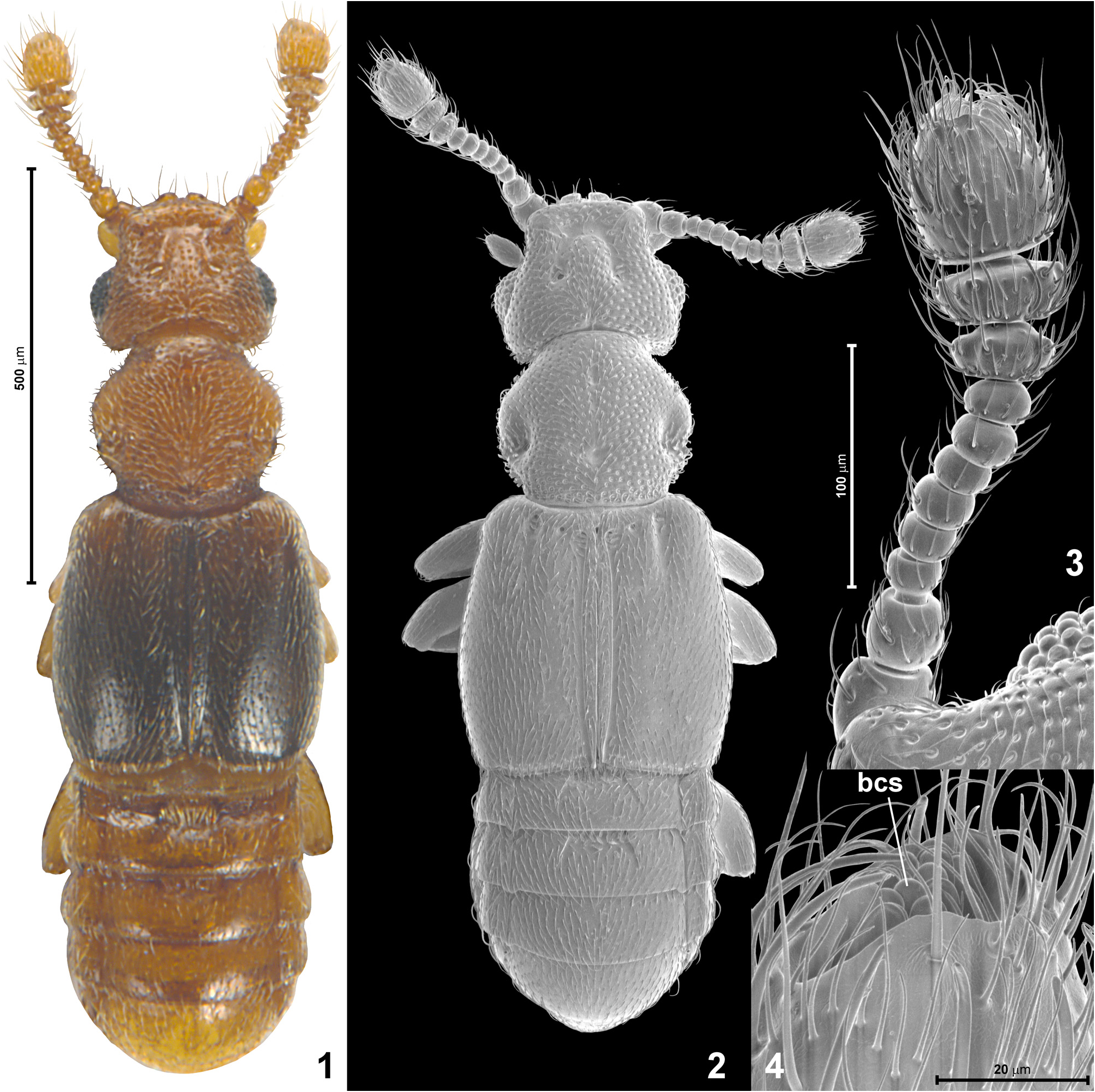
Diagnosis. Elytra in both sexes infuscate, distinctly darker than other body parts; aedeagus with several conspicuously long and slender apical projections, in lateral view forming two separate groups, distoventral and distodorsal, and with transverse row of sparse long setae, in dorsal view situated on right side of median lobe; basal capsule of aedeagus symmetrical and with basal margin evenly rounded, lacking basal projection.
Description. Body of male ( Figs 1–2 View FIGURES 1–4 ) moderately dark brown with distinctly infuscate elytra, setae slightly lighter than cuticle; BL 1.06–1.09 mm.
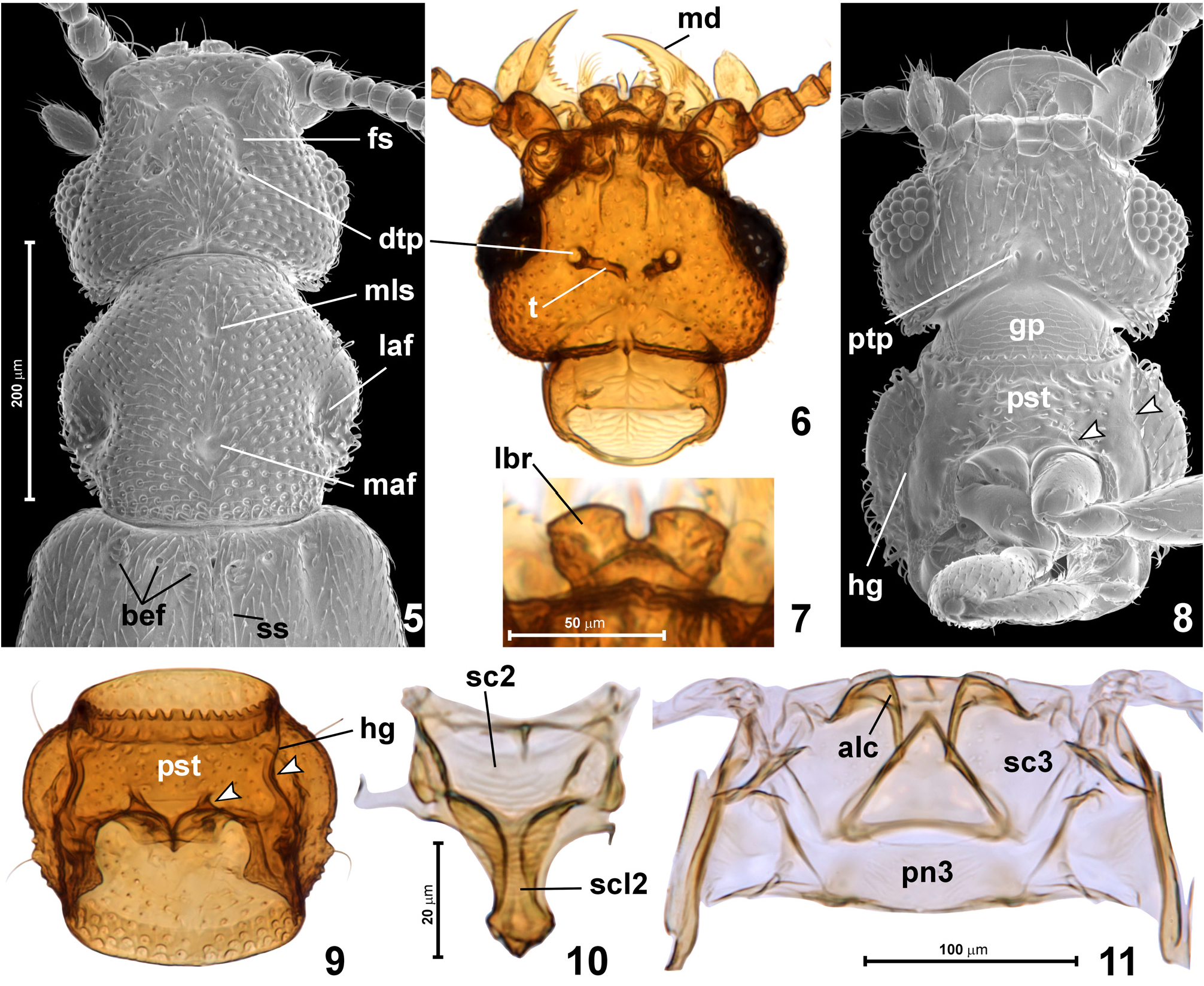
Head ( Figs 1–2 View FIGURES 1–4 , 5–6, 8 View FIGURES 5–11 ) subtrapezoidal, flattened, broadest at eyes, HL 0.16–0.18 mm, HW 0.22–0.23 mm. Vertex and sides of frons weakly convex except for distinct subtriangular median impression on posterior margin of vertex and well-developed U-shaped frontal sulcus ( Fig. 5 View FIGURES 5–11 ; fs) restricted to anterior half of head dorsum, complete and equally deep on entire length, posteriorly ending in dorsal tentorial pits ( Figs 5–6 View FIGURES 5–11 ; dtp); eyes large, strongly convex and coarsely faceted; tempora in dorsal view subequal to eye length. Setiferous punctures on frons and vertex large but shallow, evenly, densely distributed except for impunctate frontal sulcus and small impunctate area on each supra-antennal tubercle, spaces between punctures equal to 1–2 diameters of punctures. Setae on frons and vertex dense, short and weakly suberect, one pair of lateral setae on posterior margin of vertex and one lateral pair within anterior, transverse portion of frontal sulcus distinctly longer and slightly thicker than remaining ones. Antennae ( Fig. 3 View FIGURES 1–4 ) distinctly shorter than head and pronotum together; AnL 0.26–0.29 mm; scape about as long as broad; pedicel indistinctly elongate; antennomeres 3–8 each distinctly transverse and subequal in width; antennomeres 9–11 forming distinct club, 9 and 10 strongly transverse, 11 only slightly longer than broad, distinctly longer than 9–10 combined. Scape and pedicel sparsely and unevenly covered with long erect setae; antennomeres 3–8 each with one ring of sparse erect setae; antennomeres 9–10 each with two rings of similar setae and additionally with sub-basal ring of several long digitiform sensilla; antennomere 11 with sides densely covered with evenly distributed, long erect setae, sub-median ring of several long digitiform sensilla, and deeply, asymmetrically excavated apex bearing cluster of several (8 or 9) large basiconic sensilla with rounded apices ( Fig. 4 View FIGURES 1–4 ; bcs).
Tentorium ( Fig. 6 View FIGURES 5–11 ; t) reduced to separate vertical arms connecting with dorsal wall of head between anterior margins of eyes, and with ventral wall between posterior margins of eyes.
Gular plate ( Fig. 8 View FIGURES 5–11 ; gp) with transverse microreticulation; posterior tentorial pits ( Fig. 8 View FIGURES 5–11 ; ptp) small, each slightly elongate and situated distinctly in front of neck region; entire ventral surface from anterior submental margin to posterior margins of genae and postgenae covered with punctures and setae similar to those on frons and vertex; additionally tempora with modified, flattened setae near occipital constriction; transverse impressions demarcating neck region from anterior portion of head capsule and area adjacent to posterior tentorial pits impunctate and asetose. Area at mesoventral margin of each eye with arcuate impression step-wise delimited from median region of head.
Submentum ( Fig. 8 View FIGURES 5–11 ) with concave anterior margin; mentum subrectangular, with pair of anterior setae; labial palps with strongly elongate palpomere 2 and conspicuously long apical seta; cardines conspicuously large, quadrangular; lacinia and galea each elongate and with dense brush of setae along mesal and anterior margins; each mandible ( Fig. 6 View FIGURES 5–11 ; md) subtriangular, strongly elongate, with broad base, slender and sharply pointed distal portion and serrate mesal margin up to distal third; labrum ( Fig. 7 View FIGURES 5–11 ; lbr) with median emargination U-shaped, broad, and well-defined, with slightly obtuse-angled anteromesal and anterolateral corners; posterior, broadly subtriangular portion of labrum delimited by angulate, anteriorly convex dorsal transverse carina.
Pronotum ( Fig. 5 View FIGURES 5–11 ) sub-pentagonal, broadest indistinctly in front of middle; PL 0.20–0.21 mm, PW 0.23–0.24 mm. Anterior margin slightly arcuate; lateral margins in anterior half rounded and smooth, in posterior half slightly sinuate and with several denticles ( Fig. 9 View FIGURES 5–11 ); posterior margin nearly straight at middle and weakly arcuate laterally. Pronotal disc with median longitudinal sulcus ( Fig. 5 View FIGURES 5–11 ; mls) developed as small and only slightly elongate impression near anterior 1/4; pair of large but shallow lateral antebasal foveae ( Fig. 5 View FIGURES 5–11 ; laf) slightly behind middle; and small, shallow median antebasal fovea ( Fig. 5 View FIGURES 5–11 ; maf) in posterior third; lateral and median foveae connected by barely discernible, diffuse transverse impression (not visible in SEM micrograph, but visible under stereomicroscope under certain lighting angles). Pronotal disc nearly evenly covered with punctures similar to those on frons and vertex, except for narrow area along posterior margin, covered with distinctly larger and denser punctures; rudiment of median longitudinal sulcus and median antebasal fovea impunctate; openings of lateral antebasal foveae covered with small, indistinct punctures. Basic vestiture of pronotal disc composed of dense, short and weakly suberect setae; additionally each side with two long, erect setae (visible in Fig. 9 View FIGURES 5–11 ), and sides in anterior and posterior thirds with modified, flattened setae with truncate apices.
Prosternum ( Figs 8–9 View FIGURES 5–11 ; pst) longer than procoxae, laterally fused with hypomera (i.e., notosternal sutures obliterated); prosternal carina between procoxae narrow and weakly elevated; anteroventral margin of prothorax deeply serrate; hypomeron with deep longitudinal hypomeral groove ( Figs 8–9 View FIGURES 5–11 ; hg) bearing rudiment of lateral fovea in precoxal region; distinct opening of fovea is also situated on arcuate carina demarcating procoxal rests from basisternal region ( Figs 8–9 View FIGURES 5–11 ; foveae indicated by arrowheads).
Mesonotum ( Fig. 10 View FIGURES 5–11 ) with transverse, subrectangular mesoscutum ( Fig. 10 View FIGURES 5–11 ; sc2) and narrow, elongate mesoscutellum ( Fig. 10 View FIGURES 5–11 ; scl2) not delimited by scutoscutellar suture; scutellar shield exposed between elytral bases, but extremely small, barely discernible.
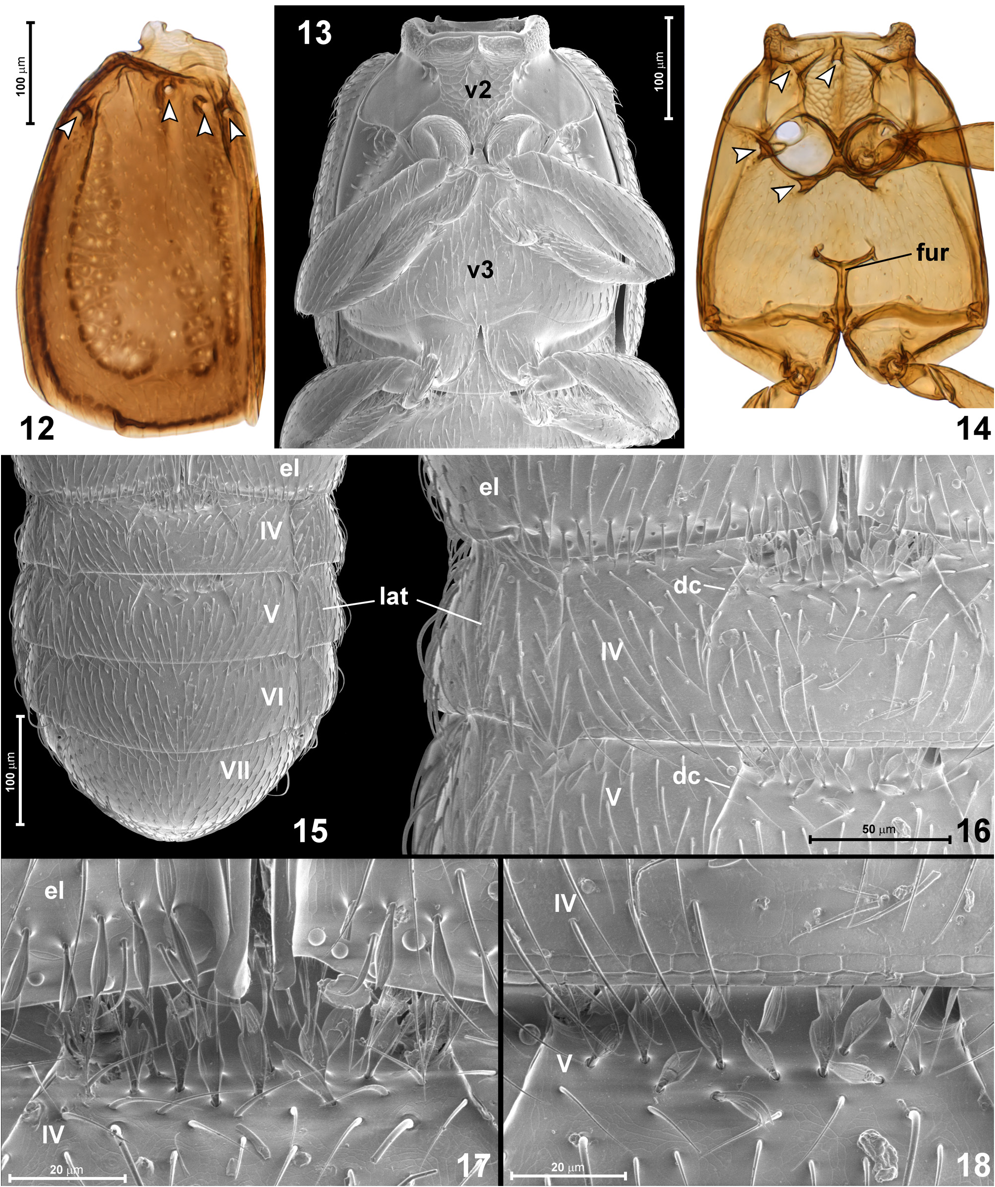
Elytra ( Figs 1–2 View FIGURES 1–4 , 5 View FIGURES 5–11 , 12 View FIGURES 12–18 ) together subrectangular with rounded sides, broadest slightly behind middle; EL 0.33 mm, EW 0.30–0.32 mm. Each elytron with complete sutural sulcus ( Fig. 5 View FIGURES 5–11 ; ss), one short and diffuse discal sulcus in anterior 1/4, three dorsal basal elytral foveae ( Fig. 5 View FIGURES 5–11 ; bef), and one lateral fovea not visible in dorsal view ( Fig. 12 View FIGURES 12–18 , indicated by arrowheads). Posterolateral elytral corner obtuse-angled; posterior elytral margin rounded; posterior sutural corner with small rounded projection ( Figs 12, 17 View FIGURES 12–18 ). Punctures on elytra much smaller than those on head and pronotum; setae similarly dense, short and suberect as those on pronotum; additionally area behind adsutural and discal basal foveae with 3–5 modified, flattened setae with pointed apices; similar setae are also distributed in one row just in front of posterior elytral margin ( Fig. 17 View FIGURES 12–18 ).
Hind wings well developed, functional.
Mesoventrite ( Figs 13–14 View FIGURES 12–18 ; v 2 View FIGURES 1–4 ) transverse, with lateral regions fully demarcated from metaventrite. Mesoventral intercoxal process subtrapezoidal, elongate, posteriorly overlapped by anterior metaventral process. Anteromedian region of mesoventrite with pair of shallow transverse procoxal rests with transverse rows of microtrichia; subtrapezoidal area posterior to procoxal rests and mesoventral intercoxal process covered with scale-like microsculpture and several setae; sides of mesoventrite smooth and asetose. Three foveae are situated on mesoventrite (indicated by arrowheads in Fig. 14 View FIGURES 12–18 ): pair of deep lateral foveae, and median fovea directed anterad; openings of lateral foveae with 4–5 modified, flattened setae with pointed apices; opening of median fovea asetose.
Metanotum ( Fig. 11 View FIGURES 5–11 ) transverse, largely membranous, with short alacristae ( Fig. 11 View FIGURES 5–11 ; alc) restricted to anterior half of metascutum ( Fig. 11 View FIGURES 5–11 ; sc3); metascutellum not differentiated; metapostnotum ( Fig. 11 View FIGURES 5–11 ; pn3) long and broad.
Metaventrite ( Figs 13–14 View FIGURES 12–18 ; v 3 View FIGURES 1–4 ) distinctly transverse and weakly convex at sides, flattened at middle, with distinct anterior metaventral process, which, together with mesoventral process, separates mesocoxae; narrow area between metacoxae distinctly notched. Metaventrite with two pairs of lateral foveae, one just behind and laterad mesocoxal cavities, and one behind arcuate carina demarcating posteriorly mesocoxal rest (foveae indicated by arrowheads in Fig. 14 View FIGURES 12–18 ). Metaventrite virtually impunctate and densely covered with short, nearly recumbent setae, except for asetose anterolateral regions laterad mesocoxae; additionally opening of each metaventral fovea and area lateroposterad each mesocoxa with several modified, flattened setae with pointed apices.
Metafurca ( Fig. 14 View FIGURES 12–18 ; fur) nearly T-shaped, with long stalk and strongly divergent lateral arms.
Legs short and robust, lacking any peculiar characters.
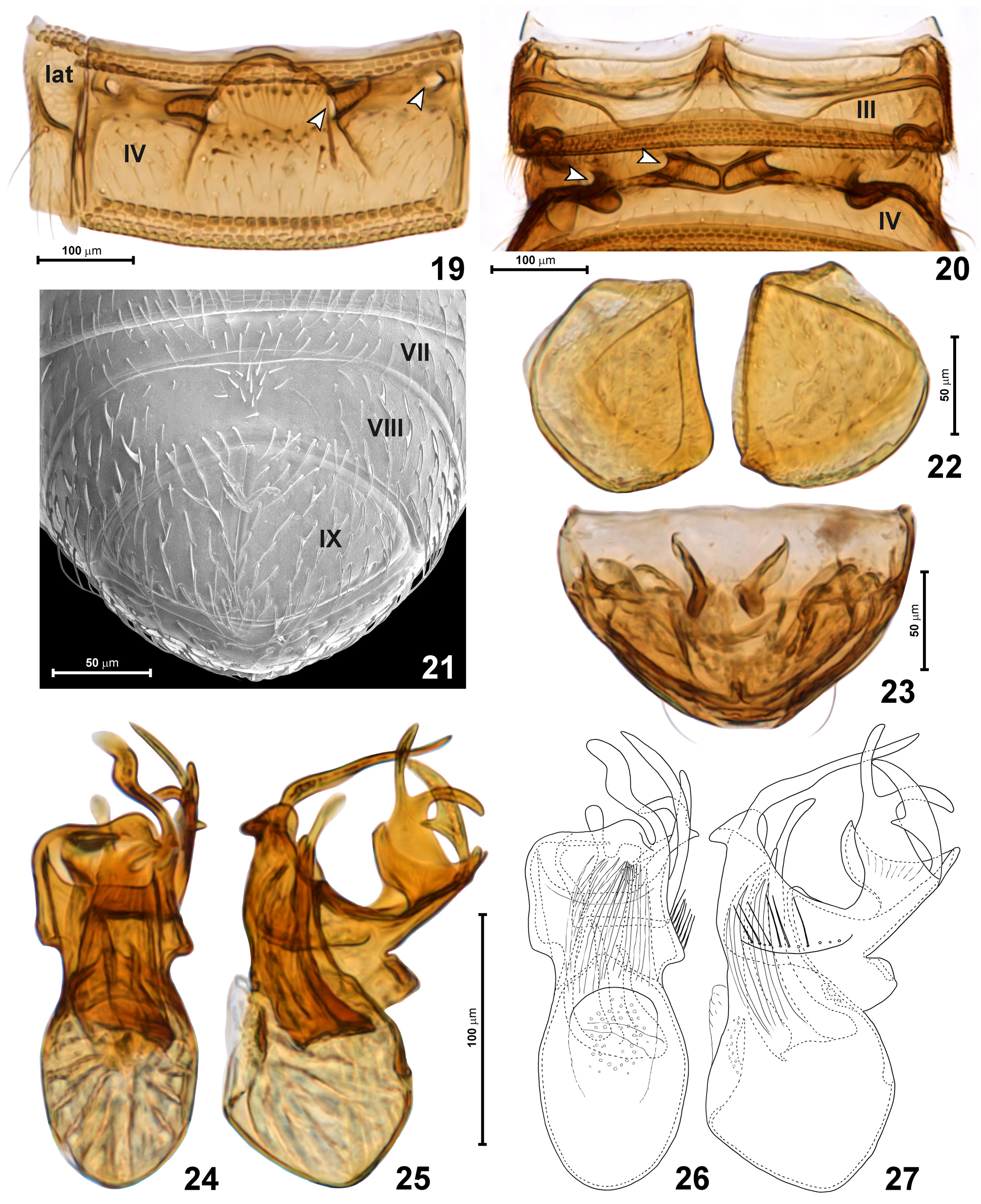
Abdomen ( Figs 15–22 View FIGURES 12–18 View FIGURES 19–27 ) only slightly longer than elytra, AbL 0.35–0.38 mm, AbW 0.28 mm. Tergites IV–VI subequal in length, VII about 1.5 × as long as VI; tergites IV and V each with sharply marked and posteriorly strongly divergent discal carinae ( Fig. 16 View FIGURES 12–18 ; dc) reaching one-half length of tergite, area between anterior ends of carinae distinctly narrower than 1/3 width of segment, only weakly impressed and at base with two irregular rows of modified, flattened setae with pointed apices ( Figs 17–18 View FIGURES 12–18 ); tergites IV and V with pair of deep foveae situtated at base of mesal margin of discal carinae and directed laterad ( Fig. 19 View FIGURES 19–27 ; indicated by arrowheads); additionally tergite IV with one pair of shallow anteroateral foveae near border with laterotergite ( Fig. 19 View FIGURES 19–27 ; lat); the latter foveae are covered by elytra. Surface of tergites and laterotergites virtually impunctate and densely covered with short, nearly recumbent setae. Sternite III ( Fig. 20 View FIGURES 19–27 ) with distinct metacoxal rests posteriorly demarcated by arcuate carinae; sternite IV ( Fig. 20 View FIGURES 19–27 ) with two pairs of foveae: submedian pair with broadly separated openings but deep subcuticular pockets meeting at middle; and shallow lateral foveae. Surface of all sternites with punctures and setae similar to those on tergites, except for sternite VIII ( Fig. 21 View FIGURES 19–27 ), which bears broad median impression without defined margins, oval lateral asetose regions of impression are separated at middle by group of short setae directed anterad and anterolaterad. Sternite IX ( Figs 21–22 View FIGURES 19–27 ) completely divided into lateral and slightly asymmetrical halves, division slightly slanted in relation to long axis of abdomen.
Aedeagus ( Figs 24–27 View FIGURES 19–27 ) elongate; AeL 0.20 mm; median lobe in dorsal view with symmetrical basal capsule, indistinctly demarcated diaphragm; distal portion of aedeagus demarcated by distinct sub-median constriction, strongly asymmetrical, bearing elaborate complex of elongate projections; in lateral view projections form distodorsal and distoventral groups; in dorsal view the right side of median lobe with transverse row of long, sparsely distributed setae; endophallus with weakly sclerotized group of densely assembled, needle-like sclerites.
Female. Externally differs from male only in abdominal sternite VIII lacking median impression and undivided sternite IX. Sclerotized postabdominal structures composed of pair of elongate, curved elements ( Fig. 23 View FIGURES 19–27 ); BL 1.06–1.08 mm; HL 0.18 mm, HW 0.21–0.23 mm, AnL 0.28 mm; PL 0.21–0.23 mm, PW 0.23 mm; EL 0.33 mm, EW 0.30 mm; AbL 0.35 mm, AbW 0.29 mm.
Distribution. Okinawa Island, Ryukyu Archipelago, Japan.
Etymology. In Okinawan language, Kijimunaa (Japanese: kijimuna; noun in apposition) are forest spirits taking a form of red-haired little children living in trees. As there is a tradition to give Japanese names to the Pselaphinae species that occur in this country, we name the new Leptoplectus “Kijimunaa-konaga arizukamushi”.
Remarks. Leptoplectus kijimunaa has the most complicated aedeagus of all members of this genus, with several elongate and variously curved distal projections. This character alone allows for unambiguous identification. The elytra darker than remaining body parts can be found in L. similis , and also in a paratype of L. illex (elytra of holotype are not infuscate). The new species differs from each previously known Leptoplectus (in alphabetical order) in the following structures:
- from Leptoplectus anguinus (aedeagus not illustrated in literature) in head and pronotum approximately equal in width (in L. anguinus “prothorax capite paulo angustior”, i.e., prothorax slightly narrower than head); elytra together much broader than head (in L. anguinus “elytra capiti latitudine aequalis”, i.e., elytra equal in width to head);
- from Leptoplectus annamita (aedeagus illustrated by Jeannel 1952: fig. 149) in sides of pronotum bearing a tiny denticle at the level of lateral antebasal foveae (lacking in L. annamita ); and aedeagus with elongate, rod-like distal projections (lacking in L. annamita );
- from Leptoplectus falcifer (aedeagus illustrated by Kurbatov 1992a: fig. 2.7) in frontal sulcus complete, Ushaped, unmodified (in L. falcifer lateral arms of sulcus are short, demarcated from anterior transverse portion of sulcus by transverse ridges, and anterior portion of sulcus developed as large and deep impression); discal carinae of tergites IV and V equal in length and not exceeding one-half length of tergite (in L. falcifer carinae of tergite IV reach 2/3 length of tergite, and those on V reach half of its length); basal capsule of aedeagus lacking long median proximal projection (present in L. falcifer ); setae on median lobe of aedeagus much closer to constriction demarcating basal capsule than to aedeagal apex (in L. falcifer setae are situated near middle between constriction and aedeagal apex); and apex of median lobe in dorsal view sub-truncate (in L. falcifer with deep, asymmetrically situated U-shaped emargination);
- from Leptoplectus illex (aedeagus illustrated by Kurbatov 1992a: fig. 2.5) in head not broader than pronotum (broader in L. illex ); complete frontal sulcus (in L. illex incomplete, with longitudinal arms not connected with transverse anterior portion); large eyes subequal in length with tempora (in L. illex eyes distinctly shorter than tempora); antennomere 3 distinctly transverse (in L. illex as long as broad); antennomere 11 distinctly shorter than 7–10 combined (in L. illex antennomere 11 as long as 7–10 together); discal carinae of tergites IV and V not exceeding one-half length of tergite (in L. illex carina on tergite IV as long as 3/4 length of tergite, and that on tergite V as long as 2/3 length of tergite); basal capsule of aedeagus lacking long asymmetrically placed proximal projection (present in L. illex ); aedeagus in dorsal view lacking rod-like apical projection on the right side bent lateroproximad (such projection is present in L. illex );
- from Leptoplectus nipponensis (aedeagus illustrated by Jeannel 1958: fig. 7) in head as broad as pronotum (in L. nipponensis head broader than pronotum); basal capsule of aedeagus narrower than distal half of median lobe (in L nipponensis clearly broader); and aedeagus in dorsal view lacking apical projections on the left side (with three projections in L. nipponensis ), and with four on the right side (one in L. nipponensis );
- from Leptoplectus perraulti (aedeagus illustrated by Besuchet 1993: fig. 1) in the labrum bilobed (not emarginate in L. perraulti ; see Discussion); basal capsule of aedeagus lacking proximal subconical projection (present in L. perraulti ); in dorsal view, a row of long setae on median lobe of aedeagus situated on its right side (on the left side in L. perraulti ); and several apical projections of aedeagus (only one, short and sinuate in L. perraulti );
- from Leptoplectus pertenuis (aedeagus illustrated by Grigarick & Schuster 1980: plate 39, fig. 8) in head as broad as pronotum (in L. pertenuis head clearly broader than pronotum); lateral foveae on hypomeral grooves extremely shallow, barely discernible (deep, with long subcuticular pockets in L. pertenuis ); lateral lobes of labrum nearly 3 times as broad as median notch between them (in L. pertenuis notch broader than each lobe); mesoventral intercoxal process posteriorly overlapped with anterior metaventral process (in L. pertenuis processes between mesocoxae separated by a gap); basal capsule of aedeagus lacking asymmetrical subconical proximal projection (present in L. pertenuis ); and distal half of aedeagus in dorsal view with subparallel sides (in L. pertenuis sides form large rounded lobes strongly projecting laterad);
- from Leptoplectus pumilio (aedeagus illustrated by Kurbatov 1992a: fig. 2.11) in frons in front of frontal sulcus with similar punctures as those on sides and on vertex (in L. pumilio this area is virtually impunctate, contrasting with densely punctate sides of frons and vertex); eyes subequal in length to tempora (in L. pumilio distinctly longer than tempora); basal capsule of aedeagus lacking asymmetrically placed, proximal projection (present in L. pumilio ); distal portion of median lobe in dorsal view (excluding distal projections) not much broader than basal capsule, and with sub-parallel sides (in L. pumilio distal region much broader than basal capsule, with sides strongly divergent distad);
- from Leptoplectus remyi (aedeagus illustrated by Jeannel 1961: fig. 6) in antennomeres 3–5 each strongly transverse (in L. remyi about as long as broad); basal capsule of aedeagus narrower than distal half of median lobe (in L. remyi broader); and in having four elongate apical projections of the aedeagus (one with two arms, and one with additional short projection, so six separate lobes can be seen) (vs. only two projections in L. remyi );
- from Leptoplectus sarawakensis (aedeagus illustrated by Besuchet 1956: figs 1–2) in head not broader than pronotum (broader in L. sarawakensis ); head dorsum and pronotum distinctly, densely punctate (with small and sparse punctures in L. sarawakensis ); basal capsule of aedeagus lacking proximal subconical projection (present in L. sarawakensis ); and a group of several slender asymmetrically placed apical projections of median lobe (one thick and nearly straight median apical projection in L. sarawakensis );
- from Leptoplectus similis (aedeagus illustrated by Kurbatov 1991a: fig. d) in antennomere 3 strongly transverse (in L. similis nearly as long as broad) and antennomeres from 3 to 8 subequal in length (reducing in length in L. similis ); pronotum not narrower than head (in L. similis slightly narrower); length of discal carinae of tergites IV and V not exceeding one-half length of each tergite (exceeding in L. similis ); basal capsule of aedeagus lacking asymmetrically placed, proximal projection (present in L. similis ); distal portion of median lobe in dorsal view (excluding distal projections) not longer and not much broader than basal capsule, and with sub-parallel sides (in L. similis distal region much longer and broader than basal capsule, with sides divergent distad);
- from Leptoplectus solivagus (aedeagus illustrated by Kurbatov 1991b: figs e, i) in antennomere 3 strongly transverse (in L. solivagus nearly as long as broad); antennomere 11 distinctly shorter than 7–10 combined (in L. solivagus antennomere 11 as long as 7–10 together); discal carinae of tergite IV strongly divergent posterad and as long as one-half length of tergite (in L. solivagus only slightly divergent and conspicuously long, only slightly shorter than tergite); aedeagus in dorsal view with the longest apical projection on the right side (in L. solivagus the longest apical projection on the left side, the right side lacking projections);
- from Leptoplectus spinolae (aedeagus illustrated by Besuchet 1974: p. 323 fig. 7.1) in head as broad as pronotum (in L. spinolae head clearly broader than pronotum); pronotum slightly transverse and broadest slightly in front of middle (in L. spinolae pronotum as long as broad and broadest in anterior third); elytra indistinctly longer than broad and with distinctly rounded sides (in L. spinolae distinctly elongate and nearly parallel-sided); basal capsule of aedeagus lacking proximal subconical projection (present in L. spinolae ); and several apical projections of aedeagus (only one, bent lateroproximad in L. spinolae );
- from Leptoplectus vagans (aedeagus illustrated by Löbl 1975: figs 4–5) in head as broad as pronotum (in L. vagans head clearly broader than pronotum); frons in front of anterior margins of eyes distinctly longer than tempora (in L. vagans frons equally long as tempora); antennomere 3 strongly transverse (in L. vagans as long as broad); pronotum slightly transverse and broadest slightly in front of middle (in L. vagans pronotum as long as broad and broadest in anterior third); area between discal carinae of abdominal tergite IV and V distinctly narrower than 1/3 width of each segment (in L. vagans equal to 1/3); and aedeagus with elongate apical projections (lacking in L. vagans ).
| NSMT |
National Science Museum (Natural History) |
No known copyright restrictions apply. See Agosti, D., Egloff, W., 2009. Taxonomic information exchange and copyright: the Plazi approach. BMC Research Notes 2009, 2:53 for further explanation.