
Neocervinia itoi Lee & Yoo, 1998
|
publication ID |
https://doi.org/ 10.5281/zenodo.209382 |
|
DOI |
https://doi.org/10.5281/zenodo.6175412 |
|
persistent identifier |
https://treatment.plazi.org/id/D469FB21-4A07-FF88-FF1A-FD7DFD3BFF71 |
|
treatment provided by |
Plazi |
|
scientific name |
Neocervinia itoi Lee & Yoo, 1998 |
| status |
|
Neocervinia itoi Lee & Yoo, 1998
( Figures 9–12 View FIGURE 9 View FIGURE 10 View FIGURE 11 View FIGURE 12 )
Synonymy. Neocervinia itoi Lee & Yoo, 1998 , p. 165–175, figs. 1–6.
Specimens examined. A total of ten females and seven males were examined. Two males dissected on one slide each (CR00179984-5). Four females in 70% ethanol (CR00179986-9), six females and five males (CR00179990-CR0018000) were dissected on from two to eleven slides respectively. All specimens are from Sagami bay, collected by Dr. M. Shimanaga on May 20 2002.
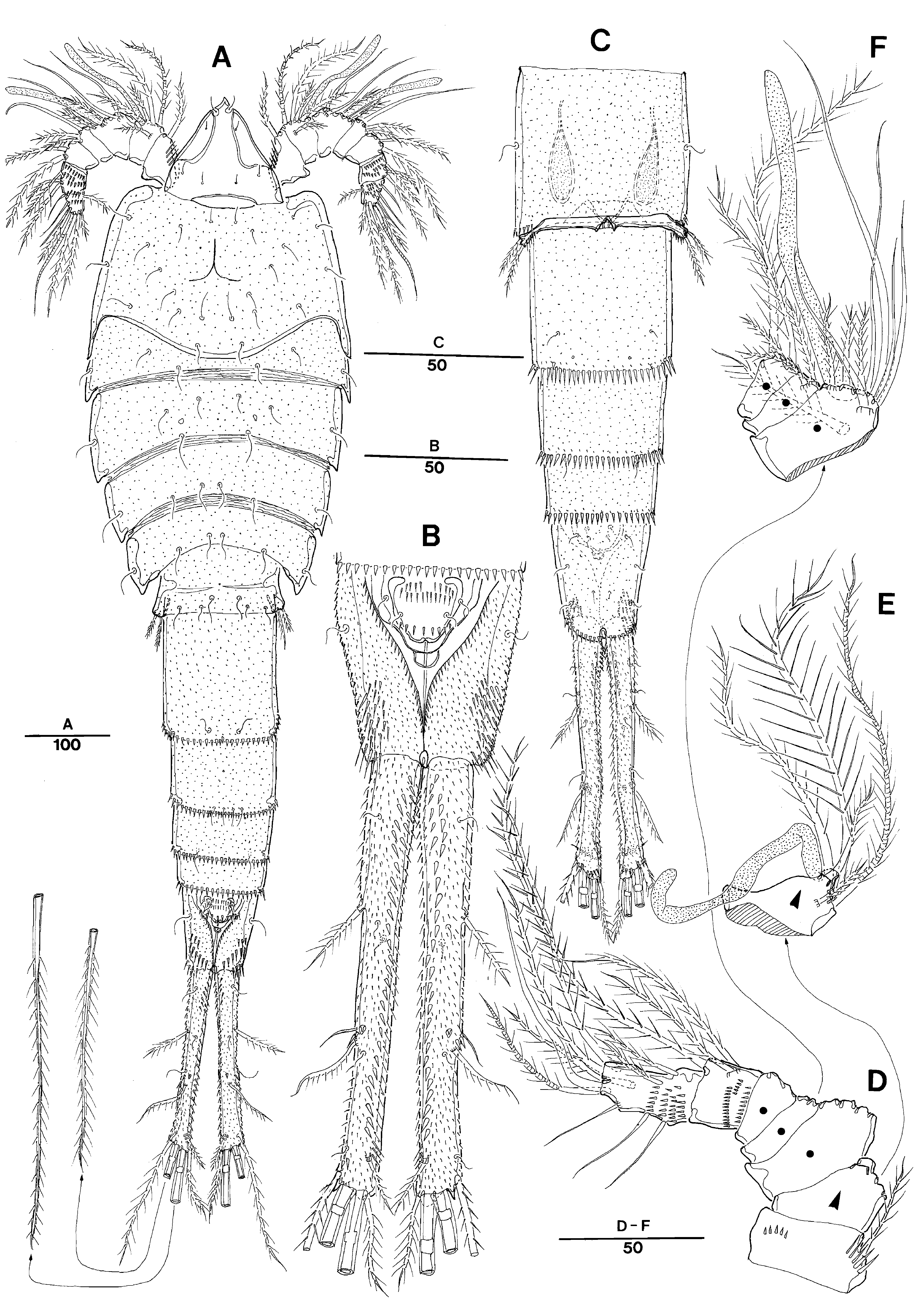
Description. Male. Total body length 1,038 µm (measured from tip of rostrum to posterior margin of caudal rami). Maximum width 311µm measured at posterior margin of P2-bearing somite. Entire body surface armed with minute denticles ( Fig. 9 View FIGURE 9 A–C).
Prosome ( Fig. 9 View FIGURE 9 A) 5-segmented, comprising cephalosome and 4 free pedigerous somites. P1 bearing somite clearly separated from cephalosome. Cephalothorax denticulated ( Fig. 9 View FIGURE 9 A), with few sensilla and smooth posterior margin. Three branched wrinkle present in middle of cephalosome. Pleural areas of cephalic shield narrow and posterolateral angles rounded. Succeeding four prosomites without distinct hyaline frills and with smooth posterior margin.
Rostrum well developed, elongated and triangular-shaped with pointed anterior apex, clearly defined at base ( Fig. 9 View FIGURE 9 A). Dorsal surface smooth with 4 pairs of sensilla.
Urosome ( Figs. 9 View FIGURE 9 B –C) 6-segmented, comprised of P5-bearing somite, 4 free abdominal, and anal somites. All urosomites denticulated, with spinulated posterior margins except for smooth P5-bearing somite.
Anal somite ( Fig. 9 View FIGURE 9 B) ornamented with several row of spinules on whole surface and with well-developed operculum with spinulate posterior margin and accompanied by 2 pairs of sensilla.
Caudal rami ( Fig. 9 View FIGURE 9 A–B) slightly divergent, about 8 times longer than wide and with several row of spinules and sensilla on whole surface; seta I located at proximal 1/3, pinnate; seta II located at distal 1/3, and pinnate; seta III about twice longer than seta II and pinnate; caudal setae V and IV very long and pinnate; seta VI shorter than seta III and located on distal inner corner; seta VII pinnate, shortest, close to seta VI and tri-articulated at base.
Antennule ( Fig. 9 View FIGURE 9 D–F) 7-segmented, segments 1, 6, and 7 with dorsal surface ornamented with rows of spinules. Armature formula: 1-[1 pinnate], 2-[5 pinnate + 1 ae], 3-[5 bare + 4 pinnate + 1 ae], 4-[2 pinnate], 5-[3 pinnate], 6- [3pinnate], 7-[3 bare + 7 pinnate]. Aesthetasc on segment 2 and 3. No clear apical aesthetasc on segment 7.
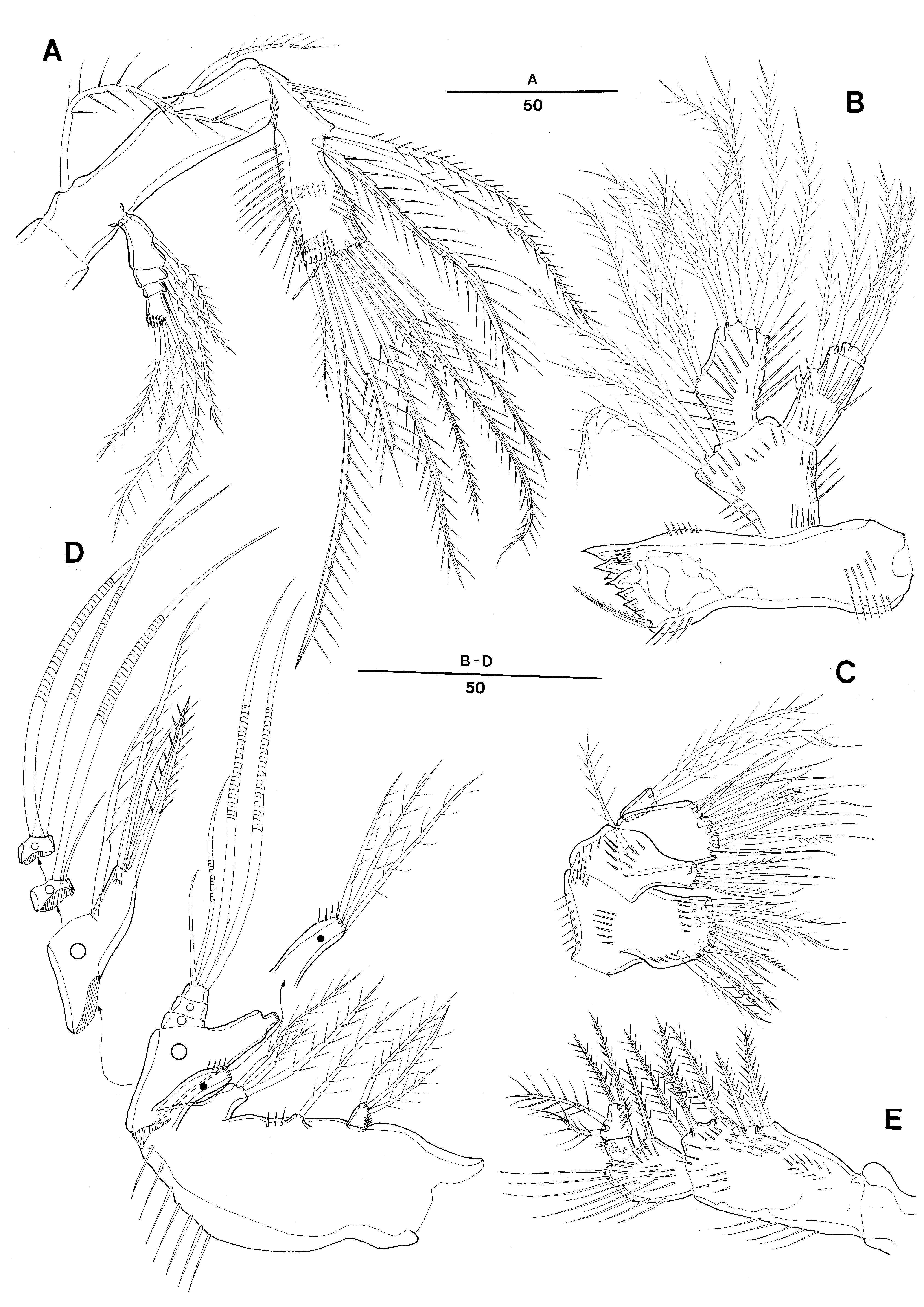
Antenna ( Fig. 10 View FIGURE 10 A) 3-segmented, comprising coxa, allobasis and free 1-segmented endopod. Allobasis with 2 plumose abexopodal setae. Exopod 4-segmented with seta formula 2.1.1.120, respectively; all setae pinnate. Free endopodal segment with strong spinules along inner proximal margin and with 3 strong pinnate spines laterally and 4 geniculate pinnate setae and 2 pinnate spines apically; outermost pinnate spine fused to 1 small pinnate seta and 1 long tube pore proximally.
Mandible ( Fig. 10 View FIGURE 10 B) coxa bearing reduced gnathobase rather than one in female, presumably non-functional; cutting edge with 7 major blunt teeth overlapping each other; accessory seta pinnate; with rows of spinules on anterior surface. Mandibular palp ornamented with rows of spinules on anterior surface; all setae less pinnate than those in female. Basis with 3 plumose setae. Endopod 1-segmented, with 3 pinnate lateral and 6 naked apical setae. Exopod 1-segmented, small with 2 long pinnate setae.
Maxillule ( Fig. 10 View FIGURE 10 C), arthrite small with 2 surface and 11 apical setae; all setae not rigid as those in female, presumably non-functional. Coxa with epipodite represented by 1 plumose seta; endite short with 2 plumose and 4 naked slender setae. Basis and endopod completely fused forming maxillulary allobasis, with 4 pinnate and 10 slender setae apically. Endopod 1-segmented, with 1 pinnate and 3 naked setae apically. Exopod 1-segmented, with 2 plumose setae.
Maxilla ( Fig. 10 View FIGURE 10 D), syncoxa with row of spinules on outer lateral margin and 4 endites (2 praecoxal, 2 coxal); enditic setal fomula [3, 1, 3, 3]; all enditic setae flexible, plumose and not rigid like those in female; distal praecoxal endite largely incorporated. Allobasis produced into strong pinnate spine; accessory armature consisting of 2 pinnate and 2 slender setae. Endopod 3-segmented without geniculated seta; enp-1 with 1 striated and 1 slender setae; enp-2 with 2 striated spiniform setae; enp-3 with 1 short and 2 long striated and 1 slender apical setae.
Maxilliped ( Fig. 10 View FIGURE 10 E) comprising syncoxa, basis and 2-segmented endopod. Syncoxa elongate, with row of spinules on anterior surface and 6 strong pinnate spines distally. Basis with 1 strong bipinnate, and 1 minute naked setae. Endopod 2-segmented; enp-1 with 1 short pinnate seta on anterior median surface; enp-2 with 2 lateral and 2 apical pinnate setae.
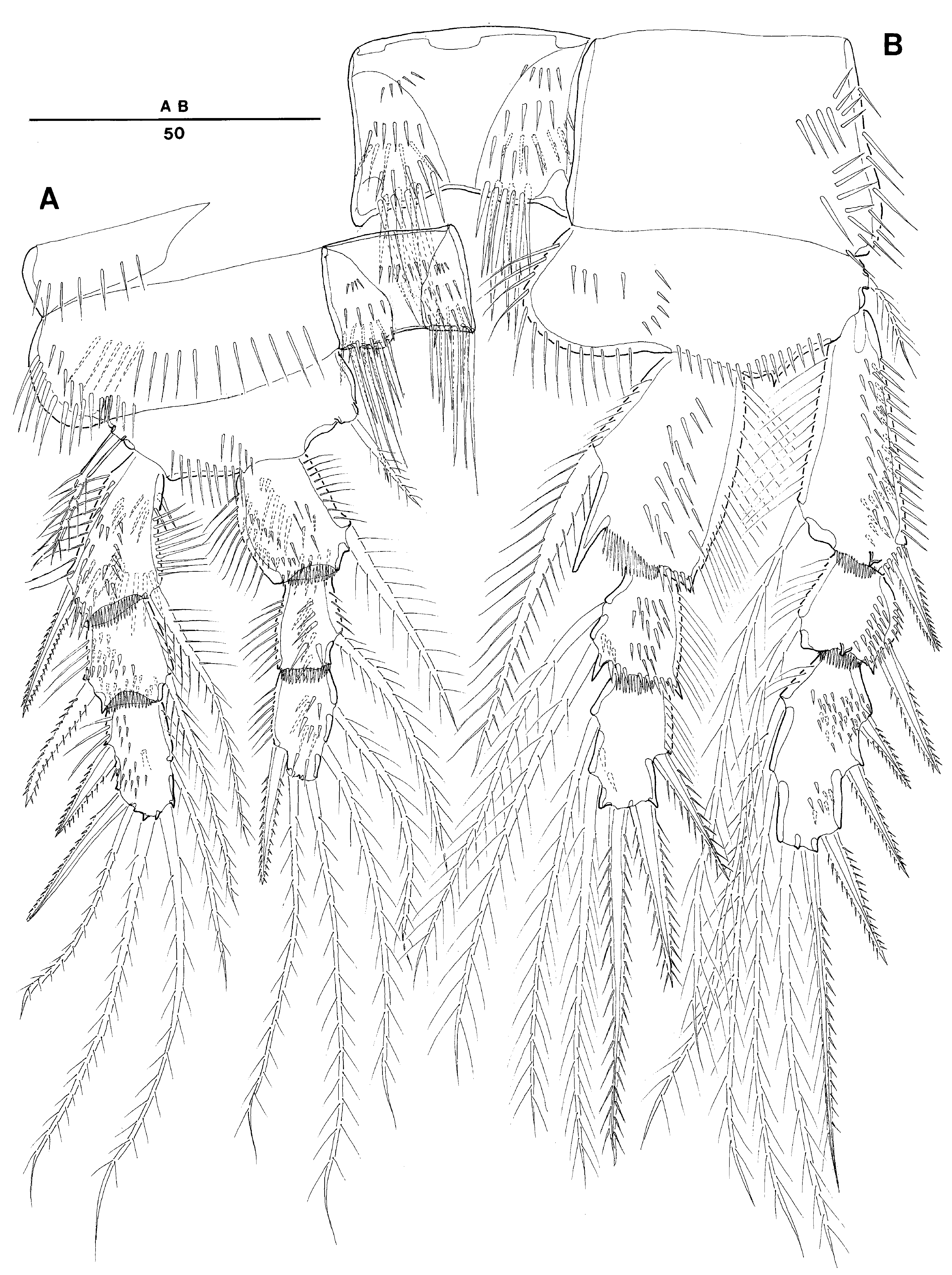
Swimming legs 1–4 ( Figs. 11 View FIGURE 11 A, B; 12A, B) biramous, P1–P4 with 3-segmented exopod and 3-segmented endopod, each ramus ornamented with setules and spinules along inner and outer margins as illustrated. Intercoxal sclerites well developed and ornamented with several rows of spinules on anterior and posterior surface.
P1 ( Fig. 11 View FIGURE 11 A), praecoxa with spinules on distal margin. Coxa wider than long with row of spinules along round outer lateral margin. Basis with 1 outer and 1 inner strong pinnate spine. Endopod 3-segmented, enp-1 longest, enp-2 shortest; enp-3 not reaching to tip of exopod. Exopod 3-segmented, exp-1 longest and exp-2 shortest.
P2 ( Fig. 11 View FIGURE 11 B). Coxa with row of spinules on anterior surface and along outer margin. Basis with 1 pinnate outer seta and row of long spinules on inner lateral lobe and row of spinules along distal margin between endopod and exopod. Endopod 3-segmented, not extending to distal end of exopod; row of long setules along outer margin of each segment; enp-1 largest, and with elongated inner distal end forming sharp triangular tip; enp-2 shortest. Exopod 3-segmented. Each segment with row of spinules along outer margins; exp-1 longest and exp-2 shortest.
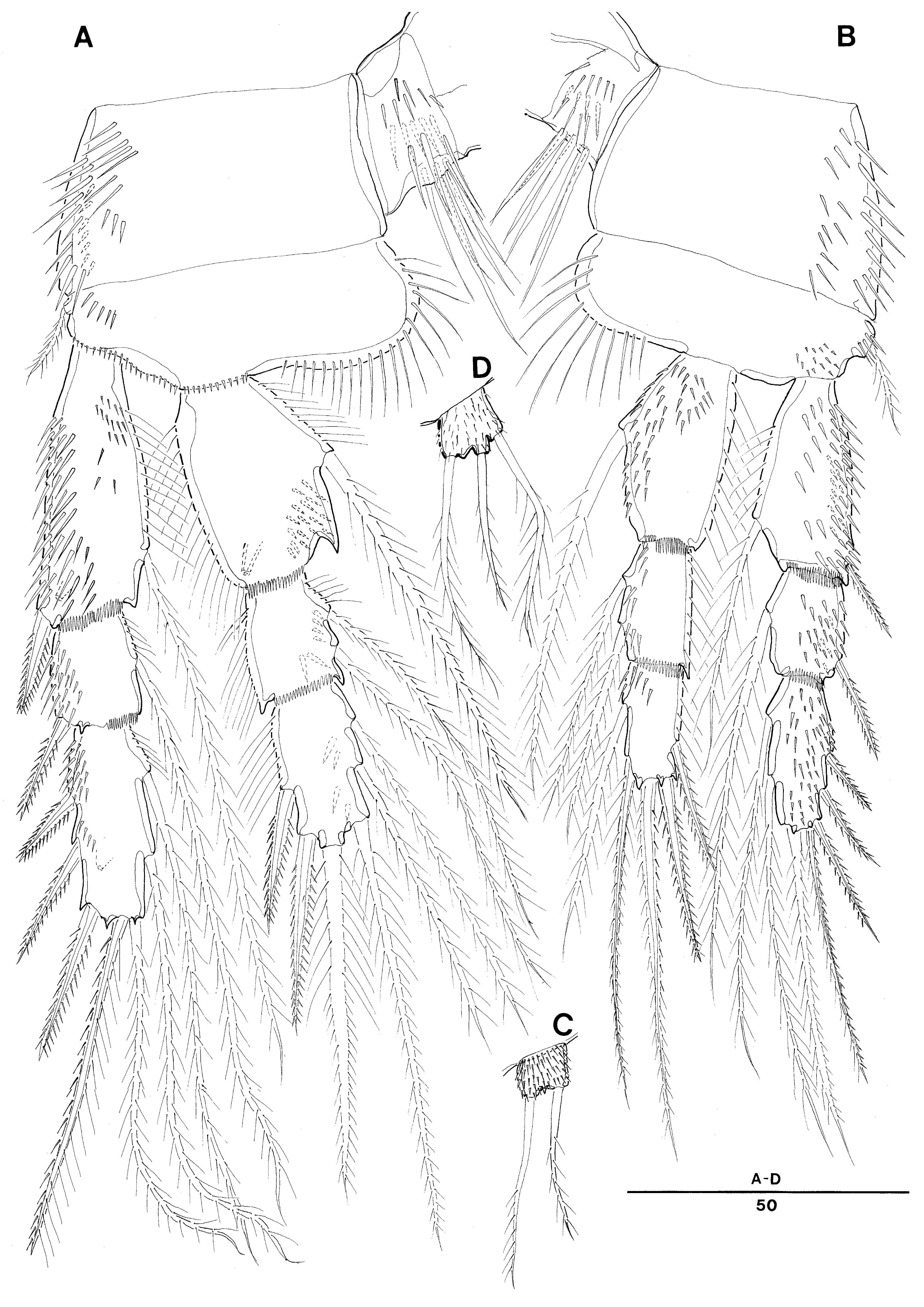
P3 ( Fig. 12 View FIGURE 12 A). Coxa with row of spinules on anterior surface and along outer margin. Basis with 1 pinnate outer seta and row of long spinules on inner lateral lobe and row of spinules along distal margin including articulation area with endopod and exopod. Endopod 3-segmented, not extending to distal end of exopod; row of long setules along outer margin of each segment; enp-1 largest, and with elongated inner distal end forming sharp triangular tip; enp-2 shortest. Exopod 3-segmented. Each segment with row of spinules along outer margins; exp-1 longest and exp-2 shortest.
P4 ( Fig. 12 View FIGURE 12 B). Coxa with row of spinules on anterior surface and along outer margin. Basis with 1 pinnate outer seta and row of long spinules on inner lateral lobe and patch of spinules near articulation with exopod. Endopod 3-segmented, not extending to distal end of exopod; row of long setules along outer margin of each segment; enp-1 largest; enp-2 and enp-3 subequal in lenghs. Exopod 3-segmented. Each segment with row of spinules along outer margins and anterior surfaces; exp-1 longest and exp-2 shortest.
Armature formula as follows:
P5 ( Fig. 12 View FIGURE 12 C) laterally displaced, largely incorporated into somite, not defined at base, represented by small subrectangular lobe densely spinulose on its surface with 2 plumose setae distally
Sixth pairs of legs ( Fig. 9 View FIGURE 9 C) not fused medially, symmetrical. Each P6 forming single genital slit covered on both sides by opercula derived from sixth legs; internal spermatophores in Fig.9 View FIGURE 9 C indicating activeness of both opercula. P6 with small protuberance bearing 2 pinnate setae; inner seta longest.
Female. Total body length of examined samples ranged 1,044–1,183 µm (n=3, mean = 1,093µm, measured from tip of rostrum o posterior margin of caudal rami). All characteristics agreeing with those in Lee & Yoo (1998), except for entire body surface covered by minute denticles as in male. Especially P5 distinctly covered with minute denticles as in male; with 3 setae ( Fig. 12 View FIGURE 12 D).
No known copyright restrictions apply. See Agosti, D., Egloff, W., 2009. Taxonomic information exchange and copyright: the Plazi approach. BMC Research Notes 2009, 2:53 for further explanation.