
Microvelia inguapi, Padilla-Gil & Moreira, 2013
|
publication ID |
https://doi.org/ 10.11646/zootaxa.3745.5.7 |
|
publication LSID |
lsid:zoobank.org:pub:632DC433-CE22-4216-B348-FF129DDDA896 |
|
persistent identifier |
https://treatment.plazi.org/id/D62F87CD-FFA7-FFA5-FF5C-821706ABA2E7 |
|
treatment provided by |
Felipe |
|
scientific name |
Microvelia inguapi |
| status |
sp. nov. |
Microvelia inguapi View in CoL sp. n.
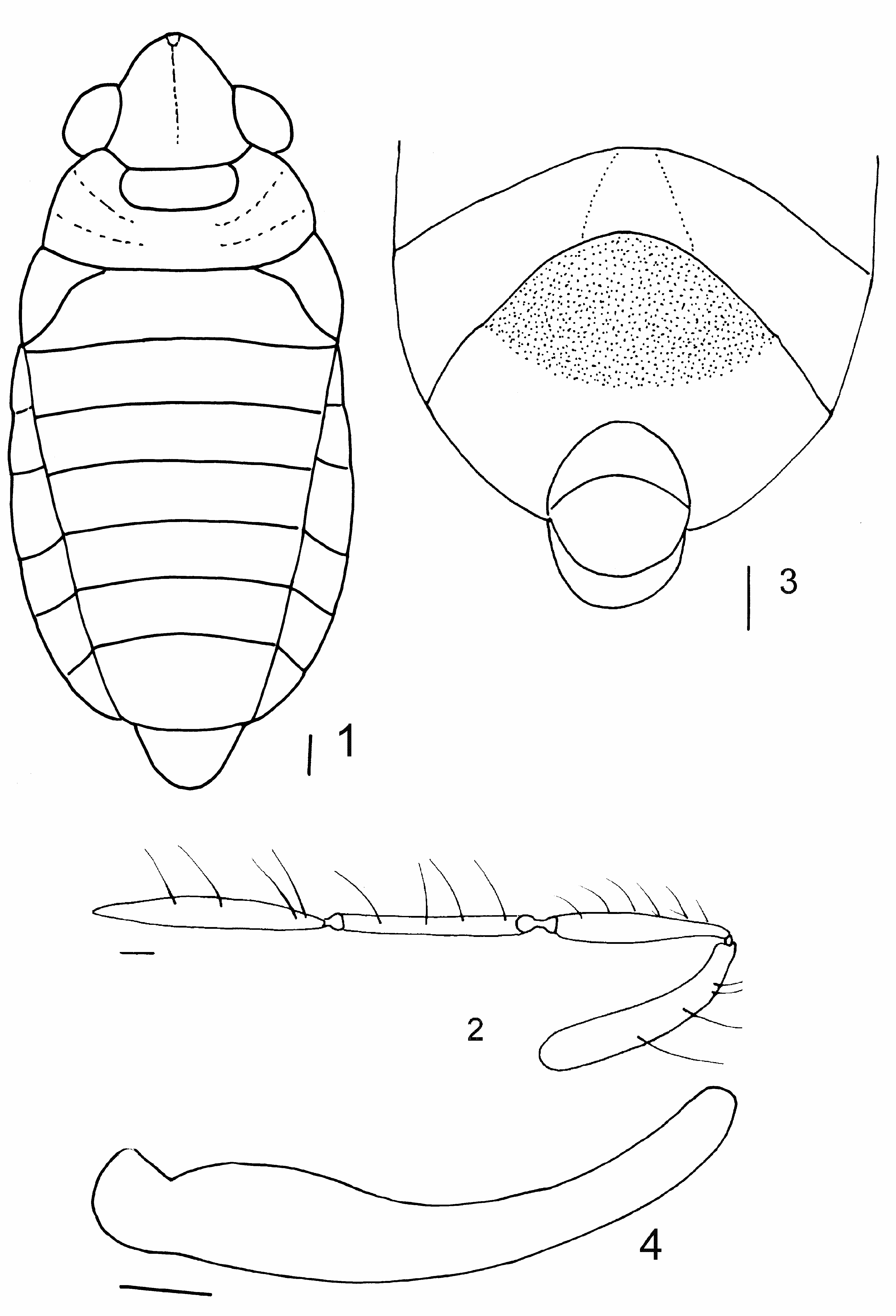
( Figs. 1–9 View FIGURES 1–4. 1 View FIGURES 5–9. 5 )
= Microvelia inquilina ; Padilla-Gil & Arcos (2011), nec Polhemus & Hogue, 1972
= Microvelia inquilina ; Padilla-Gil (2012), nec Polhemus & Hogue, 1972
Type material. HOLOTYPE, ♂ apt: COLOMBIA, Nariño Department, Tumaco Municipality, Inguapi , 17.XI.2010, D.N. Padilla, leg. ( ICN) . PARATYPES, same data as holotype, 3 apt ♀, 1 apt ♂, ( PSO-CZ); 2 apt ♀ ( ICN); 22. VI .2010, 1 apt ♀ ( PSO-CZ). Same municipality, Estero Chilvi, Mar Agrícola, 17.XI.2010, D.N. Padilla, leg, 2 apt ♀, 1 apt ♂ ( ICN) .
Color and pilosity. Body covered by short, brown pubescence and semi-recumbent golden setae. Head dorsally and ventrally dark brown. Eyes red. Antennae and legs shining yellow, except tibiae and tarsi brown. Rostrum shining brown except for segment IV dark black; genital segments shining brown. Pronotum with anterior transverse yellow band. Venter of abdomen dark grey; abdominal sternite VII centrally with black pubescence.
Apterous male ( Fig. 1–7 View FIGURES 1–4. 1 View FIGURES 5–9. 5 ) BL: 1.86; HL: 0.34; HW: 0.56; ANT I–IV: 0.24, 0.22, 0.28, 0.30; INT: 0.32; EYE: 0.12; PL: 0.32; PW: 0.78; FORELEG: FEM: 0.52, TIB: 0.44, TAR I: 0.04, TAR II: 0.14; MIDLEG: FEM: 0.62, TIB: 0.60, TAR I: 0.10, TAR II: 0.22; HINDLEG: FEM: 0.64, TIB: 0.72, TAR I: 0.06, TAR II: 0.18.
Head with midline shining black ( Fig. 1 View FIGURES 1–4. 1 ). Antennae reaching well beyond pronotum. Antennomere I thickest, curved outward, II widening to apex, III cylindrical, IV fusiform ( Fig. 2 View FIGURES 1–4. 1 ). Rostrum extending slightly past middle of mesosternum. Pronotum covering mesonotum and metanotum; with scattered subcircular punctuations, lacking a median carina ( Fig. 1 View FIGURES 1–4. 1 ). Abdominal connexiva slightly elevated, with posterolateral angles rounded; last abdominal tergite trapezoidal, with posterior margin slightly rounded. Genital segment I rounded ( Fig. 1 View FIGURES 1–4. 1 ). Abdominal sternite VII with a broad medial depression ( Fig. 3 View FIGURES 1–4. 1 ). Venter of genital segment I with posterior margin rounded ( Fig. 3 View FIGURES 1–4. 1 ). Genital segment II hidden. Parameres elongate ( Fig. 4 View FIGURES 1–4. 1 ).
Fore tibia triangular with well developed grasping comb extending beyond apex, black, apex rounded ( Fig. 5 View FIGURES 5–9. 5 ). Middle tibia with a pair of long, black setae at posteroventral angle ( Fig. 6 View FIGURES 5–9. 5 ). Hind trochanter angled upward and bearing a small tubercle projecting beyond posterior margin ( Fig. 7 View FIGURES 5–9. 5 ).
Apterous female ( Figs. 8–9 View FIGURES 5–9. 5 ) BL: 2.08; HL: 0.32; HW: 0.60; ANT I–IV: 0.26, 0.22, 0.30, 0.40; INT: 0.36; EYE: 0.12; PL: 0.30; PW: 0.80; FORELEG: FEM: 0.52, TIB: 0.44, TAR I: 0.04, TAR II: 0.20; MIDLEG: FEM: 0.62, TIB: 0.60, TAR I: 0.10, TAR II: 0.24; HINDLEG: FEM: 0.64, TIB: 0.74, TAR I: 0.10, TAR II: 0.24.
Abdomen widened, with tergites I– V concave ( Fig. 8 View FIGURES 5–9. 5 ) and sternites slightly convex; connexiva slightly turned upward (20º); last abdominal tergite trapezoidal, with posterior margin slightly concave. Genital segment I with divergent lateral margins and posterior margin broadly rounded. Sternite VII bearing two pits on anterolateral margin ( Fig. 9 View FIGURES 5–9. 5 ). Venter of genital segment I with posterior margin broadly rounded ( Fig. 9 View FIGURES 5–9. 5 ). Genital segment II smaller and rounded ( Fig. 9 View FIGURES 5–9. 5 ). Grasping comb absent; all tibiae with grooming structures on posteroventral angles; hind trochanter not raised, lacking a tubercle.
Comparative notes. Specimens of Microvelia inguapi sp. n. can be diagnosed by the long pronotum which covers the meso- and metanotum; the male with grasping comb extending beyond apex of fore tibia ( Fig. 5 View FIGURES 5–9. 5 ); all femora without spines; the male hind trochanter raised and bearing a tubercle ( Fig. 7 View FIGURES 5–9. 5 ); and by the modifications found on the venter of the male and female abdomens ( Figs. 3 View FIGURES 1–4. 1 , 9 View FIGURES 5–9. 5 ).
Based on the original description, M. inguapi sp. n. is similar to M. inquilina J. Polhemus & Hogue, 1972 . They share similar habitats, and general shape of body, antennae, apical abdominal segments of the male, and the shape of the male paramere are similar. However, M. inquilina is smaller (male BL: 1.15; female BL: 1.33); the metanotum is not covered by the pronotum; no modifications are found on the legs; and the grasping comb of the fore tibia is short.
Habitat. Microvelia inguapi sp. n. has been collected in two estuaries. Chilvi was characterized by an air temperature of 25 ºC, a water temperature of 26.5 ºC, pH 6, dissolved oxygen 65%, conductivity 1 S/m, depth> 1 m, and substrate with numerous crab holes. Inguapi had an air temperature of 25.5 ºC, a water temperature of 27 ºC, pH 6, dissolved oxygen 108%, conductivity 1 S/m, and depth> 1 m.

| ICN |
Instituto de Ciencias Naturales, Museo de Historia Natural |
| VI |
Mykotektet, National Veterinary Institute |
| PL |
Západoceské muzeum v Plzni |
| PW |
Paleontological Collections |
| V |
Royal British Columbia Museum - Herbarium |
No known copyright restrictions apply. See Agosti, D., Egloff, W., 2009. Taxonomic information exchange and copyright: the Plazi approach. BMC Research Notes 2009, 2:53 for further explanation.
|
Kingdom |
|
|
Phylum |
|
|
Class |
|
|
Order |
|
|
Family |
|
|
Genus |