
Argestes angolaensis, George, Kai Horst, 2008
|
publication ID |
https://doi.org/ 10.5281/zenodo.183720 |
|
DOI |
https://doi.org/10.5281/zenodo.6235380 |
|
persistent identifier |
https://treatment.plazi.org/id/D70E87E9-8820-FFF4-F6AE-FB01402FFB0E |
|
treatment provided by |
Plazi |
|
scientific name |
Argestes angolaensis |
| status |
sp. nov. |
Argestes angolaensis sp. nov.
Figures 2–14 View FIGURE 2 View FIGURE 3 View FIGURE 4 View FIGURE 5 View FIGURE 6 View FIGURE 7 View FIGURE 8
Locus typicus . Northern Angola Basin, #346 (16°17.0’S, 05°27.0’E, depth: 5389m). Holotype (#346/512) (i.e. station 346, replicate no. 5, MUC core no. 12): female, placed on 11 slides, coll. nos. SMF 32027/111. Seven paratypes (PT) were put on slides and used for species description: PT1 (#346/81): female, coll. no. SMF 32028, PT2 (#346/82): female, coll. no. SMF 32029, PT3 (#346/87): female, fixed on 10 slides, coll. nos. SMF 32030/110, PT 4 (#346/85): female, put on 10 slides, coll. nos. SMF 32031/110, PT5 (#346/81): female, coll. no. SMF 32032, PT6 (#346/61): male, placed on 3 slides, coll. nos. SMF 32033/13, PT7 (#346/ 710): male, fixed on 8 slides, coll. no. SMF 32034/18. The remaining 90 specimens are preserved in alcohol and distributed in eight vials with respect to the corresponding MUC cores, coll. nos. SMF 32035–SMF 32042.
Female.
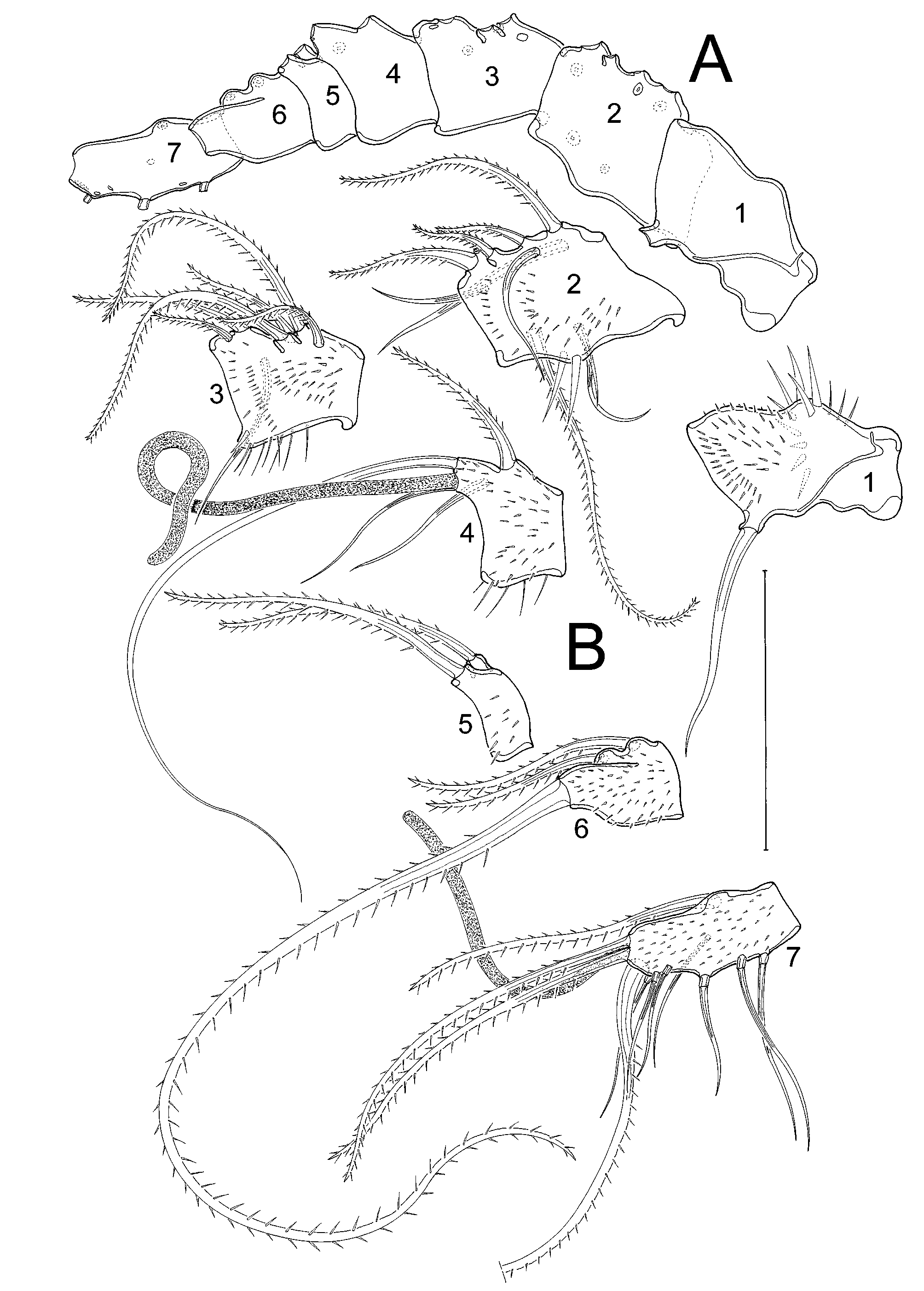
Habitus ( Fig. 2 View FIGURE 2 A, B) slender, body length including FR of approximately 545µm. Cphth less than 1/3 of total body length. Whole body densely covered with small spinules, except the P5bearing thoracic somite, the second and the third abdominal somite, and the telson, which show fewer but bigger spinules. Body with several remarkably long sensilla arising from tubercles. In particular, the sensilla of the P5bearing thoracic somite and abdominal somites are very long.
Telson ( Fig. 3 View FIGURE 3 ) as large as 2 preceding abdominal somites together, almost square from lateral view, but tapering slightly from dorsal/ventral view. Ventrally with a pair of small tube pores at inner margin close to FR, and with some strong spinules near the proximal margin. Anal operculum small, with row of spinules at its distal margin and accompanied by 2 sensillate tubercles.
FR ( Fig. 3 View FIGURE 3 ) long and slender, varying remarkably in length (cf. Figs 2 View FIGURE 2 B, 3), and covered with many spinules of different sizes. All 7 setae concentrated at terminal part: I and II subterminally on outer margin, I smaller than II. III ventrally at terminal margin. IV and V largest setae, inserting terminally. VI as long as III, arising at the inner terminal margin. VII dorsally, arising from knoblike projection. Setae I–III and VI in many specimens of “rattail” appearance.
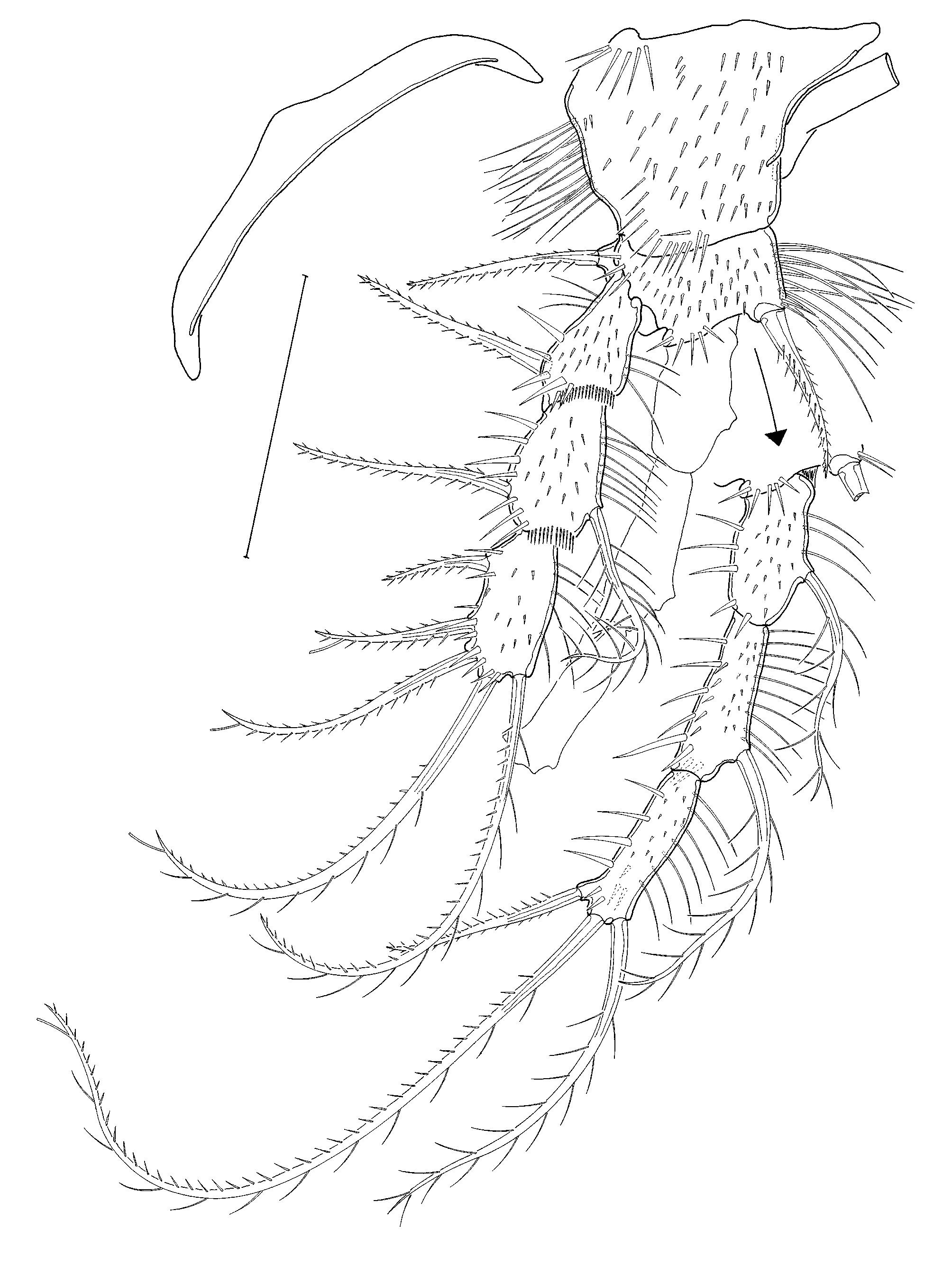
A1 ( Fig. 4 View FIGURE 4 ) 7segmented, fifth segment smallest, all segments with several bare and bipinnate setae. Second segment at its posterior margin with few long spinules. Aes on fourth segment, arising together with 2 setae from protrusion. Sixth segment terminally with 1 very long bipinnate seta. Seventh segment terminally with small aes, and at posterior terminal margin with remarkable long seta. All segments covered with small spinules.
Setal formula: 1/1; 2/9; 3/7; 4/4 + aes; 5/2; 6/3; 7/11 + aes.
A2 ( Figs 5 View FIGURE 5 A, A’) with allobasis and 1segmented exp bearing 1 bare seta and 3 long spinules. Enp and allobasis covered with small spinules. Enp laterally with 2 bipinnate setae, the second subterminal with tube pore. Terminally, enp carries 5 geniculate setae and 1 unipinnate seta. Subterminally with long tube pore, fused with longest seta.
Md ( Fig. 6 View FIGURE 6 ) with gnathobase ( Fig. 6 View FIGURE 6 A, A’) formed by several toothlike projections. Subterminal seta not discernible. Md palp ( Fig. 6 View FIGURE 6 B) strong, covered with spinules, basis with 2 bipinnate setae. Enp 1segmented, with 1 outer bipinnate seta, additionally with 3 subterminal and 2 terminal bipinnate setae. Exp smaller than enp, with 1 outer and 2 terminal bipinnate setae.
Mxl ( Fig. 5 View FIGURE 5 B, B’) with distinct exp. Precoxal arthrite terminally with 6 spines, subterminally with another spine. At its proximal margin with 1 unipinnate seta, and on its surface with 2 bare setae. Coxa terminally with 3 setae. Basis terminally with 2 setae. Enp represented by 1 bare seta. Exp long and spinulose, terminally with 2 multipinnate setae.
Mx ( Fig. 5 View FIGURE 5 C) unfortunately broken at its distal part that is therefore not described. Syncoxa with 2 endites, the proximal one bearing 1 bare seta. Distal endite with 3 setae, the biggest one fused to segment, bipinnate. Basis fused to syncoxa, with 3 setae. Enp distinct, with 2 bare setae.
Mxp ( Fig. 5 View FIGURE 5 D) prehensile, syncoxa slightly shorter than basis, with many spinules of different sizes, distally with 2 long multipinnate setae. Basis covered with small spinules, additionally with 2 rows of long spinules. Enp produced into long unipinnate claw, with 1 bare seta at its base.
P1 ( Fig. 7 View FIGURE 7 ) with 3segmented exp and enp. Coxa considerably bigger than basis, with several spinules and 1 row of setules at its outer margin. Intercoxal sclerite transversely long and bowlike. Basis with inner and outer spine, covered with small spinules and long setules at its inner margin. Both exp and enp covered with small spinules on anterior side. Exp1 without, exp2 with inner seta. Exp3 with 5 setae/spines, three of which are subterminal with tube pores. Enp1 and 2 with 1 inner seta each, enp3 with 2 terminal setae, the longer one subterminally with tube pore, and 1 outer spine. For setal formula see table 1.
P2–P4 ( Figs. 8–10 View FIGURE 8 ) with 3segmented exps and enps. Coxae approximately 2.5 times bigger than bases, showing decreasing covering with small spinules from P2 to P4. Each coxa on outer side with 3 strong spinules. Bases much broader than long, exps and enps turned outwardly. Bases with outer spines, at inner margin with long setules, covered with small spinules as in coxae. Appendages covered with small spinules, in most specimens showing a decrease from P2 to P4 (but cf. Fig. 10 View FIGURE 10 B: female P4!). Setation of exp and enp as in table 1. Exp3 as long as exp1 and exp2 together. Enps showing increasing length of segments, enp1 being the shortest, and enp3 the longest segment. As shown in Fig. 10 View FIGURE 10 B, few females show a variability in the setation of P4 exp3 (as also in the covering with spinules). Instead of only 1, it bears 2 inner setae, the first being considerably smaller than the second.
TABLE 1: Argestes angolaensis sp. nov., female, setation of P1–P4 (no. outer spines in roman numbers).
P5 ( Fig. 11 View FIGURE 11 A) benps fused together, forming a single plate. Outer basal seta arising from moderately produced setophore. Endopodal lobe strongly reduced, represented by 1 longer biplumose and 1 shorter bare seta. Exp distinct, long and slender, with 2 outer, 2 terminal, and 1 inner seta. Additionally, outer distal margin with extremely long tube pore.
GF ( Fig. 11 View FIGURE 11 B) strongly cuticularized, with several small and long spinules surrounding the gonoporus. P6 reduced, detectable as paired small processes without setae, flanking the GF.
Male.
Habitus ( Fig. 12 View FIGURE 12 A, 13) smaller than female, body length including FR approximately 480µm. Coverage of body with small cuticular spinules not reaching density as in female. Tubercles bearing sensilla longer than in female.
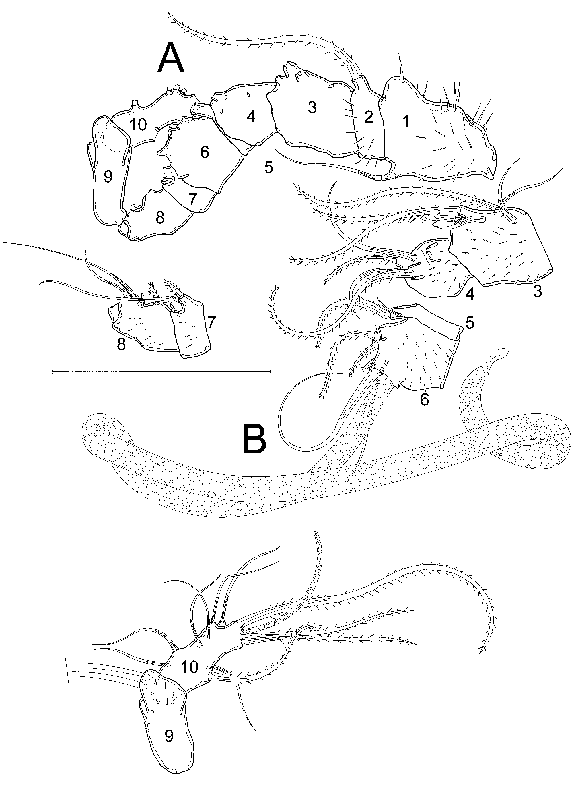
A1 ( Fig. 14 View FIGURE 14 A, B) haplocer, 10segmented. All segments except fifth and tenth with small spinules. Sixth segment with strong aes. Eighth segment additionally with toothlike spinule. Ninth segment terminally with strong seta (broken in Fig. 14 View FIGURE 14 B).
Setal formula: 1/1; 2/1; 3/6; 4/5; 5/2; 6/6+aes; 7/2; 8/3; 9/1; 10/11+aes.
P4 ( Fig. 12 View FIGURE 12 B) sexually dimorphic. General shape as in female, but exp3 always bears 2 welldeveloped inner setae.
P5 ( Fig 11 View FIGURE 11 C) exp distinct, shorter than in female, with 6 setae and 1 long tube pore. Benps fused, with 1 small seta.
Remarks. With 98 adult individuals, Argestes angolaensis sp. nov. was by far the most abundant argestid taxon collected in the Angola Basin during DIVA 1 expedition in 2000. The high number of collected specimens, usually a highly uncommon occurrence for deepsea sampling, provides a good insight into the intraspecific morphological variability, which certainly exists, but whose study is not the object of the present contribution. However, comparison of the specimens revealed remarkable variability in body and furcal length (male bodies varying between 380 and 480µm length), in body ornamentation (density and size of spinules covering body and appendages), and even in setation (cf. P4 exp, Fig. 10 View FIGURE 10 B). Nevertheless, an assignment of the corresponding specimens to different new species seems rather implausible, because the mentioned variability shows a quite heterogeneous distribution over the individuals, preventing the recognition of any morphological pattern. Moreover, all specimens share a series of apomorphic characteristics (cf. discussion), justifying their allocation into one single species. Taking into account similar conditions in other taxa e.g. Ancorabolidae ( George 2006; George, personal observation), it has to be stated, that intraspecific variability in deepsea Harpacticoida is greatly underestimated.


No known copyright restrictions apply. See Agosti, D., Egloff, W., 2009. Taxonomic information exchange and copyright: the Plazi approach. BMC Research Notes 2009, 2:53 for further explanation.