
Haemoproteus copsychi, Paperna & Keong & May, 2008
|
publication ID |
https://doi.org/ 10.5281/zenodo.5340123 |
|
persistent identifier |
https://treatment.plazi.org/id/D81E2B70-A862-B94B-9B99-DAB3FEF725CF |
|
treatment provided by |
Diego |
|
scientific name |
Haemoproteus copsychi |
| status |
sp. nov. |
Haemoproteus copsychi , new species
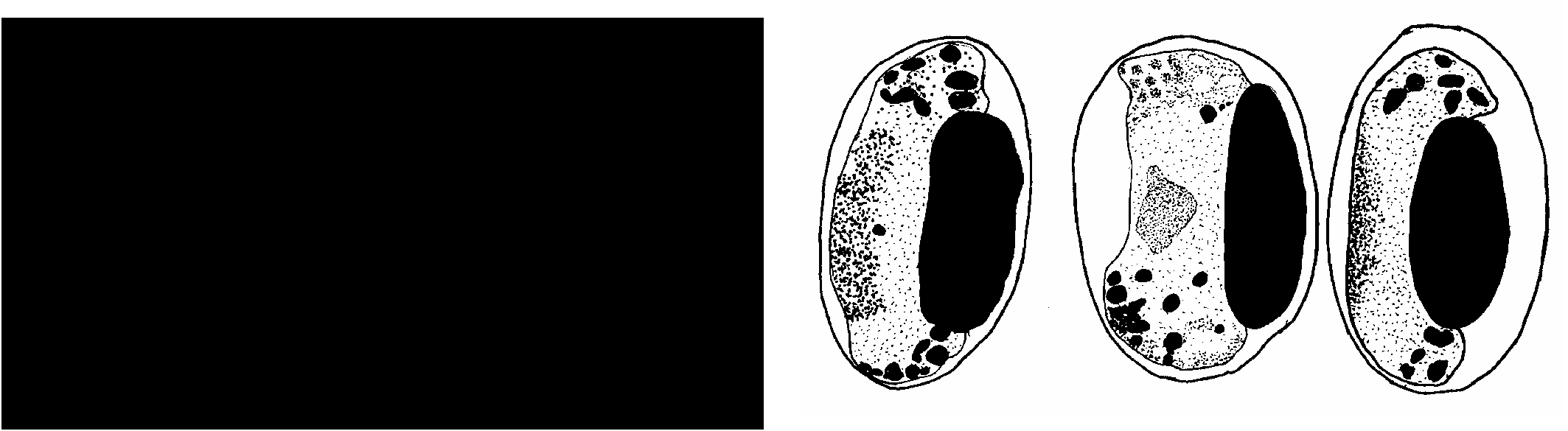
( Fig. 8 View Fig ) (Table 3)
Locality. – Malaysia: Sarawak.
Description. – Found only in mature erythrocytes. Gametocytes are closely applied to the host cell nucleus, causing slight swelling in the erythrocyte as well as moderately or extremely displacing its nucleus (NDR–macrogametocytes: <0.2, microgametocytes: 0.15, 0.40, 0.78). The ends of the gametocytes slightly or moderately extend beyond the erythrocyte nucleus, ending bluntly and only occasionally gripping the host nucleus. The macrogametocyte cytoplasm stains deep-blue and is coarsely granular. The nucleus is pink-staining, discrete and in a median position. The pigment granules, either small (fragmented) and numerous (<25) or fewer and larger (> 10), usually of mixed sizes (15–20), are more often aggregated at the distal ends and adjoined by a few vacuoles. The microgametocyte cytoplasm stains blue, while the large nucleus occupies the major volume of the cell. The pigment granules are located at the gametocyte extremities and accompanied by one or two vacuoles. They are either coarse (approximately 10) or fine (15–20). The juvenile stages were not found.
Etymology. – Named after the generic name of the parasite’s host.
Remarks. – Haemoproteus copsychi differs from Hae. alcippe found in sylviid birds in considerably displacing the erythrocyte nucleus. Bennett & Campbell (1972) reported Hae. fallisi from Copsychus malabaricus . These authors considered species of Haemoproteus found in blood of various species from the Turdidae family from both Holarctic regions (North America and Eurasia) and from the tropics (SE Asia) to be conspecific with Hae. fallisi . They acknowledge considerable variations in the dimensions of parasites as well as the infected erythrocytes. The differences between the presently described species and Hae. fallisi as described from different species from the Turdidae are as follows: in Hae. copsychi , the host erythrocyte nucleus is radically displaced. The surface of the gametocytes do not form amoeboid extensions or invaginations. The macrogametocyte nucleus is in a median rather than in a subterminal position. With only few exceptions (one microgametocyte), the gametocytes end bluntly and do not embrace the erythrocyte nucleus. Gametocytes reported by Bennett & Campbell (1972) from Copsychus malabaricus are considerably larger (13.9 ± 0.8 × 3.0 ± 0.5), cause an elongation rather than a swelling of the erythrocyte (infected: 14.3 × 7.3, non-infected 13.0 × 8.2) and displace the erythrocyte nucleus to a lesser extent (NDR = 0.64 ± 0.26).
No known copyright restrictions apply. See Agosti, D., Egloff, W., 2009. Taxonomic information exchange and copyright: the Plazi approach. BMC Research Notes 2009, 2:53 for further explanation.