
Tapinesthis Simon, 1914
|
publication ID |
https://doi.org/ 10.5852/ejt.2014.82 |
|
publication LSID |
lsid:zoobank.org:pub:BA6707B1-9DAD-4448-9431-47B7E9B63980 |
|
DOI |
https://doi.org/10.5281/zenodo.6137739 |
|
persistent identifier |
https://treatment.plazi.org/id/D85987E5-FFA7-CA24-D1F8-FD50A7FB5C38 |
|
treatment provided by |
Jeremy |
|
scientific name |
Tapinesthis Simon, 1914 |
| status |
|
Tapinesthis Simon, 1914 View in CoL View at ENA
Diagnosis
Soft-bodied, pale Oonopidae with strongly sloping cephalothorax provided with dark net-shaped pattern, without leg spines, with unipectinate tarsal claws; pedicel with meshed texture; male palp with short scleriFed sperm duct and female genitalia with wide median T-shaped structure visible by transparency, Fanked by two rounded apodemes.
Description
Male
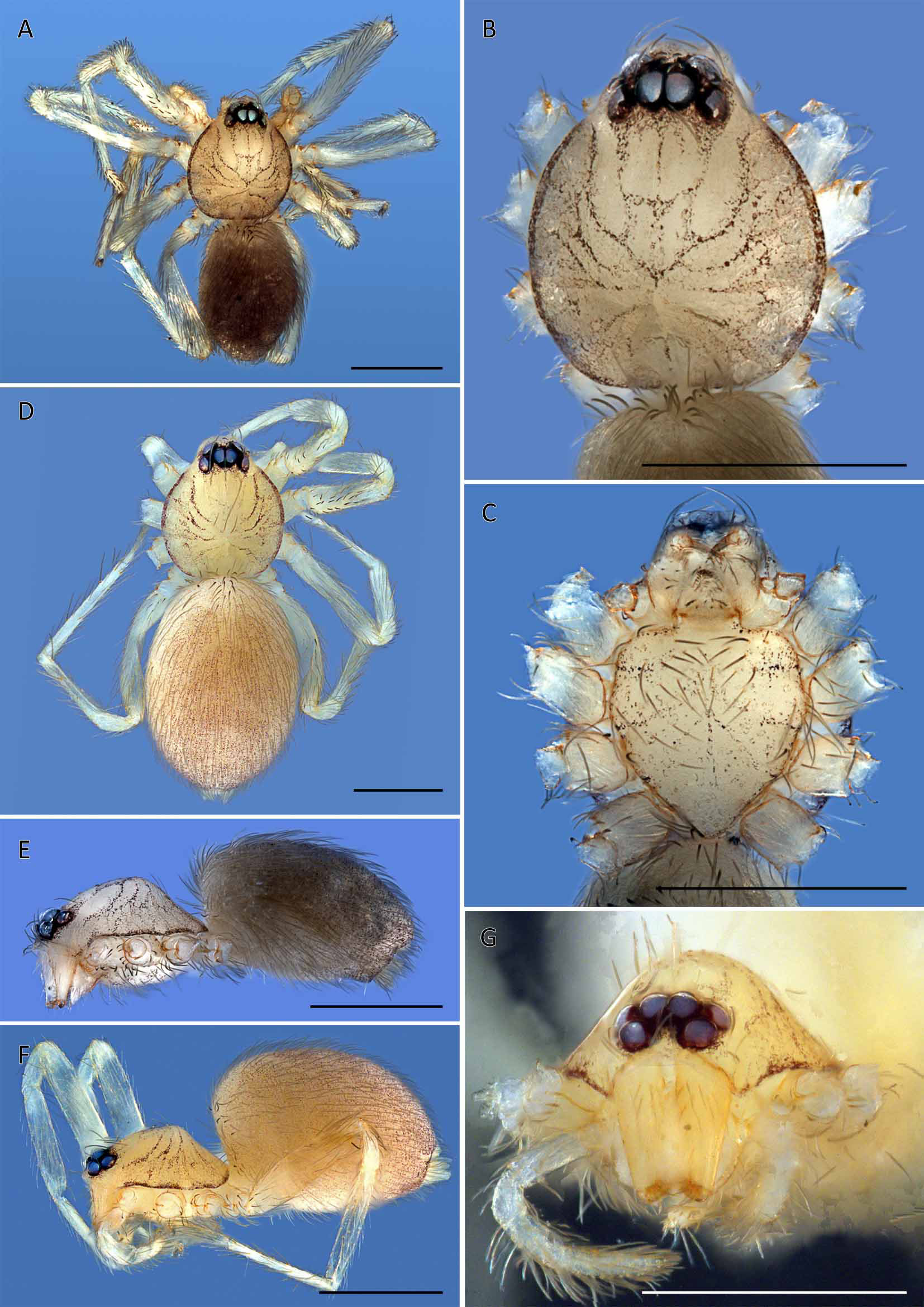
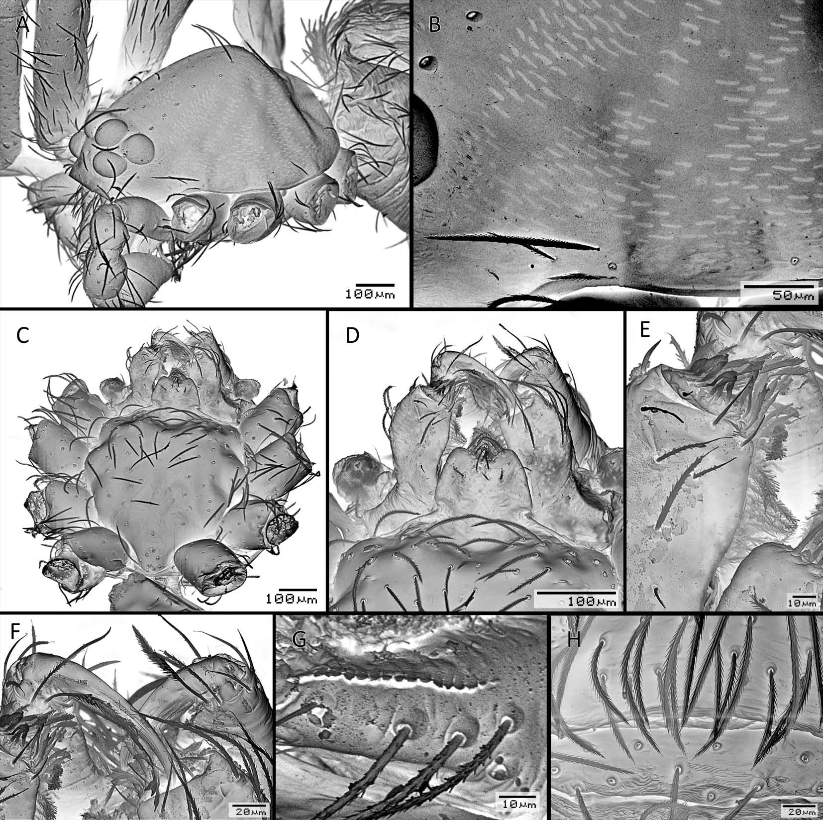
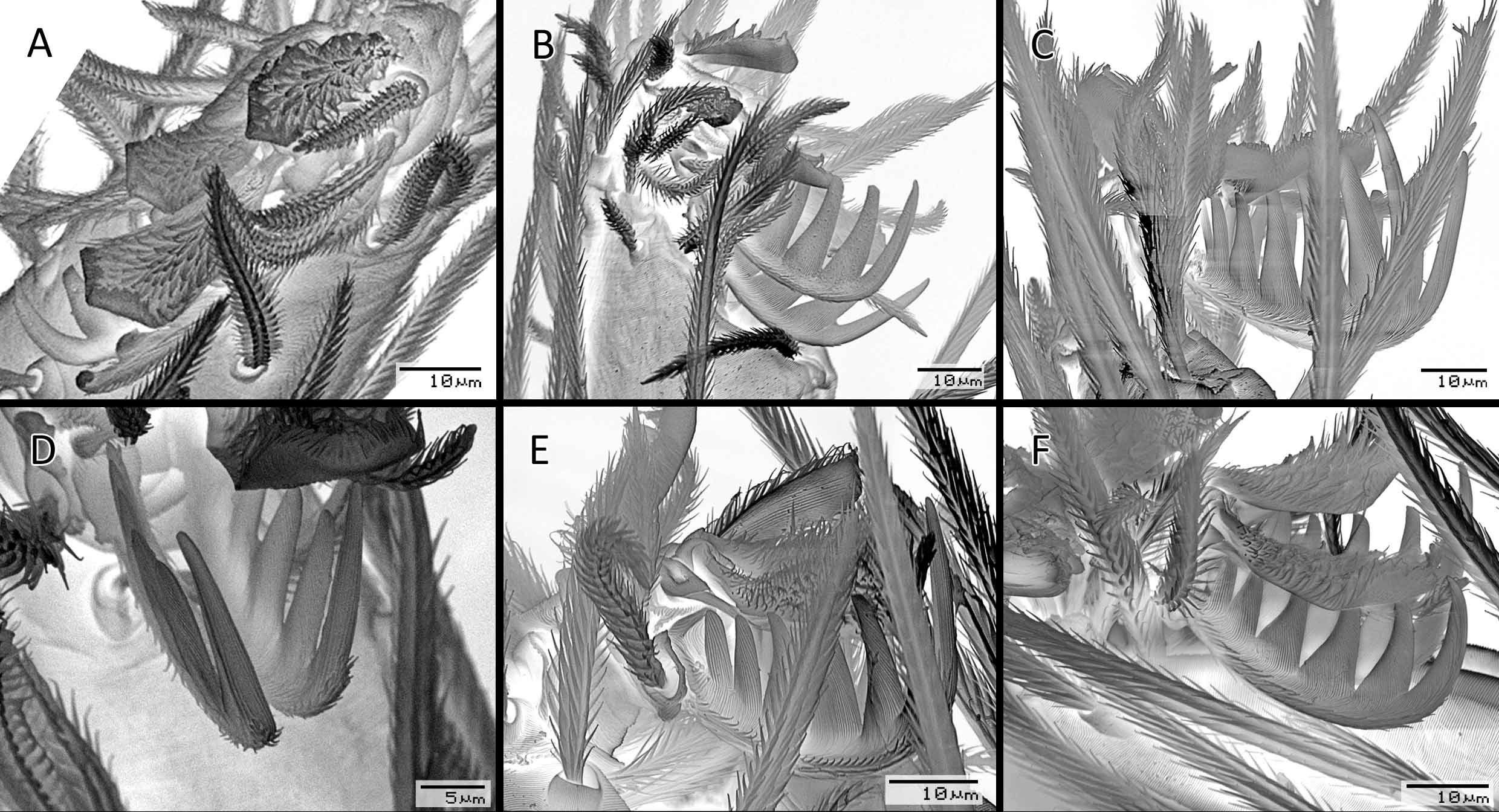
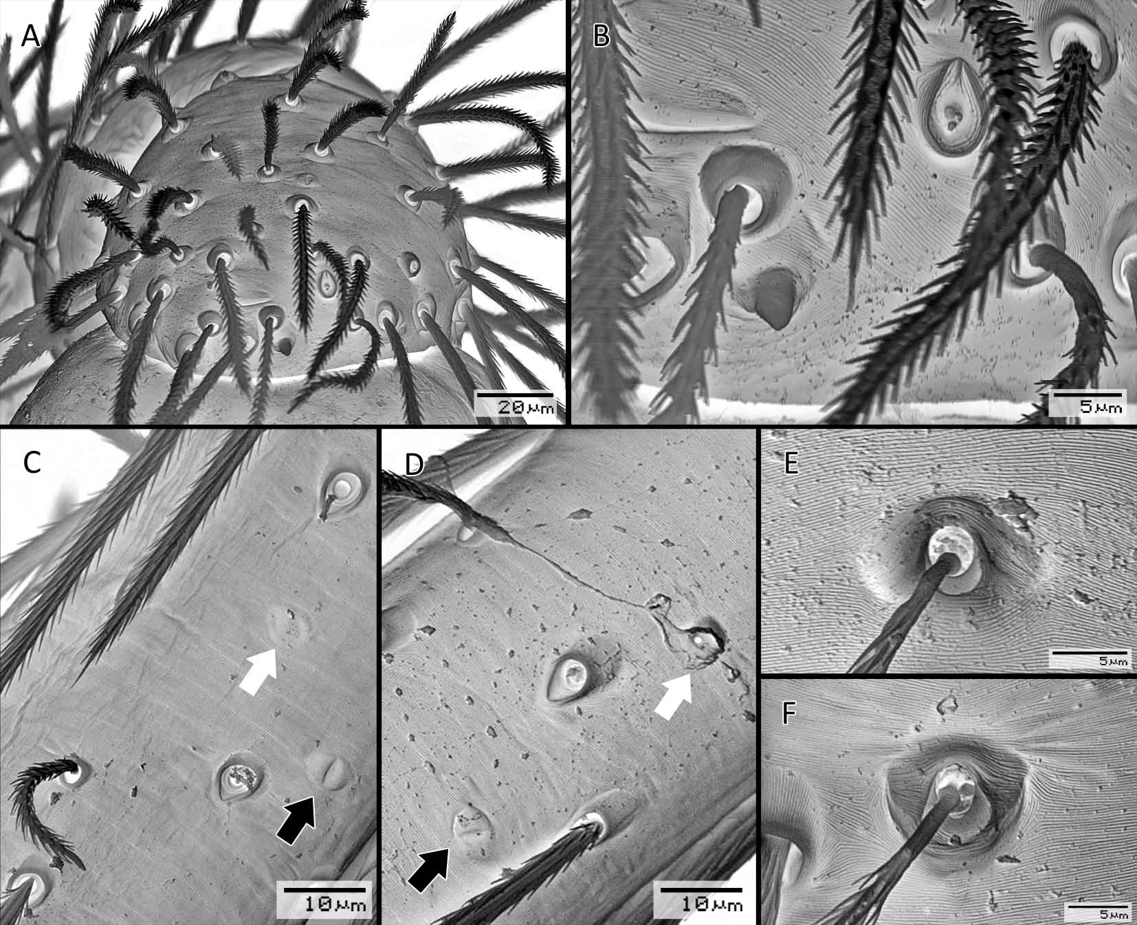
CEPHALOTHORAX. Carapace in vivo pale yellowish-orange (Fig. 1A–B) to pinkish, in ethanol yellowishwhite to pale orange ( Figs 2A–B View Fig. 2 , 3A–D), with dark net-shaped pattern and dark margins; non-marginal pars cephalica setae present in three rows, ovoid in dorsal view, pars cephalica strongly sloping in lateral view, reaching highest point at level of second coxa ( Fig. 2E View Fig. 2 ), anteriorly narrowed to 0.49 times its maximum width or less, with rounded posterolateral corners ( Fig. 2A–B View Fig. 2 ), surface of elevated portion of pars cephalica smooth, sides smooth, with radiating patches of smooth platelets ( Fig. 6A–B View Fig. 6 ); thorax without depressions, fovea absent, lateral margin undulate, smooth; pars cephalica setae needle-like. Clypeus margin curved downwards in front view ( Fig. 2G View Fig. 2 ), sloping forward in lateral view ( Fig. 2E View Fig. 2 ), high, ALE separated from edge of carapace by their radius or more. Eyes ( Fig. 2A, E View Fig. 2 ): six, well developed, all subequal, ALE oval, PME circular, PLE circular; posterior eye row recurved from both above and front; ALE separated by their radius to diameter, ALE–PLE separated by less than ALE radius, PME touching, PLE–PME separated by less than PME radius, ALE–PME separated by less than PME radius. Sternum ( Fig. 2C View Fig. 2 ) yellowish white, with faint radiating dark stripes, longer than wide, with radial smooth furrows between coxae I–II, II–III and III–IV, anterior margin with semicircular depression on the middle half, posterior margin extending posteriorly beyond anterior edges of coxae IV as single extension, distance between coxae approximately equal; setae abundant, evenly scattered, without hair tufts. Mouthparts: chelicerae, endites and labium yellowish white. Chelicerae ( Fig. 6F View Fig. 6 ) straight; without teeth on both promargin and retromargin; fangs directed medially, paturon inner margin with scattered setae. Labium ( Figs. 2C View Fig. 2 , 6C–D, F) rectangular, fused to sternum, anterior margin indented at middle, basal corners with small circular depression, sclerotization as in sternum; with 6 or more setae on anterior margin, subdistal portion with unmodiFed setae. Endites ( Fig. 6C–G View Fig. 6 ) distally not excavated, serrula present in single row, anteromedian tip unmodiFed, posteromedian part unmodiFed, sclerotization as in sternum; anterior margin with ventral row of spatulate setae and dorsal row of distally pectinate setae, median margin with Fnely barbed setae.
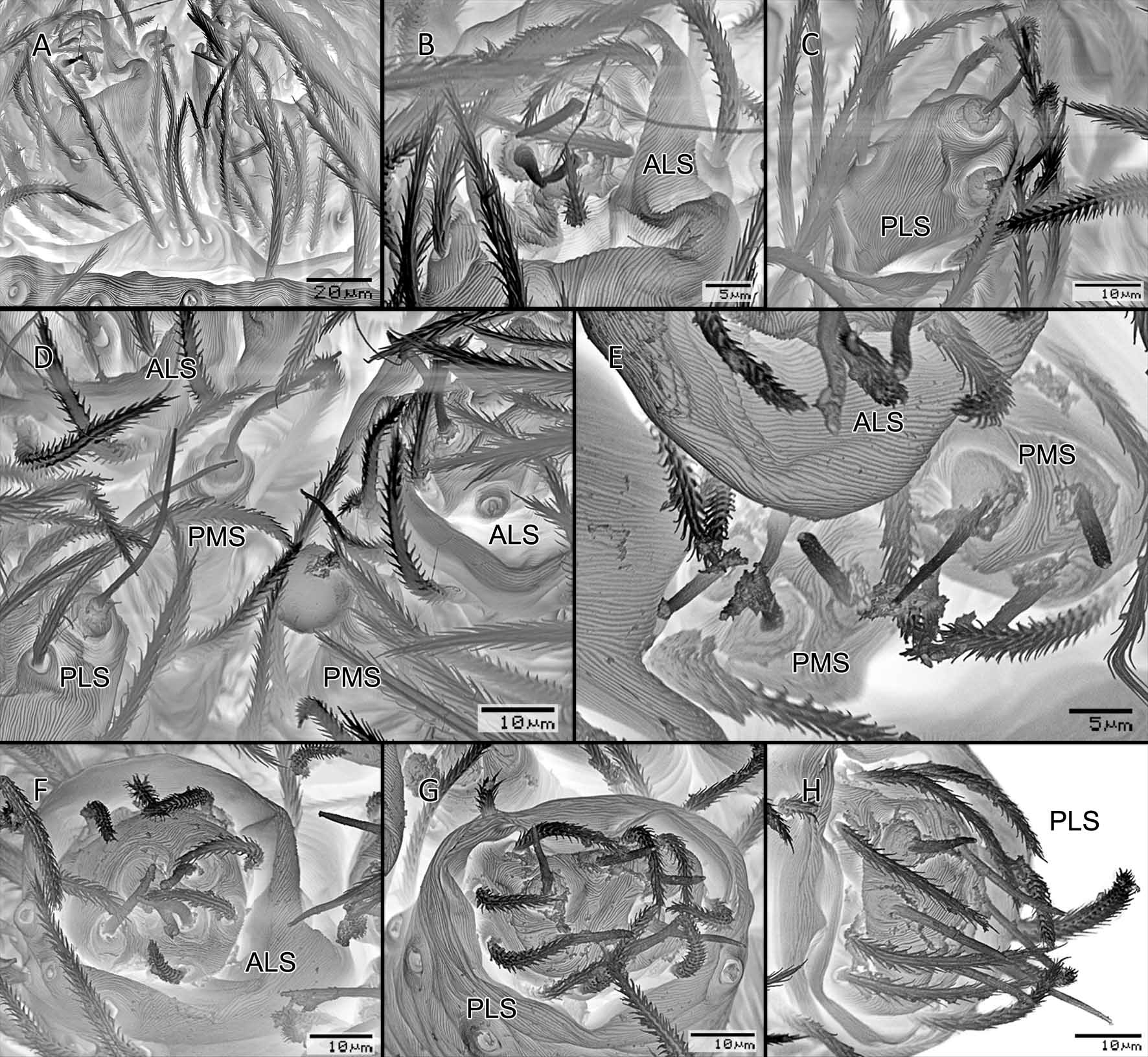
ABDOMEN ( Fig. 2A–B, D–F View Fig. 2 ). Ovoid, rounded posteriorly,without scuta; dorsum soft portions yellowbrown, without color pattern. Dorsum setae present, needle-like. Epigastric area setae uniform, needlelike. Postepigastric area setae present, needle-like. Pedicel with meshed texture ( Fig. 6C View Fig. 6 ) as in Orchestina (see Henrard & Jocqué 2012, Fgs. 21, 113, 114, 404), abdomen extending anteriad of pedicel. Colulus present ( Fig. 9A View Fig. 9 ), with four setae. Spinnerets ( Fig. 9B–D View Fig. 9 ): ALS with one major ampullate gland spigot emerging from cylindrical tubercle and two piriform gland spigots emerging from shallow tubercle; PMS with one minor ampullate gland spigot emerging from cylindrical tubercle; PLS with two aciniform gland spigots emerging from cylindrical tubercle.
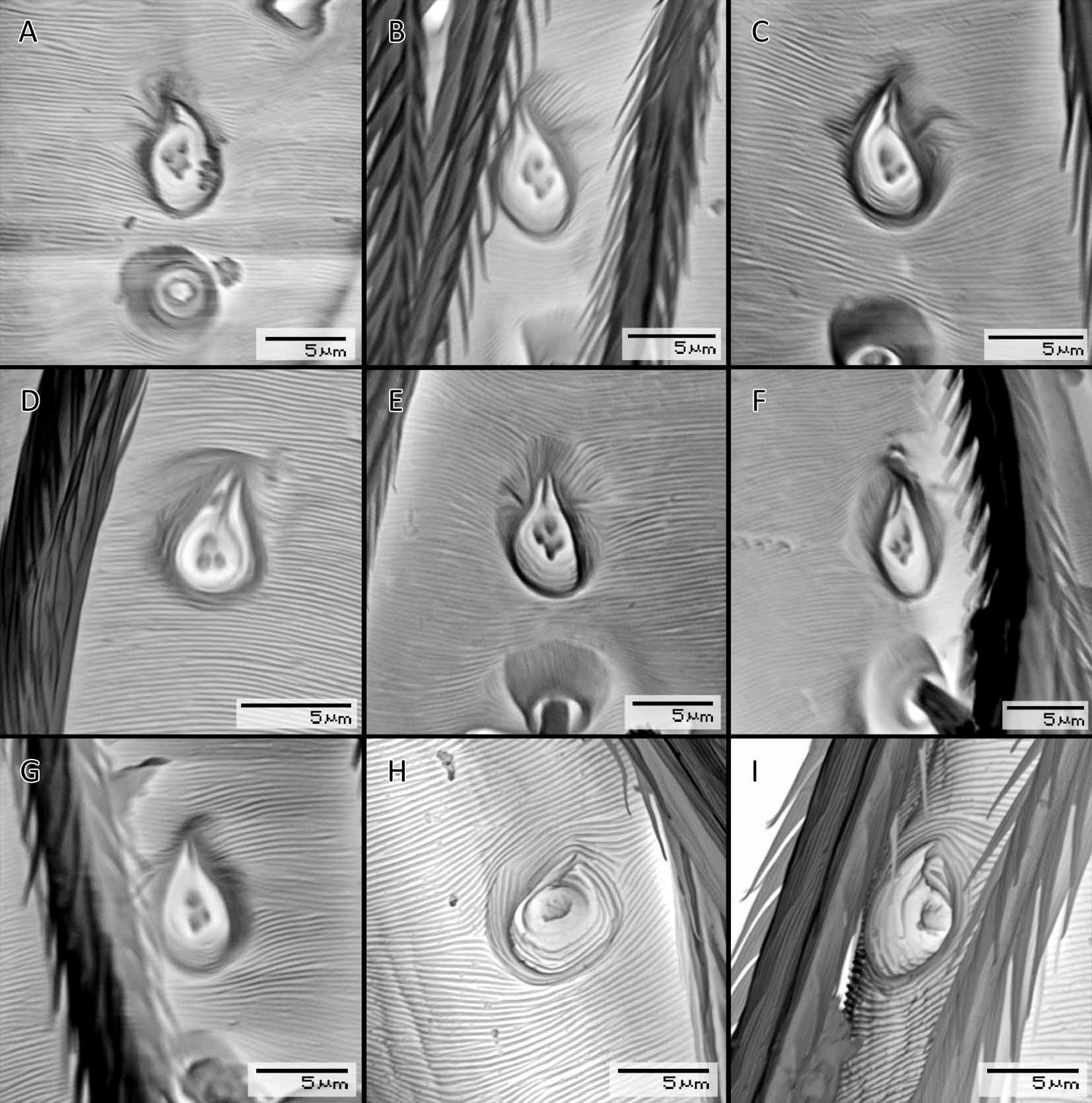
LEGS. White, without color pattern; femur IV not thickened, same size as femora I–III, patella plus tibia I longer than carapace, tibia I unmodiFed, metatarsi of at least leg II in the middle and IV at basal quarter, with eye-shaped smooth gland outlet with single pore ( Fig. 12C–E View Fig. 12 ). Leg spines absent. Tarsal claws ( Fig. 10A–F View Figs. 10 ) unipectinate, tarsal proclaws and retroclaws inner face smooth; tarsus I–IV superior claws with six teeth on lateral surface of proclaw, six teeth on lateral surface of retroclaw. Tarsi claw tuft with three carpeted setae. Tarsi I to IV without inferior claw. Trichobothria ( Fig. 12D–E View Fig. 12 ) with rounded bothrium, internal texture of aperture not grate-like, hood covered by numerous low, closely spaced ridges; sometimes accompanied by eye shaped structure with unknown function ( Fig. 12C–D View Fig. 12 ). Tarsal organ ( Fig. 11A–D, H–I View Fig. 11 ) pear-shaped, margin raised above surroundings, with 4 receptors on legs I and II, with 3 receptors on legs III and IV. In a probably unusual case, the tarsal organ has only 1 receptor on both legs IV ( Fig. 11H–I View Fig. 11 ), whereas I–III of the same specimen have the usual 4-4-3 formula (not illustrated). According to observations made on other oonopids the formula appears to be 3-3-2-2, since the distalmost receptor is distally biFd, but both parts stem from a common base as in Stenoonops Simon, 1891 ( Platnick & Dupérré 2010) .
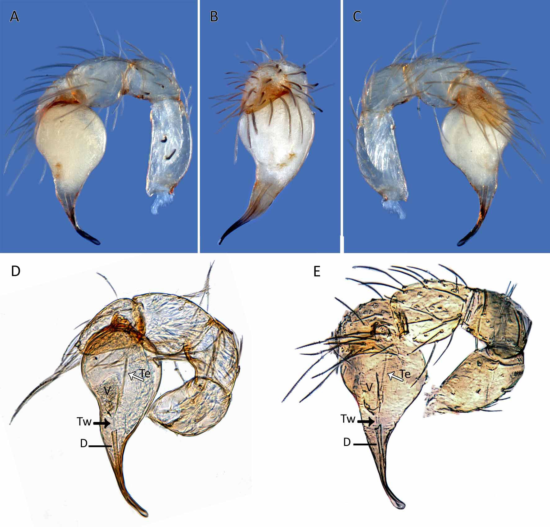
GENITALIA ( Figs. 4A–E View Fig. 4 , 7A–D). Epigastric region with sperm pore small, narrow, slit-like, situated at level of anterior spiracles. Palp normal size, not strongly sclerotized, right and left palps symmetrical, proximal segments white; trochanter normal size, unmodiFed; femur normal size, two or more times as long as trochanter, without posteriorly rounded lateral dilation, attaching to patella basally; patella shorter than femur, not enlarged, without prolateral row of ridges, setae unmodiFed; tibia ventral margin swollen; cymbium white, ovoid in dorsal view, not fused with bulb, not extending beyond distal tip of bulb, plumose setae absent, without stout setae, without distal patch of setae; bulb white, 1 to 1.5 times as wide as cymbium, stout, piriform with frontal margin slightly concave, tapering apically with long, medially bent embolus, distal sclerotized part of seminal duct (D) short, restricted to distal part, abruptly interrupted, internally with poorly deFned vesicle (V), opening into embolus through duct with basal part thin-walled (Tw), distal part sclerotized; tendon (Te) running from base of cymbium to distal end of vesicle (V); embolus simple, dark, tube-shaped, Fattened apically, tip blunt, grooved, appearing pale.
Female (Figs 1C–E, 2D, F–G)
As male except as noted.
CEPHALOTHORAX. Female palpal claw absent; spines absent. Tarsal organs ( Fig. 11A–D View Fig. 11 ) as in male.
ABDOMEN. Spinnerets ( Fig. 9E–H View Fig. 9 ):ALS with one major ampullate gland spigot emerging from cylindrical tubercle and three piriform gland spigots emerging from shallow tubercle; PMS with minor ampullate gland spigot and three aciniform gland spigots; PLS with six aciniform gland spigots.
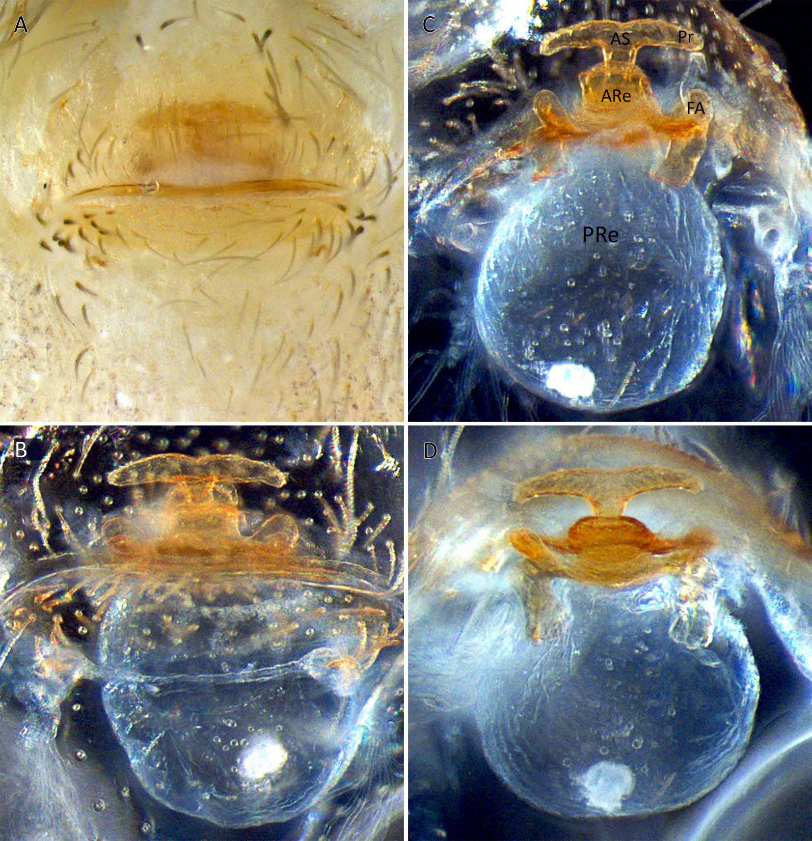
GENITALIA ( Figs 5A–D View Fig. 5 , 8A–G). Shape: anterior epigastric margin strongly sclerotized, preceded by T-shaped structure visible in transparency (AS), with larger, square part (ARe) in between, the latter provided at base with Fne transverse sclerite (T) ending in two faint procurved lobes (FA) extending as far as lateral protrusions (Pr) of frontal T-shape (AS). SEM view, details: anterior sclerite (AS) short, T-shaped, with lateral protrusions (Pr) well developed and provided with ramiFed ridges on anterior side and dorsally near extremities; AS connected with anterior receptaculum (ARe) antero-ventrally; ARe about half as wide as AS including Pr, square, provided dorsally and apically with rows of gland ducts (GD) conFned in frontal groove; ARe adjacent to transverse sclerite (T) ending laterally in well developed, bilobed, Fattened apodeme (FA), its dorsal extremity anchoring into the posterior receptaculum (PRe); PRe well developed, egg-shaped, emerging from base of ARe. Uterus externus not visible.
No known copyright restrictions apply. See Agosti, D., Egloff, W., 2009. Taxonomic information exchange and copyright: the Plazi approach. BMC Research Notes 2009, 2:53 for further explanation.