
Strongylostoma devleeschouweri, Steenkiste, Niels Van, Tessens, Bart, Krznaric, Kathleen & Artois, Tom, 2011
|
publication ID |
https://doi.org/ 10.5281/zenodo.201106 |
|
DOI |
https://doi.org/10.5281/zenodo.6182203 |
|
persistent identifier |
https://treatment.plazi.org/id/D9138784-697F-0067-FF41-FE4EFDEEFAAC |
|
treatment provided by |
Plazi |
|
scientific name |
Strongylostoma devleeschouweri |
| status |
sp. nov. |
Strongylostoma devleeschouweri View in CoL n. sp.
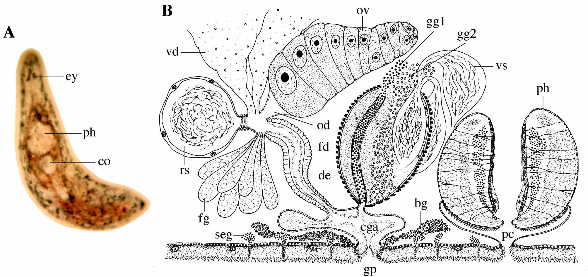
( Figs. 6 View FIGURE 6 A–6B)
Localities. Doñana National Park, Provincia de Huelva, Spain (36°58’48.80”N, 6°28’55.60”W). Laguna de Santa Olalla: submersed parts of sedges along the western shore of the lake (19/03/2008) (type locality).
Doñana National Park, Provincia de Huelva, Spain (36°49’27”N, 6°21’40”W). Llanos del Taraje near Ecomuseo Robledo de la Plancha: muddy temporal pond with Ranunculus aquatilis and sedges (25/03/2008).
Doñana National Park, Provincia de Huelva, Spain (37°04’23”N, 6°22’28”W). Laguna de la FAO: permanent water body with submersed vegetation (05/04/2008).
Material. Observations on several live animals. Eleven serially-sectioned specimens, one of which designated holoype ( MNCN, no. 4.01/55) and four designated paratype (HU, nos. 423–426). Six whole mounts.
Etymology. Species name in honour of Prof. Dr. Steven De Vleeschouwer, neurosurgeon at UZ Leuven, as a token of gratitude by the senior author (TA).
Description. The animals are 0.6–0.8 mm long measured in the serial sections. Brown, semilunar eyes are present at about 10%. The body shape is bluntly rounded anteriorly and more pointed posteriorly. The habitus is highly variable. Animals from Laguna de Santa Olalla are characterized by a conspicuous colouration. In general these specimens are relatively transparent, but the region coalescing with the position of the intestine has a red colouration. A very striking feature is the dense pattern of green spots, present throughout the entire animal. In contrast, the animals from Llanos del Taraje and Laguna de la FAO lack this green colouration.
The ciliated epidermis is syncytial, relatively thick (± 15 µm) and packed with small vacuoles. Nuclei are present in the basal part of the epidermis. The cilia measure about 7 µm. Tiny round dermal rhabdites lie on the apical side of the epidermis. Circular and longitudinal muscle layers are present under the basal membrane.
In the rostral part of the body, large rhabdite glands, originating just in front of the pharynx, form rod tracts between the eyes. They seem to secrete two different types of adenal rhabdites: small, darkly staining oblong rhabdites and larger eosinophilic rhabdites with a variable shape. In the specimens from Laguna de Santa Olalla subepidermal, darkly staining glands are also present dispersed over the entire body (seg in Fig. 6 View FIGURE 6 B). They secrete a basophilic granular secretion through the epidermis and are especially concentrated around the gonopore. These glands are absent in the specimens from Llanos del Taraje and Laguna de la FAO. Possibly they cause the green spots present in the animals from Laguna de Santa Olalla.
The mouth and pharynx are situated at about 30%. The organization and structure of the pharynx is identical with that of the other species of Strongylostoma Ørsted, 1844 (see e.g. Meixner 1915; Luther 1963). The nephridiopores are combined with the mouth.
Except for the position and structure of the testes, the internal organization strongly resembles that of Strongylostoma simplex Meixner, 1915 .
The paired testes are visible as two broad lateral bands that run from the front of the pharynx to the caudal body end. In nearly all specimens, both testes coalesce caudally, forming one large U-shaped mass. At about 60%, the vasa deferentia emerge from both testes to run anteriorly. Just behind the pharynx they broaden to form two large extracapsular seminal vesicles, lined with a membranous epithelium and filled with sperm. When entering the copulatory organ, both seminal vesicles narrow and join to form an intracapsular seminal vesicle.
The rest of the genital system strongly resembles that of Strongylostoma simplex simplex Meixner, 1915 (see Meixner 1915 for a detailed description). The copulatory organ consists of a large copulatory bulb, containing an intracapsular seminal vesicle, a sclerotized ejaculatory duct and two types of prostate glands. The intracapsular seminal vesicle is surrounded by granular, eosinophilic prostate glands, originating extracapsularly (gg 2 in Fig. 6 View FIGURE 6 B). The ejaculatory duct has smooth, sclerotized walls and is filled with a granular, basophilic secretion of prostate glands (gg 1 in Fig. 6 View FIGURE 6 B) and surrounded by plasmatic tissue containing nuclei in its proximal part. The distal part of the ejaculatory duct (described as “male genital duct” by Meixner 1915) penetrates the bulbus wall and empties into the common genital atrium through its dorsal wall. The copulatory bulb is surrounded by well-developed, spirally-running muscles.
The common genital atrium is relatively large and lined with a high, nucleated epithelium. Caudally a sack-like protrusion, variable in size, is present, resembling a sort of atrial bursa. It is, however, not separated from the rest of the common genital atrium, not by a sphincter nor by a different constitution or musculature of its wall, and therefore it cannot be considered as a true atrial bursa. In the specimens from Laguna de Santa Olalla, coarse-grained, basophilic glands (bg in Fig. 6 View FIGURE 6 B) empty into the gonopore, which is situated at around 50%.
Dorsocaudally the common genital atrium receives the female duct (described as “common duct” by Meixner 1915). This female duct is a relatively long, broad tube lined with the same epithelium as the common genital atrium and provided with strongly-developed circular muscles. Proximally it receives the oviduct, the vitelloducts, the seminal receptacle and the eosinophilic secretion of the female glands. The single ovary is relatively large in the animals from Laguna de Santa Olalla and smaller in the animals from the other localities. In live animals it was observed on the right side of the body, next to the centrally-located copulatory organ. The oviduct is broad and lined with a thin epithelium. The large, spherical seminal receptacle is filled with sperm and has a relatively high, nucleated epithelium. A short stalk, surrounded by a strong sphincter of several circular muscles, connects the seminal receptacle with the female duct.
Diagnosis. Species of Strongylostoma Ørsted, 1844 with very large, elongated testes, mostly coalescing at the rear end. Ejaculatory duct smooth, without spines. Seminal receptacle with strongly-developed sphincter around the stalk. Common genital atrium without division into upper and lower parts and having a large caudal protrusion. Subepidermal glands and green spots in one of the three populations.
Discussion. This species clearly belongs to the taxon Strongylostoma . According to different authors (e.g. Luther 1904, 1963; Graff 1913), Strongylostoma is characterized within the Typhloplanidae by the following features: nephridiopores combined with the mouth, absence of a copulatory atrium and presence of a seminal receptacle with a muscular stalk. However, S. simplex simplex (see further) lacks the muscular stalk of the seminal receptacle and complies therefore with the diagnosis of Typhloplanella Sekera, 1912 . It is nevertheless placed in Strongylostoma because of the presence of dermal rhabdites, a feature lacking in Typhloplanella (see Meixner 1915).
Several species within Strongylostoma are uncertain (“species dubiae”: S. gonocephalum , S. coecum , S. lanceolatum Sekera, 1912 , S. levandovskii Nasonov, 1924 , S. rocaseum Higley, 1918 ; see Marcus 1946). The welldescribed species of Strongylostoma ( S. cirratum , S. dicorymbum , S. elongatum , S. radiatum , S. simplex ) can be distinguished from each other by a number of conspicuous features: overall body form, construction and position of the testes and vitellaria, partition of the common genital atrium into an upper part receiving the male and female genital system and a lower part emptying into the gonopore, presence or absence of a copulatory bursa and construction of the copulatory organ (armature of the ejaculatory duct, prostate glands). S. devleeschouweri n. sp. is easily distinguished from the above-mentioned species (except for S. simplex ), by the lack of spines in the ejaculatory duct. Moreover, it differs from S. dicorymbum , S. elongatum and S. radiatum by lacking a typical copulatory bursa. All these species also have a bipartite common genital atrium, while S. cirratum even possesses a bipartite seminal receptacle.
The construction of the ejaculatory duct is often one of the most important diagnostic characters. In most species, this sclerotized duct is completely or partly lined with spines (see Discussion of S. elongatum ). In S. radiatum , it has a twofold construction with the longer part evacuating the prostate secretion and the minor “diverticulum” receiving sperm from the seminal vesicle (see Luther 1904). Other species ( S. dicorymbum , S. elongatum ) have a similar construction, but as opposed to Luther (1904, 1963), Marcus (1946) prefers to consider the smaller “diverticulum” as an ejaculatory duct and the larger part as a ductus granulorum. For S. cirratum , S. simplex and the new species from Andalusia, it is not clear from the descriptions or the observations on the serial sections where sperm enters the ejaculatory duct (see Meixner 1915; Luther 1963).
Considering all these diagnostic features, S. devleeschouweri n. sp. mostly resembles S. simplex simplex , which is considered a subtaxon of S. simplex together with S. simplex lapponicum Papi in Luther, 1963. The latter differs from S. s. simplex by the division of its common genital atrium into two parts. This feature is present in most other species of Strongylostoma , but is lacking in both S. s. simplex and in S. devleeschouweri n. sp. (see Luther 1963). Other similarities of S. devleeschouweri n. sp. with S. s. simplex are: body length, the smooth-walled ejaculatory duct and the absence of a copulatory bursa. However, S. devleeschouweri n. sp. differs from S. s. simplex by having a conspicuous green-spotted colouration, at least in the specimens from Laguna de Santa Olalla, the presence of granular eosinophilic prostate glands filling the copulatory bulb, the caudal protrusion of the common genital atrium and a strongly-developed sphincter surrounding the stalk of the seminal receptacle. Moreover, the specimens from Spain lack the different types of glands present in the frontal part of S. simplex simplex as described by Meixner (1915). Probably the most conspicuous difference with all known representatives of Strongylostoma is the construction of the testes. Although the position and volume varies in the different species, none of them has testes coalescing caudally. Because of the above-mentioned differences, the specimens from Spain are described as a new species.
In spite of the fact that there are some minor differences between the specimens from Laguna de Santa Olalla and those from Llanos del Taraje and Laguna de la FAO (see the description above), their internal organization is virtually identical. Therefore they are provisionally placed in the same species until additional morphological and/ or molecular data prove otherwise.
| MNCN |
Museo Nacional de Ciencias Naturales |
No known copyright restrictions apply. See Agosti, D., Egloff, W., 2009. Taxonomic information exchange and copyright: the Plazi approach. BMC Research Notes 2009, 2:53 for further explanation.