
Adelatelecrinus sulcatus
|
publication ID |
https://doi.org/ 10.11646/zootaxa.3681.1.1 |
|
publication LSID |
lsid:zoobank.org:pub:7F9B0117-90AC-471C-B98E-9001DF3BC455 |
|
DOI |
https://doi.org/10.5281/zenodo.5659109 |
|
persistent identifier |
https://treatment.plazi.org/id/D9378A50-8E5C-FFE1-FF0A-5575249C2A98 |
|
treatment provided by |
Plazi |
|
scientific name |
Adelatelecrinus sulcatus |
| status |
|
Adelatelecrinus sulcatus View in CoL AH Clark, 1912
Figure 7 View FIGURE 7
Atelecrinus sulcatus View in CoL AH Clark, 1912:152.—AH Clark, 1915:192 (fig. 123).
Atelecrinus wyvilli View in CoL : AH Clark and AM Clark, 1967: 819, 820–823 (part).—AM Clark, 1967:171 –172.
Holotype. NCB (V. ECH.C 2098), Siboga 85, Makassar Strait, 00º36’30”S, 119º29’30”E, 724 m, 17 Jun 1899.
Other material examined. Indonesia: USNM 36220, Albatross 5619, W of Halmahera I., 0º35’00”N, 127º14’40”E, 27 Nov 1909, 795 m (1 spec.). Nicobar Islands: ZSM (no catalog number), Deutschen Tiefsee- Expedition 210, 06º53’N, 93º33’E, 753 m (2; examined from photographs). Solomon Islands: MNHN IE- 2007- 7710, IE- 2007-7711, SALOMON 1 cruise, DW-1773, N of Malaita I., 08°11.0’S, 160°39.9’E, 331–397 m, 28 Sep 2001 (2 spec; [ IE- 2007-7710 dissociated for SEM]); MNHN IE- 2007-7712, SALOMON 1 cruise, CP-1802, off Guadalcanal I., 09°31.1’S, 160°35.0’E, 245–269 m, 2 Oct 2001 (1 partly crushed, dissociated for SEM).
Diagnosis. A species of Adelatelecrinus with brachitaxes and arm bases rounded and separated, with a small distolateral triangular tooth on Ibr1 and a small lateral flange or 1–2 weak teeth on Iax2; either or both may be rudimentary or absent on some rays.
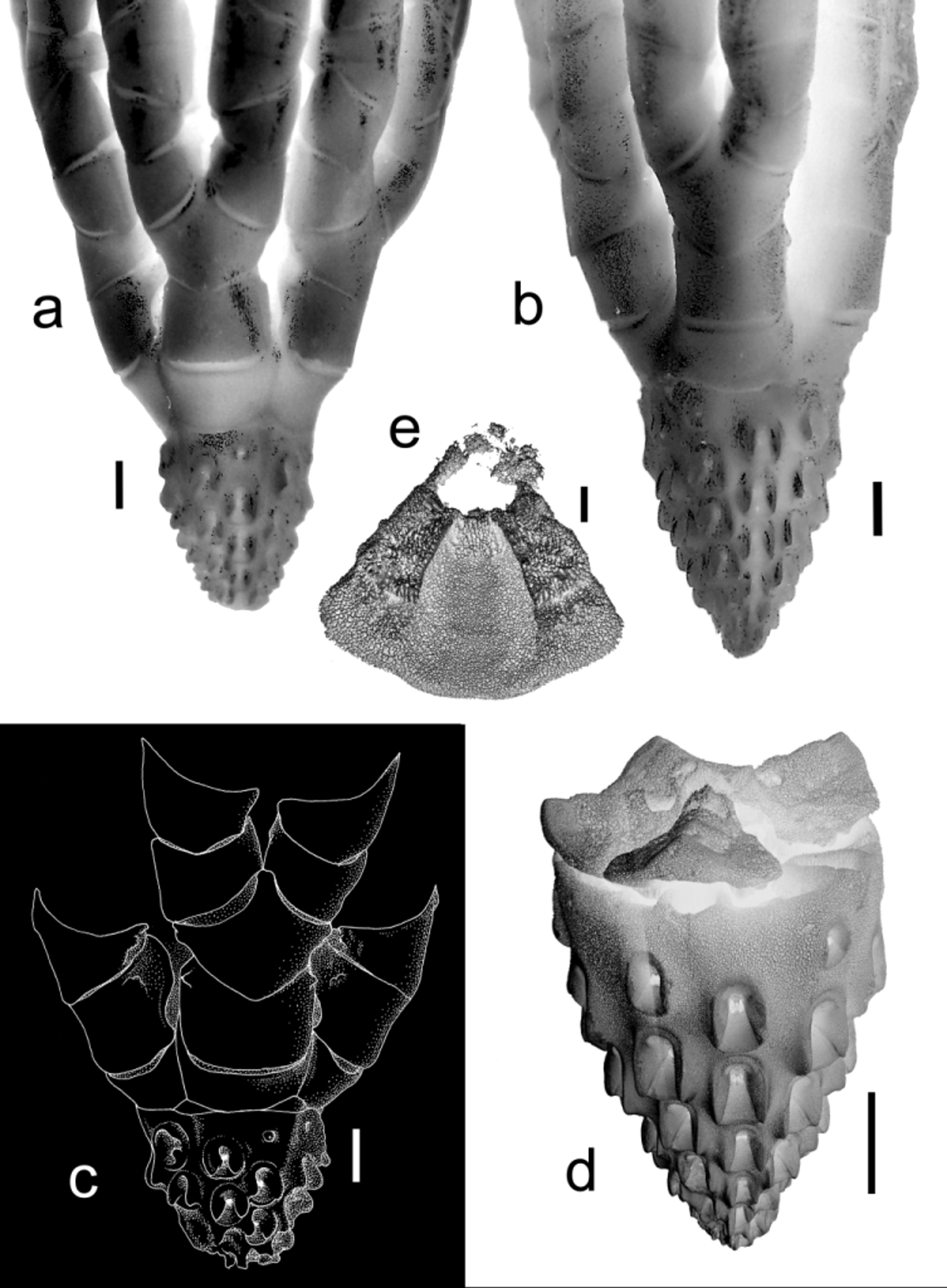
Redescription of the holotype ( Figure 7 View FIGURE 7 a). Centrodorsal conical, 2.85 mm across the base, 3.3 mm high; HD 1.17; base parallel-sided; tip broken with open hole at apex, 0.4 mm across. Cirrus sockets XXXII, 3–4 per column in 10 columns, no cirri retained. Fulcral tubercles moderately developed, triangular or very slightly hooked in profile. Interradial ridges weak but well defined, tapering toward apex, reaching level of second socket in each radial area. Centrodorsal margin very shallowly concave across radial areas, negligibly concave interradially. Centrodorsal/basal suture close; no space or ligament visible externally. Externally visible portion of basals triangular interradially, very slightly swollen; narrower and parallel-sided laterally; suture between adjacent basals barely visible, if at all. Radials oblong, slightly wider distally; radial profile ≤80º; WL 2.0; distal margin very broadly and shallowly U-shaped. IBr2 smoothly rounded aborally, with no synarthrial swelling; well separated laterally with well-developed roughly rhombic gap between adjacent rays (“water pore” in earlier terminology; Figure 2a). Ibr1 oblong with small triangular distolateral tooth; WL 1.9; distal margin shallow V-shaped. Iax2 hexagonal with diverging lateral margins; small rounded or triangular lateral tooth usually present; distal angle truncated; WL 1.1. IIbr1–2 separated interiorly by gap, slightly flattened exteriorly. IIbr1 slightly U-shaped; WL 1.6. IIbr2 irregularly quadrate, longer exteriorly; proximal margin shallow V-shaped; WL 1.3. IIbr3+4 rounded aborally, in contact interiorly, WL 1.2; diameter 1.8 mm. IIbr5 almost triangular, WL 1.7. Brachials following IIbr6+7 strongly wedge-shaped, WL 1.6–1.8. Syzygies at 6+7, 9+10.
New material. Centrodorsal conical, 3.5–3.8 mm across the base, 4.6 mm high; HD 1.2–1.3; tapering from base; apex sometimes with narrow ridges derived from obsolete apical sockets. Cirrus sockets XLV– LIIII, 4–6 per column in 10 columns. Fulcral tubercles moderately developed. Interradial ridges almost nonexistent to slight low swellings that reach apical end of first socket. Centrodorsal margin flat ( Figure 7 View FIGURE 7 c) or very shallowly concave across radial areas ( Figure 7 View FIGURE 7 b), slightly projecting and flat or shallowly V-shaped interradially. Centrodorsal/basal suture distinct, with slight to low triangular interradial gap. Externally visible portion of basals flat to slightly arched, triangular interradially, slightly swollen; narrower and parallel sided laterally; suture between adjacent basals visible externally ( Figure 7 View FIGURE 7 d) or not ( Figure 7 View FIGURE 7 c). In IE- 2007-7710, dissociated for SEM, the pair of internal curved projections meet at their tips, enclosing a circular space; those of adjacent basals are attached along their length; these projections together form a rosette-like structure roofing the centrodorsal cavity. Radials oblong; WL 2.0–2.2; radial profile 60º; distal margin almost straight to very broadly and shallowly U-shaped. IBr2 smoothly rounded aborally, with no synarthrial swelling; well separated laterally by roughly rhombic gap. Ibr1 oblong with small triangular distolateral tooth (sometimes lost); WL 1.5–1.7; distal margin shallow V-shaped. Iax2 hexagonal with diverging lateral margins, sometimes with small lateral tooth; distal angle short and rounded or truncated; WL 1.0–1.1. IIbr1–2 slightly flattened exteriorly, separated interiorly by roughly rhombic gap. IIbr1 almost oblong and longer exteriorly; WL 1.7–1.8. IIbr2 irregularly quadrate/pentagonal, longer exteriorly; proximal margin shallow Vshaped; WL 1.3. IIbr3+4 rounded aborally, in contact interiorly; WL 1.3; diameter 1.6–1.8 mm. IIbr5 almost triangular; WL 1.8–2.0. Brachials following 6+7 wedge-shaped; WL 1.4. IIbr8 and following brachials becoming less strongly wedge-shaped; IIbr19 with WL 1.1. Syzygies at 6+7, 9+10, 12+13, followed by intervals of 2 or 3 muscular articulations.
Distribution. From NW of Sumatra and the Makassar Strait to Halmahera, Indonesia, and the Solomon Islands in (?245) 269– 795 m.
Remarks. AH Clark (1912) distinguished Atelecrinus sulcatus (from At. wyvilli ) by its more sharply conical centrodorsal, with a shallow interradial furrow rather than a flat space between adjacent columns of sockets. Curiously, when he synonymized A. sulcatus under At. wyvilli (in Clark and Clark 1967), he added additional distinguishing features not mentioned in the original description: centrodorsal base lacking interradial indentations; sockets with stronger fulcral tubercles, and basals in close contact with both centrodorsal and radials. He attributed the narrow interradial gap filled with perisome [=noncalcareous integument (AH Clark 1915)] between the centrodorsal and each basal in the holotype of At. wyvilli to partial decalcification, and considered the other distinctions of minor importance. However, these gaps are natural; they expose interradial ligament bundles characteristic of a distinct genus, Paratelecrinus , described below.
The centrodorsal of the holotype (as of 15 August 2012) consists of three longitudinal fragments separated from the basal ring; two fragments bear interradial pits. The aboral surface of the basals lacks the complex articular features of Paratelecrinus , described below. Although the tip of the centrodorsal is now missing from USNM 36220, AH Clark (1915:192, figure 123) shows it entire, with cirri in columns of 4–5 sockets as in the holotype. [Although the figure legend states “from the Philippine Islands ”, it illustrates the Albatross specimen collected in northern Indonesia during the Albatross Philippine Expedition, 1907–1910.] This specimen differs from the holotype in having more strongly developed socket tubercles, basals visible externally as low triangles that do not meet midradially, a wider radial profile (~90°), proportionally shorter broader primibrachial ossicles, and some axils with a weak lateral flange or 1–2 (rather than just one) small flattened triangular teeth. USNM 36220 is also slightly larger (basal centrodorsal diameter 3 mm) than the holotype and has more complete rays with P1 on IIbr16. AH Clark (1912) and Clark and Clark (1967) described both specimens as having cirrus sockets of adjacent radial areas separated by a shallow interradial furrow. However, this is an effect of the adjacent high fulcral tubercles, not an actual groove in the centrodorsal surface. The differences are minor and the two specimens appear to belong to the same species.
AM Clark (1977) described two specimens collected NW of Sumatra by the Deutschen Tiefsee Expedition (sta. 210, 06º53’N, 93º33’E, 753 m) as At. wyvilli . Comments and photographs kindly provided by Thomas Heinzeller (Ludwig-Maximilians-Universität München) and Bernhard Ruthensteiner (Zoologische Staatssammlung München) show a slight interradial slit between the centrodorsal and basal ring as in the new material described herein. The longest remaining cirri (30 mm long with 29–31 cirrals) taper near the distal end with the penultimate segment much longer than wide, no opposing spine, and slightly bent, conical terminal claw, as in At. balanoides and At. helgae , and unlike the cirrus tips of Paratelecrinus species for which complete cirri are known (except for two specimens tentatively attributed to P. wyvilli ; see below). The specimens also agree with Adelatelecrinus sulcatus in having HD 1.2–1.3; weak interradial ridges at the centrodorsal base; the aboral or adoral rim of the peripheral cirrus sockets often raised above surrounding stereom; up to XLIII cirri; basals only slightly swollen interradially, if at all; a small triangular distolateral projection on Ibr1, and a weak lateral flange on the axil (AM Clark, 1977).
The holotype and USNM 36220 closely resemble the other specimens in all respects except in lacking any interradial gap or slit between centrodorsal and basal ring. In the Nicobar and Solomon Islands specimens, the suture is not tight and a small interradial gap exists. Dissociation of two of the latter reveals deep interradial pits in the centrodorsal rim. One has basals with a deep, smooth, aboral depression and a pair of curved internal projections that together form a rosette-like structure that roofs the centrodorsal cavity (not observable in the damaged specimen). The combination of features warrants separation of this species from Atelecrinus at the generic level.
Although the Solomon Islands specimens were collected in substantially shallower water (269–331 [possibly 245–397 m]) than the Nicobar and Indonesian material (753–795 m), no features appear to differ by depth, e.g., both the Solomon and Nicobar Islands specimens exhibit the gap between centrodorsal and basals.
No known copyright restrictions apply. See Agosti, D., Egloff, W., 2009. Taxonomic information exchange and copyright: the Plazi approach. BMC Research Notes 2009, 2:53 for further explanation.
|
Kingdom |
|
|
Phylum |
|
|
Class |
|
|
Order |
|
|
Family |
|
|
Genus |
Adelatelecrinus sulcatus
| Messing, Charles G. 2013 |
Atelecrinus wyvilli
| Clark 1967: 819 |
| Clark 1967: 171 |
Atelecrinus sulcatus
| Clark 1915: 192 |
| Clark 1912: 152 |